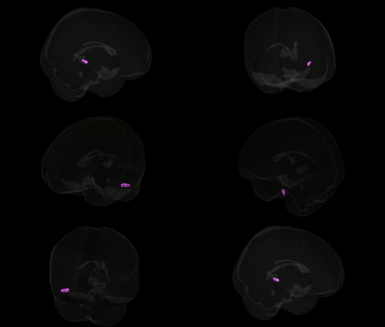
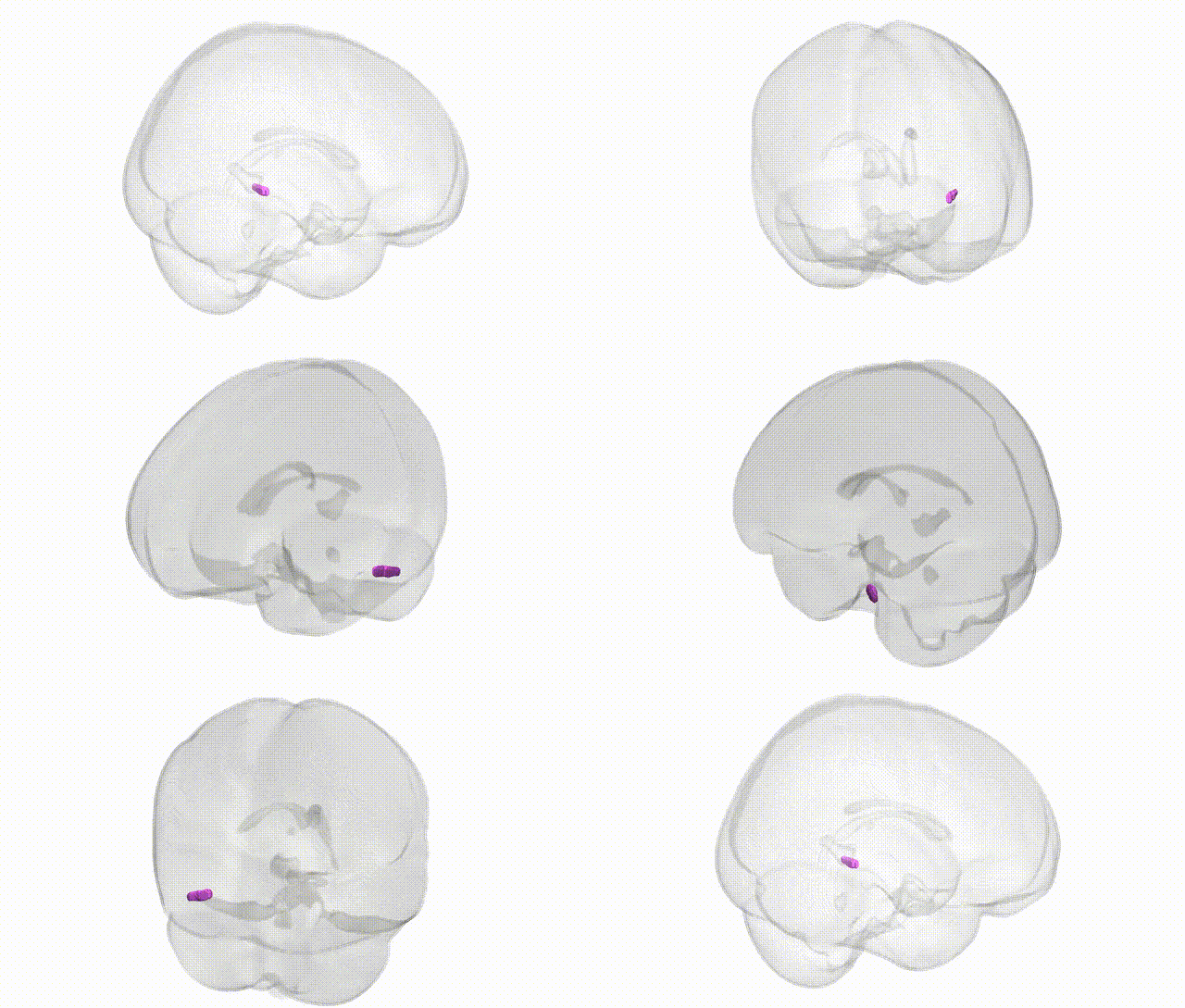
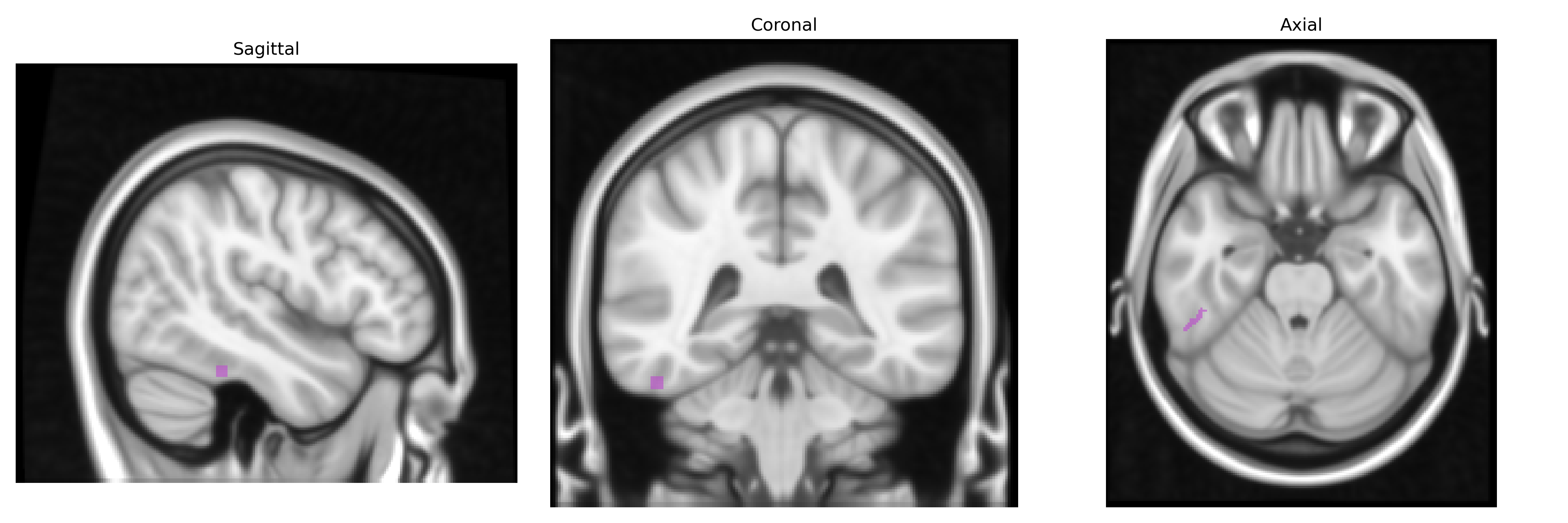
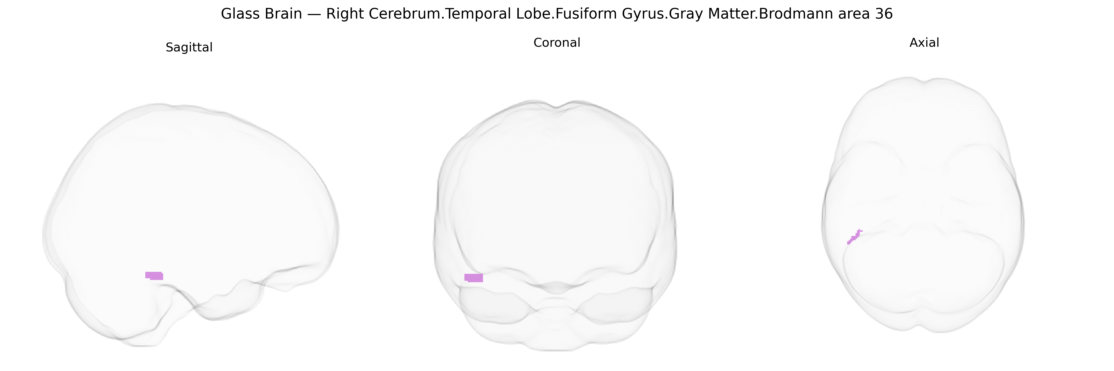
The bilateral Right Cerebrum.Temporal Lobe.Fusiform Gyrus.Gray Matter.Brodmann area 36 corresponds to a cytoarchitectonic subdivision of the parahippocampal–fusiform region located on the ventromedial surface of the temporal lobe, adjacent to the collateral sulcus and bordering Brodmann area 35 and parts of the fusiform and parahippocampal gyri. This cortex is part of the medial temporal lobe memory system and is implicated in high‑level visual processing, including object and complex scene recognition, as well as in familiarity-based recognition memory and the integration of visual input with contextual and associative information. Brodmann area 36 shows strong connectivity with the hippocampal formation, entorhinal cortex, perirhinal cortex, and higher-order visual association areas, supporting roles in declarative memory, semantic processing, and the transformation of perceptual representations into long-term memory traces. There is no direct link for Brodmann area 36; a related structure is the Parahippocampal gyrus.
Brodmann area 36 in the fusiform gyrus of the right temporal lobe, a key region for high‑level visual processing (including face and object recognition), has been implicated in several genetically influenced traits and disorders, although most GWAS do not target BA36 specifically but rather fusiform or temporal lobe gray matter volumes and activation patterns more broadly. Structural and functional variation in the fusiform gyrus has been associated in imaging‑genetics studies with common variants in genes such as BDNF (e.g., Val66Met), COMT, and DISC1, influencing cortical thickness, activation during memory and visual tasks, and risk for schizophrenia and major depressive disorder. GWAS of regional cortical thickness and surface area (e.g., ENIGMA consortium) have identified loci near genes involved in neurodevelopment and synaptic function—such as KIAA0586, PAX6, and microtubule‑related genes—associated with temporal and fusiform morphology, which partly overlap risk architecture for autism spectrum disorder, schizophrenia, and cognitive performance. Fusiform/BA36 structure and connectivity have also been tied to genetic risk for autism via rare and common variants in synaptic and neurodevelopmental genes (e.g., SHANK3, NRXN1, CNTNAP2), consistent with robust fusiform abnormalities in ASD and social cognition deficits. Additional GWAS and polygenic risk score studies link right fusiform and parahippocampal regions to face memory, general cognitive ability, and reading/word recognition, with associations to variants in genes affecting glutamatergic signaling and axon guidance; however, specific, high-confidence, BA36‑restricted genetic associations remain limited, and most evidence reflects broader fusiform/temporal lobe measures rather than fine‑grained Talairach atlas subdivisions.
Overview generated by GPT-4o (2026).
Region ID: 166
Hemisphere: bilateral
Atlas: Talairach labels 1mm
Full Quality Version: Download MP4
Full Quality Version: Download MP4
Wali Sidiqyar*, Gaurav Rudravaram*, Elyssa M. McMaster, Trent M. Schwartz, Adam M. Saunders, Kurt G. Schilling, Bennett A. Landman "Introducing SPINS: A Shared Public Visualization Library of Neuroanatomical Structures." Medical Imaging with Deep Learning- short paper
This resource is licensed under CC0 1.0 Universal (Public Domain).