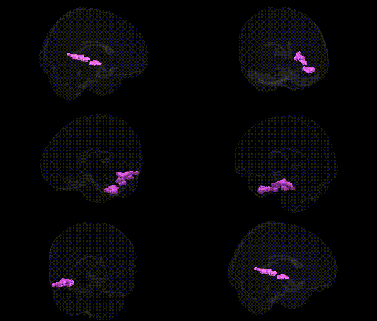
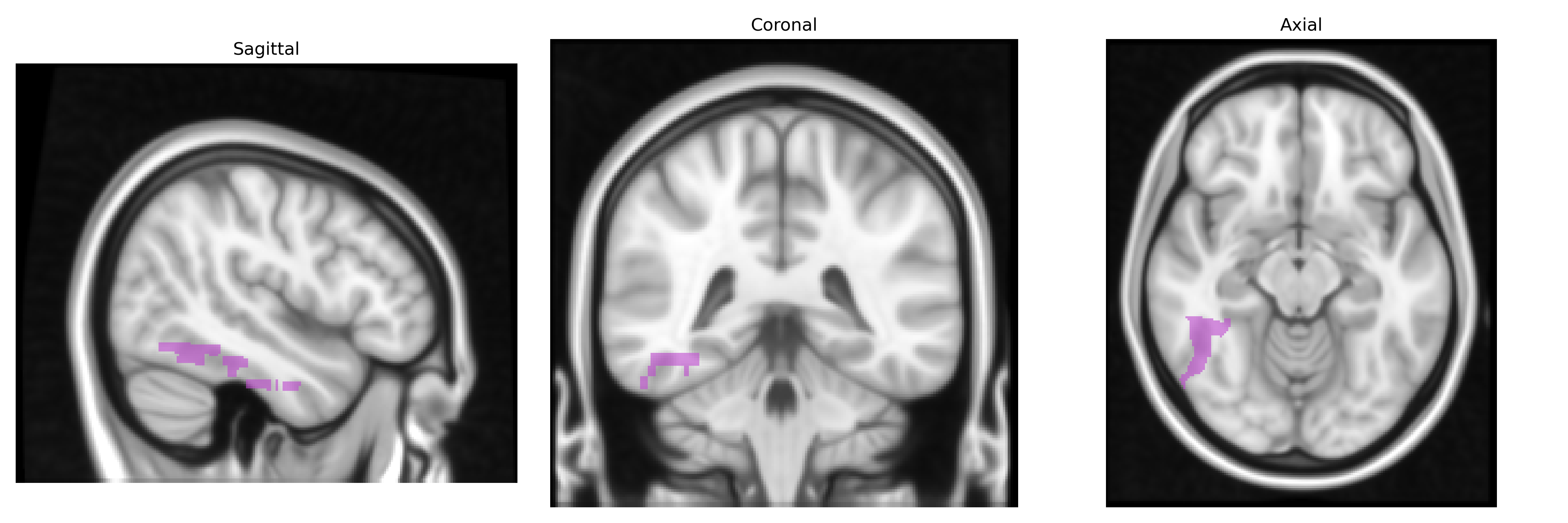
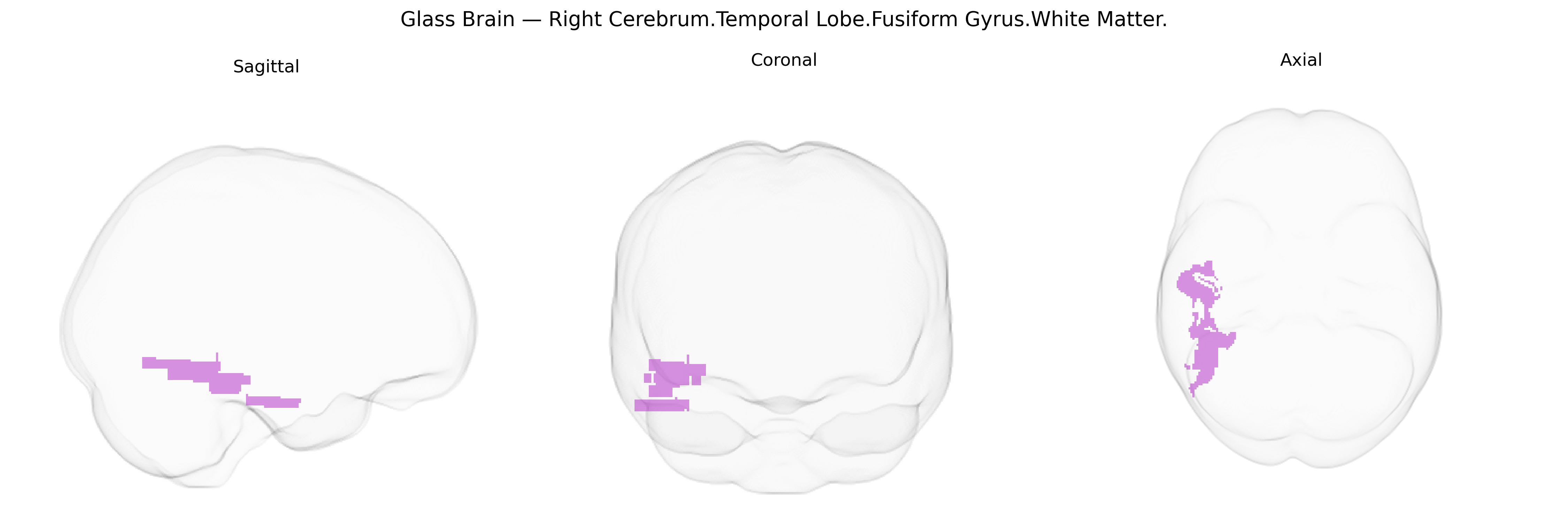
The bilateral Right Cerebrum.Temporal Lobe.Fusiform Gyrus.White Matter corresponds to the subcortical fiber pathways underlying the fusiform gyrus in the right temporal lobe, part of the ventral visual stream. This white matter region contains association fibers interconnecting fusiform cortex with adjacent temporal, occipital, and limbic areas, supporting complex visual processing such as object and face recognition, as well as higher-order integration of visual information with memory and semantic systems. It contributes to the structural connectivity that underlies functions frequently attributed to the fusiform gyrus, including aspects of visual expertise and category-specific processing. There is no direct link for this specific white matter subdivision; a related cortical structure is the fusiform gyrus: Fusiform gyrus.
Genetic associations involving the bilateral right fusiform gyrus white matter (Talairach “Right Cerebrum.Temporal Lobe.Fusiform Gyrus.White Matter”) arise mainly from imaging‑genetics and GWAS studies that link variants to fusiform structure, connectivity, and related cognitive or clinical phenotypes. Common variants in genes affecting neurodevelopment and synaptic plasticity—such as BDNF, NRG1, DISC1, and CNTNAP2—have been associated with white matter integrity and temporo‑occipital tract microstructure that encompass fusiform pathways, often via diffusion MRI metrics (e.g., fractional anisotropy) in tracts like the inferior longitudinal fasciculus. Large neuroimaging GWAS (e.g., ENIGMA, UK Biobank) report associations between polygenic risk scores for schizophrenia, bipolar disorder, autism spectrum disorder, and major depression and altered fusiform or adjacent temporal white matter, implicating shared polygenic architecture influencing cortical thickness and white matter organization in this region. Autism and dyslexia imaging‑genetics work has linked variants in genes related to neuronal migration and axon guidance (e.g., KIAA0319, DCDC2, ROBO family) to altered fusiform and ventral temporal white matter, consistent with the fusiform’s role in face and word recognition. Face‑processing and fusiform “face area” function have been tied indirectly to genetic variation through heritability studies showing substantial genetic contribution to fusiform activation patterns, and specific SNP associations (e.g., in oxytocin pathway genes such as OXTR) have been related to social perception and fusiform responsiveness. Alzheimer’s disease and general cognitive ability GWAS also point to polygenic influences on temporal and fusiform white matter atrophy, with APOE ε4 and other dementia‑risk variants contributing to degeneration of tracts passing through this region. Overall, current evidence supports a highly polygenic influence on fusiform white matter structure and function, with no single variant uniquely specific to this Talairach‑defined area, but with convergent genetic risk across neurodevelopmental, psychiatric, and neurodegenerative disorders impacting this region’s microstructure and connectivity.
Overview generated by GPT-4o (2026).
Region ID: 131
Hemisphere: bilateral
Atlas: Talairach labels 1mm
Full Quality Version: Download MP4
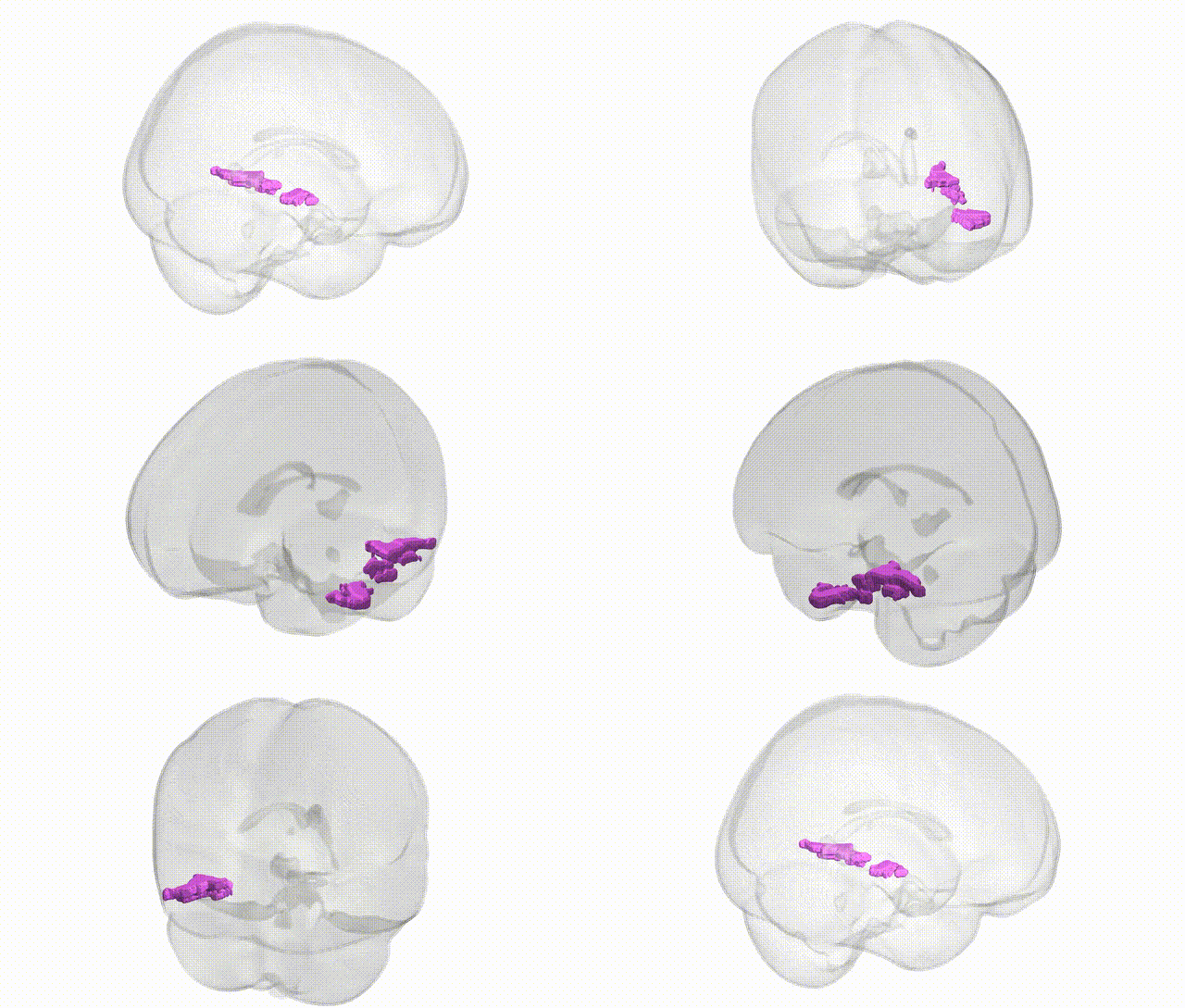
Full Quality Version: Download MP4
Wali Sidiqyar*, Gaurav Rudravaram*, Elyssa M. McMaster, Trent M. Schwartz, Adam M. Saunders, Kurt G. Schilling, Bennett A. Landman "Introducing SPINS: A Shared Public Visualization Library of Neuroanatomical Structures." Medical Imaging with Deep Learning- short paper
This resource is licensed under CC0 1.0 Universal (Public Domain).