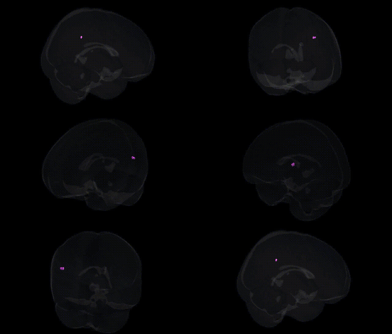
The bilateral Right Cerebrum.Temporal Lobe.Inferior Parietal Lobule.White Matter corresponds to subcortical association fiber pathways located deep to the cortical inferior parietal lobule at the parietotemporal junction in the right hemisphere. This region contains major components of the superior longitudinal/arcuate and related association fasciculi that interconnect temporal, parietal, and frontal association cortices, supporting multimodal integration of auditory, visual, and somatosensory information, as well as higher cognitive functions such as language processing, spatial attention, and aspects of working memory. As part of the inferior parietal lobule’s white matter infrastructure, it provides critical conduits for information flow between the Inferior Parietal Lobule and adjacent temporal and frontal regions.
The bilateral Right Cerebrum.Temporal Lobe.Inferior Parietal Lobule.White Matter region in the Talairach 1 mm atlas corresponds roughly to association white-matter pathways connecting inferior parietal and temporal cortices, including major tracts such as portions of the superior longitudinal fasciculus/arcuate fasciculus and nearby segments of the inferior fronto‑occipital fasciculus; GWAS and imaging‑genetics studies implicate this territory in polygenic influences on white‑matter microstructure (e.g., fractional anisotropy, mean diffusivity) and cognitive traits, with robust but distributed effects from genes involved in axon guidance, myelination, and synaptic plasticity (e.g., variants in or near genes such as CNTNAP2, NTRK1/2, MAG, and LINGO1 reported in large diffusion MRI GWAS of “g‑factor” and tract integrity). Polygenic scores for schizophrenia, bipolar disorder, major depressive disorder, and autism spectrum disorder have been associated with altered white‑matter integrity in temporo‑parietal association pathways overlapping this region, consistent with broader evidence that risk loci in pathways like neuregulin–ErbB, glutamatergic signaling (e.g., GRIN2A, GRIA genes), and immune‑related genes (e.g., MHC region) contribute to widespread association‑cortex connectivity changes rather than region‑specific effects. Additionally, GWAS of educational attainment, general intelligence, and reading/language abilities report that polygenic influences on these traits partly manifest as structural differences in temporo‑parietal association white matter, including arcuate/superior longitudinal fasciculus segments coursing through this area, though individual SNP or gene effects are small and highly polygenic. Overall, current genetic associations with this specific Talairach‑defined region are indirect and largely inferred from tract‑based or voxelwise diffusion MRI GWAS and psychiatric or cognitive imaging‑genetics studies, indicating that the region participates in widely distributed, highly polygenic networks rather than harboring unique, region‑specific genetic effects.
Overview generated by GPT-4o (2026).
Region ID: 876
Hemisphere: bilateral
Atlas: Talairach labels 1mm
Full Quality Version: Download MP4
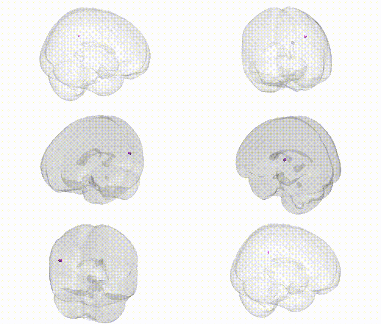
Full Quality Version: Download MP4
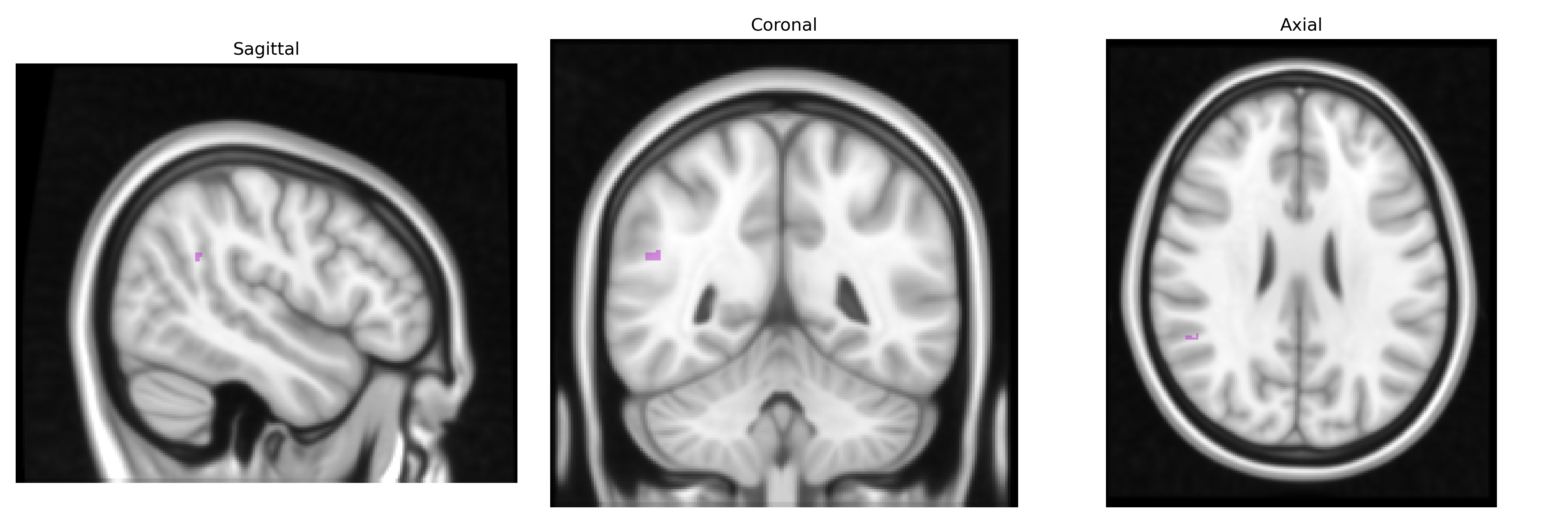
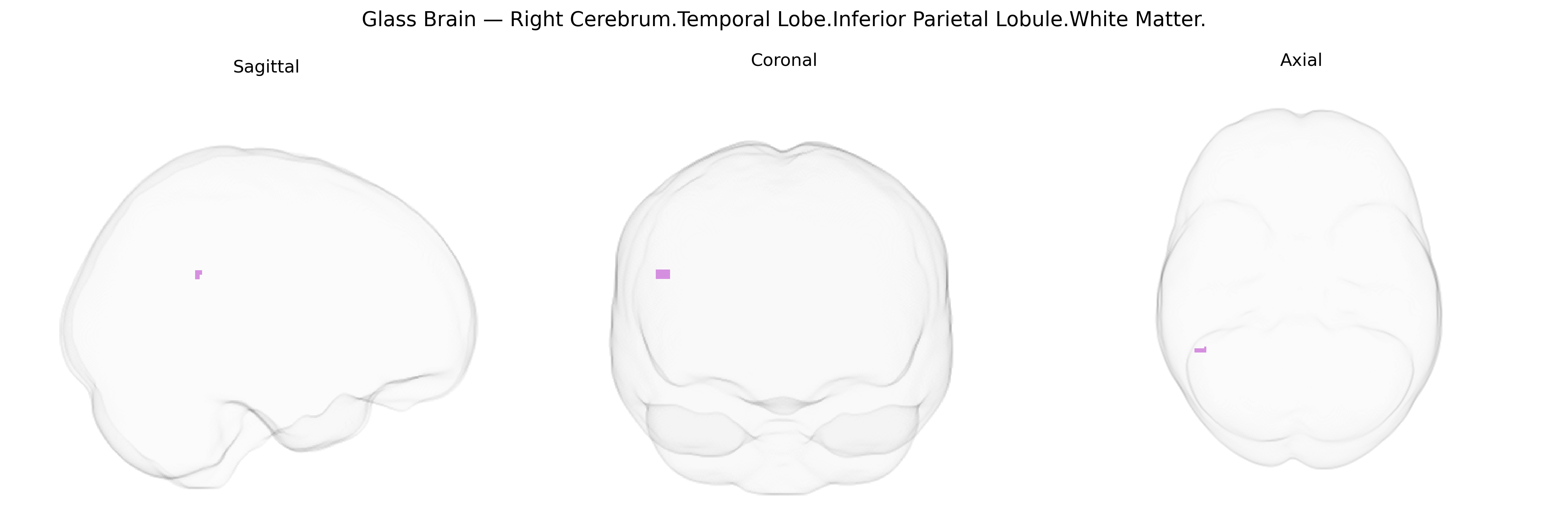
Wali Sidiqyar*, Gaurav Rudravaram*, Elyssa M. McMaster, Trent M. Schwartz, Adam M. Saunders, Kurt G. Schilling, Bennett A. Landman "Introducing SPINS: A Shared Public Visualization Library of Neuroanatomical Structures." Medical Imaging with Deep Learning- short paper
This resource is licensed under CC0 1.0 Universal (Public Domain).