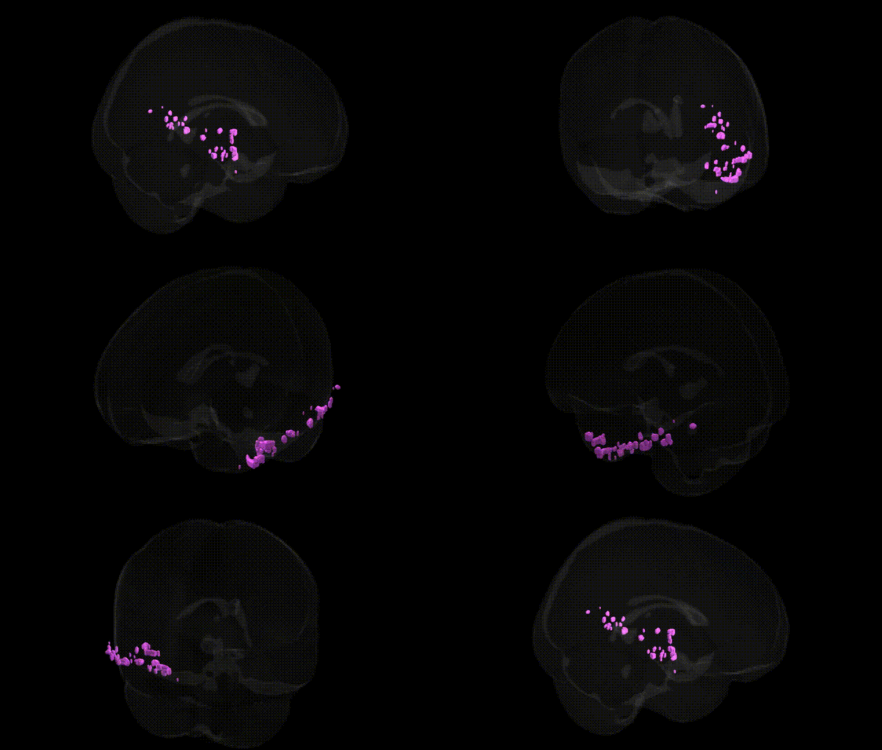
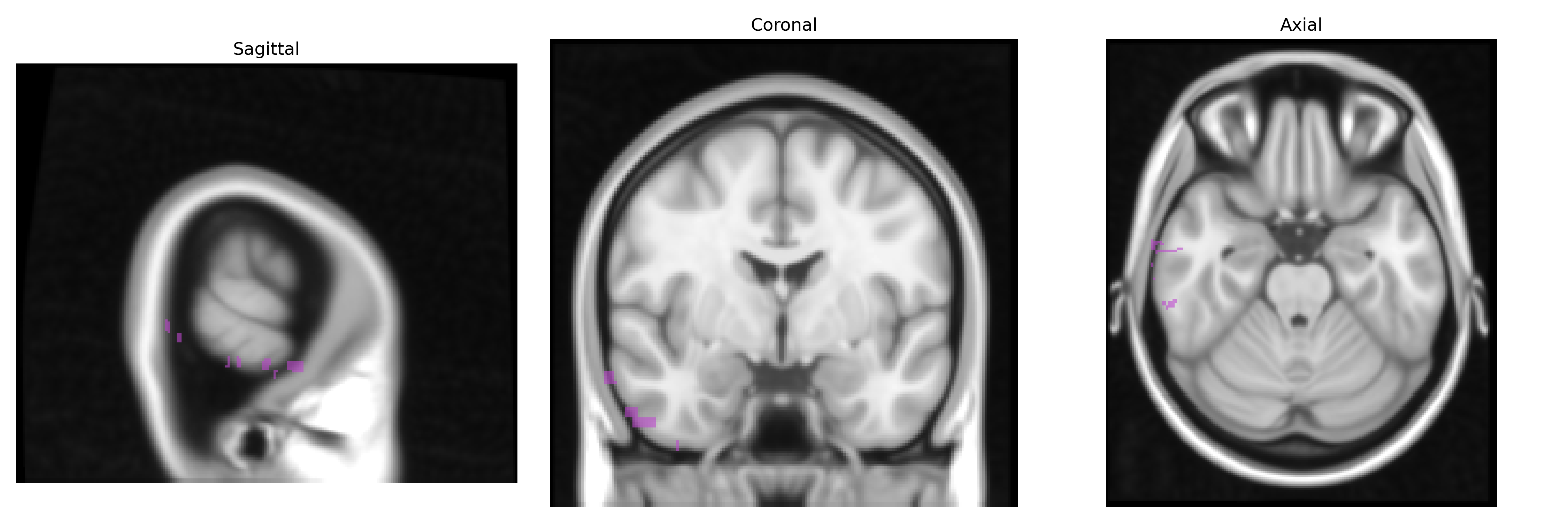
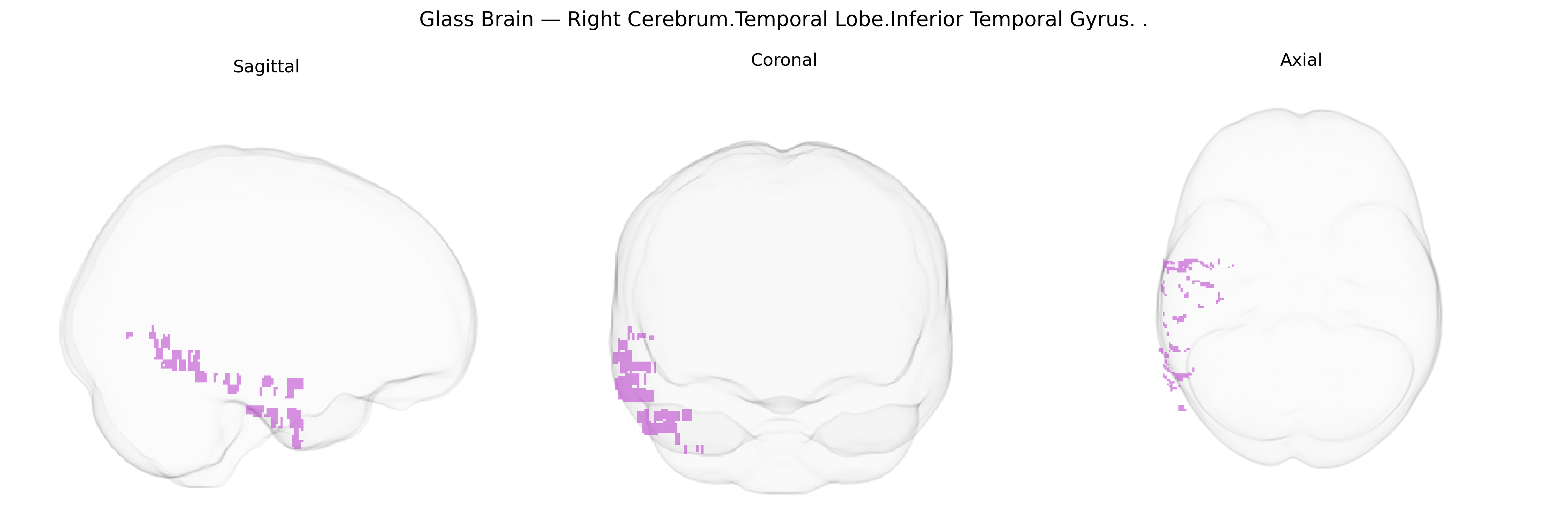
The bilateral Right Cerebrum.Temporal Lobe.Inferior Temporal Gyrus corresponds to cortex on the ventrolateral aspect of the temporal lobe, situated inferior to the middle temporal gyrus and extending from the temporal pole posteriorly toward the occipitotemporal region. This gyrus participates in high-level visual processing, including object, shape, and complex pattern recognition, and contributes to the ventral “what” visual stream. It is involved in integrating visual inputs with semantic and memory-related processes, supporting recognition of faces, objects, and categories, and is interconnected with occipital visual areas, medial temporal memory structures, and prefrontal association cortices. Functionally and anatomically, it is closely related to regions implicated in visual agnosias and category-specific recognition deficits when damaged. Inferior temporal gyrus
The inferior temporal gyrus (ITG) of the right and left temporal lobes, as defined in the Talairach 1 mm atlas, has been implicated in several genetic and GWAS findings through its roles in high-level visual processing, semantic memory, and social cognition. Large imaging-genetics consortia (e.g., ENIGMA, UK Biobank) have identified common variants influencing cortical thickness, surface area, and volume in lateral and inferior temporal regions, including ITG, with notable contributions from genes involved in neurodevelopment and synaptic function such as HMGA2, MIR548F3, and variants in loci on chromosomes 3p, 6q, and 12q linked broadly to temporal lobe morphometry. ITG structural and functional alterations have been associated with polygenic risk for Alzheimer’s disease (e.g., APOE ε4 and other AD GWAS loci), frontotemporal dementia, and schizophrenia, with temporal lobe volume and cortical thinning serving as intermediate phenotypes connecting these risk variants to disease expression. GWAS of facial recognition, reading and language-related traits, and autism spectrum conditions have identified variants in genes involved in axon guidance, neuronal migration, and synaptic plasticity that correlate with activation or morphology of inferior and lateral temporal cortex, consistent with ITG’s role in object and face processing and semantic integration. Additionally, genetic variants associated with epileptogenicity in temporal lobe epilepsy (including loci near SCN1A, LGI1, and other ion-channel or synaptic genes) show convergent effects on mesial and lateral temporal structures, with some imaging-genetic work indicating that ITG volume and connectivity patterns are partly heritable and modulated by such risk alleles, although specific ITG-focused GWAS hits remain less frequent than those reported for broader temporal or whole-cortex measures.
Overview generated by GPT-4o (2026).
Region ID: 12
Hemisphere: bilateral
Atlas: Talairach labels 1mm
Full Quality Version: Download MP4
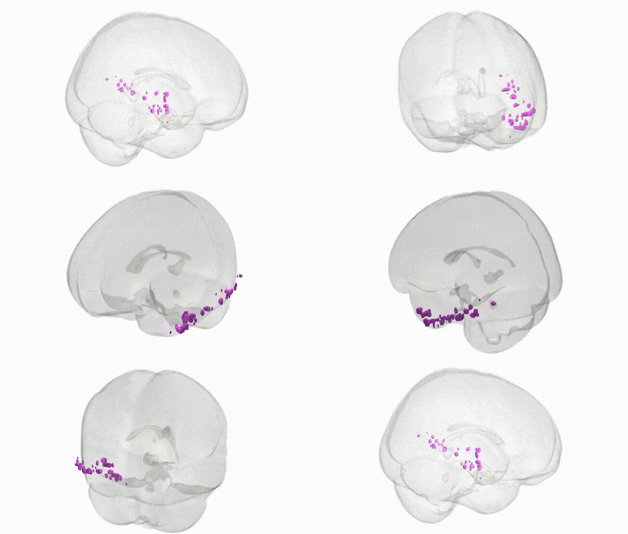
Full Quality Version: Download MP4
Wali Sidiqyar*, Gaurav Rudravaram*, Elyssa M. McMaster, Trent M. Schwartz, Adam M. Saunders, Kurt G. Schilling, Bennett A. Landman "Introducing SPINS: A Shared Public Visualization Library of Neuroanatomical Structures." Medical Imaging with Deep Learning- short paper
This resource is licensed under CC0 1.0 Universal (Public Domain).