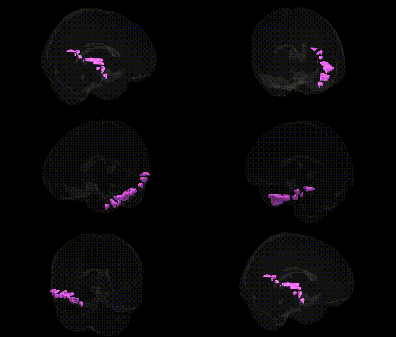
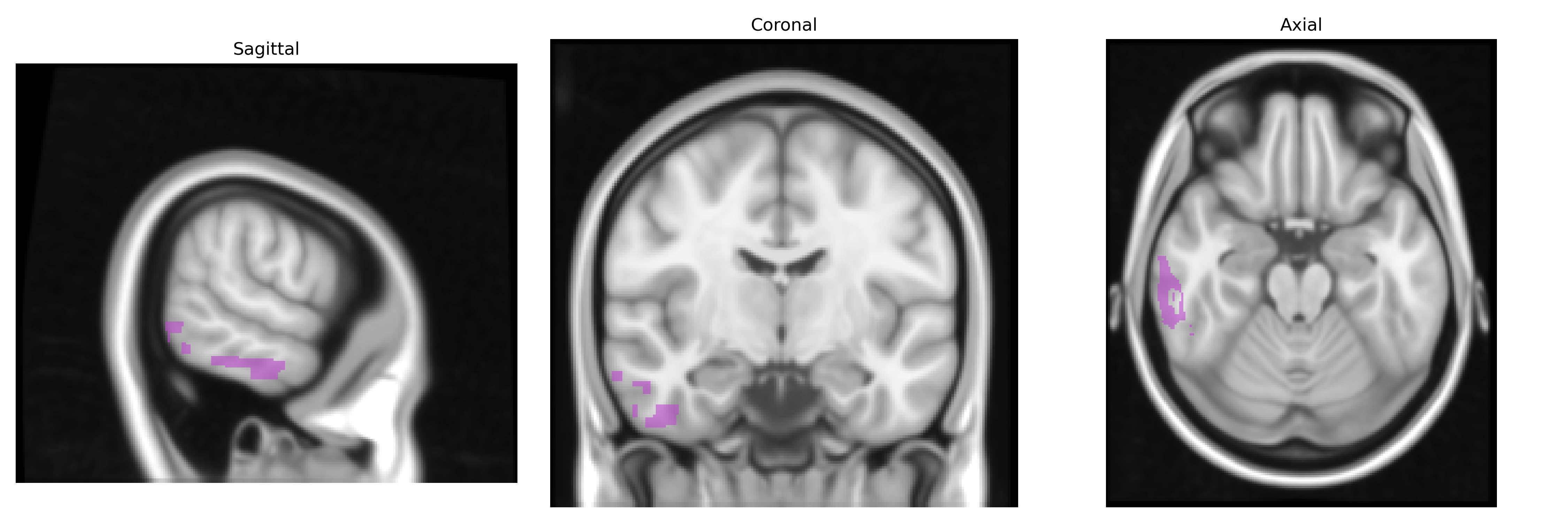
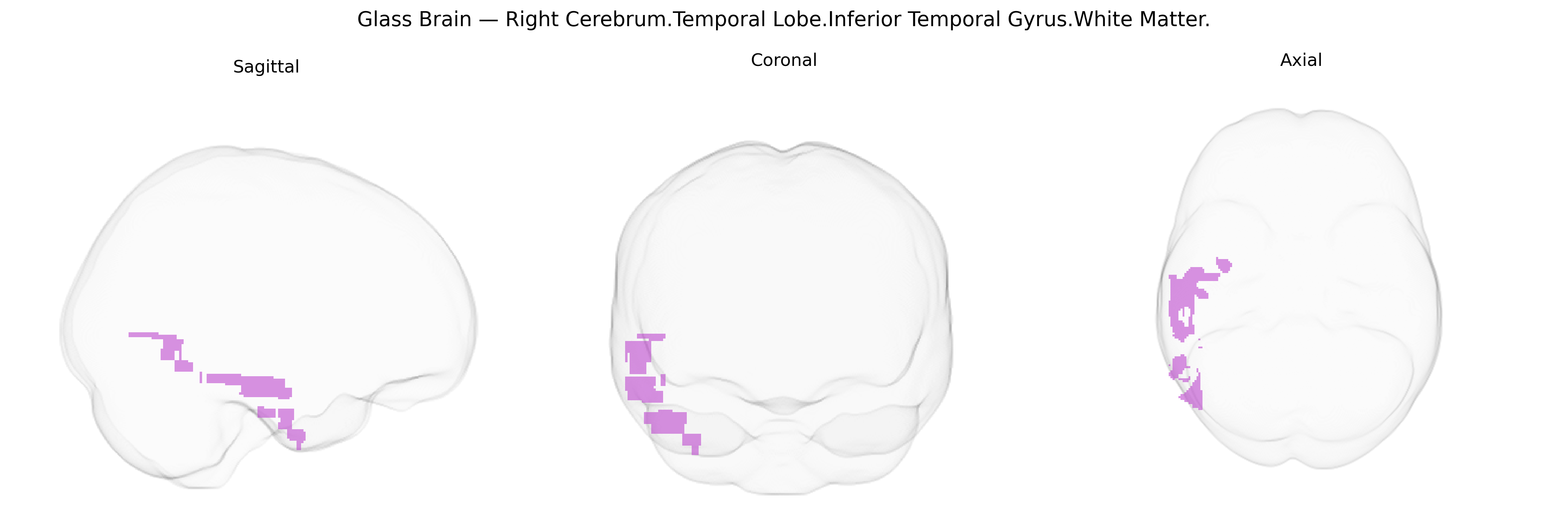
The bilateral Right Cerebrum.Temporal Lobe.Inferior Temporal Gyrus.White Matter corresponds to the subcortical fiber pathways underlying the inferior temporal gyrus in the right temporal lobe, involved in the transmission of visual, associative, and multimodal information between inferior temporal cortical areas and other regions such as the occipital lobe, limbic structures, and prefrontal cortex. This white matter includes portions of long-range association tracts (e.g., components of the inferior longitudinal fasciculus) as well as short U-fibers connecting neighboring gyri, supporting functions related to high-level visual processing, object recognition, and integration of complex sensory information. There is no direct link for this precise white matter subdivision; see the related cortical region: Inferior temporal gyrus.
The bilateral Right Cerebrum.Temporal Lobe.Inferior Temporal Gyrus.White Matter, as defined in the Talairach 1 mm atlas, lies within ventral temporal association pathways (including inferior longitudinal and occipitotemporal fasciculi) that support object, face and word recognition; while few GWAS target this precise white-matter label, multiple large-scale imaging-genetics studies (e.g., ENIGMA, UK Biobank) implicate common variants in axon guidance, myelination, and synaptic genes in the microstructure and volume of inferior temporal and adjacent ventral temporal white matter, including loci near genes such as NTRK1/2, NCAM1, CNTN4/6, and myelin-related genes (e.g., MAG, MBP) associated with diffusion tensor metrics in temporal tracts. Genetic correlations link temporal white-matter characteristics with neurodevelopmental and psychiatric disorders, including autism spectrum disorder, schizophrenia, and major depression, where risk variants in synaptic and neurodevelopmental genes (e.g., NRXN1/3, CNTNAP2, DISC1, CACNA1C) are associated with altered integrity of inferior and middle temporal white matter; similar associations have been reported for Alzheimer’s disease and other dementias, in which APOE and other AD-related loci (e.g., CLU, PICALM) are linked to temporal lobe atrophy and white-matter degeneration. GWAS of reading ability, language-related traits and face/object recognition performance also highlight polygenic influences on ventral temporal networks, including inferior temporal pathways, through variants in genes affecting cortical patterning and connectivity (e.g., KIAA0319, DCDC2, FOXP2-related networks), and more recent multivariate brain-wide association studies suggest that polygenic scores for intelligence, educational attainment and psychiatric liability correlate with structural and microstructural variation in this region’s white-matter tracts, although precise locus-level mappings to the specific Talairach-defined white-matter parcel remain limited and often derived from broader temporal or occipitotemporal tract labels.
Overview generated by GPT-4o (2026).
Region ID: 20
Hemisphere: bilateral
Atlas: Talairach labels 1mm
Full Quality Version: Download MP4
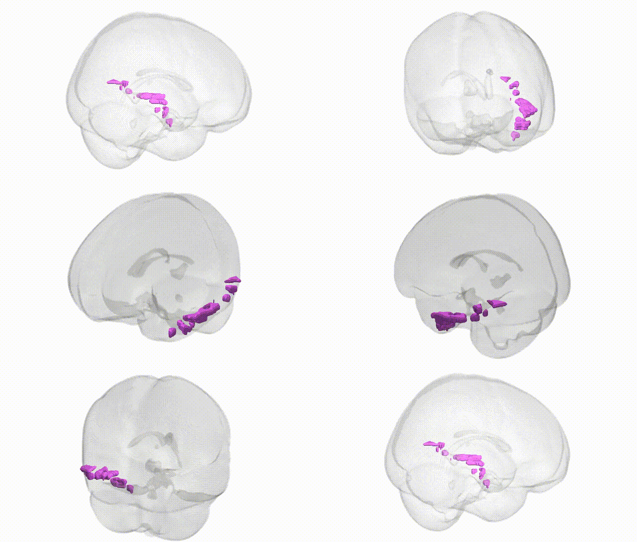
Full Quality Version: Download MP4
Wali Sidiqyar*, Gaurav Rudravaram*, Elyssa M. McMaster, Trent M. Schwartz, Adam M. Saunders, Kurt G. Schilling, Bennett A. Landman "Introducing SPINS: A Shared Public Visualization Library of Neuroanatomical Structures." Medical Imaging with Deep Learning- short paper
This resource is licensed under CC0 1.0 Universal (Public Domain).