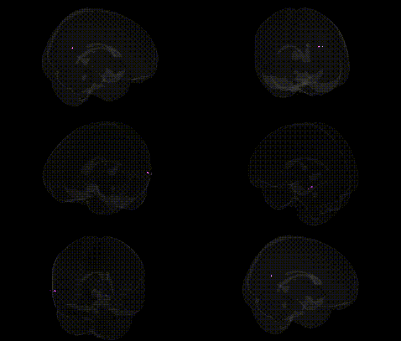
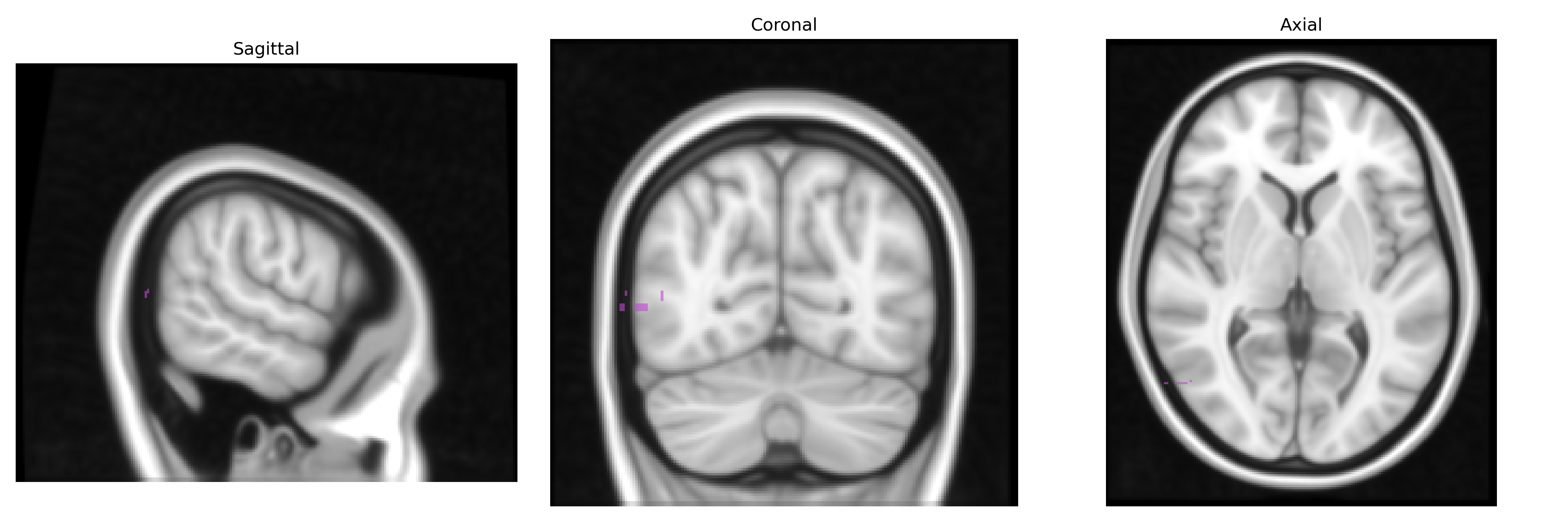
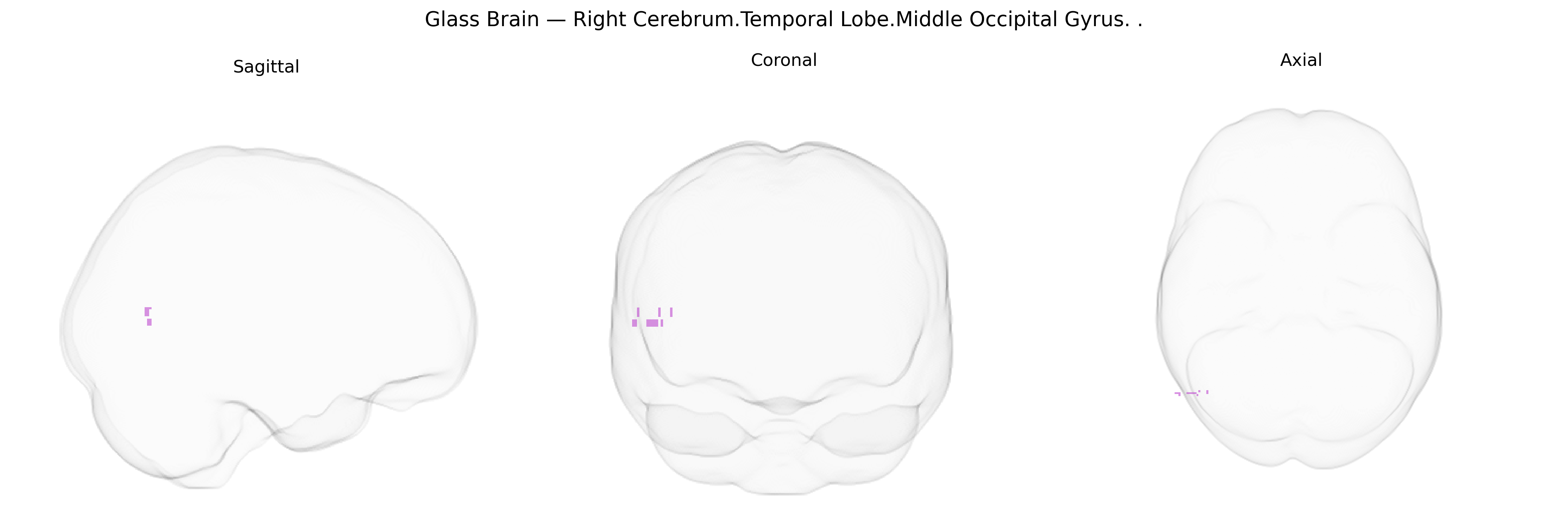
The bilateral right cerebrum temporal lobe middle occipital gyrus, as labeled in the Talairach 1 mm atlas, refers to a lateral occipital cortical strip located posterior to the temporal lobe and inferior to the superior occipital regions, typically spanning parts of Brodmann areas 18 and 19. Functionally, the middle occipital gyrus is implicated in intermediate‑level and higher‑order visual processing, including analysis of motion, shape, and complex spatial patterns, and it participates in dorsal and ventral visual streams that support object recognition and visuospatial integration. It maintains dense connections with adjacent occipital gyri, temporal visual association cortices, and parietal regions, contributing to visual attention and the integration of visual inputs with memory and semantic representations. There is no direct Wikipedia article dedicated specifically to the middle occipital gyrus; a closely related and encompassing structure is the Occipital lobe.
The middle occipital gyrus in the temporal-occipital region (Talairach: Right Cerebrum.Temporal Lobe.Middle Occipital Gyrus, bilaterally) has been implicated in multiple genetically influenced traits and disorders through imaging-genetics and GWAS-based studies, though often under broader labels such as “occipital cortex,” “visual association cortex,” or “occipitotemporal regions.” Large MRI GWAS consortia (e.g., ENIGMA, UK Biobank) have identified common variants in genes involved in neurodevelopment, synaptic signaling, and myelination—such as HMGA2, MAPT, WNT pathway genes, and multiple loci in 3p, 6q, 17q, and 19p—that associate with occipital cortical thickness, surface area, and volume, which include the middle occipital gyrus. Polygenic risk for schizophrenia, autism spectrum disorder, ADHD, and major depression has been linked to structural and functional variation in the middle occipital and adjacent occipitotemporal regions, including altered gray matter volume and connectivity with temporal and parietal networks. GWAS of visual processing traits (e.g., face and object recognition, reading-related measures such as dyslexia) and resting-state networks have pointed to genes affecting visual cortex development and dorsal visual stream function, with consistent activation and structural differences in the middle occipital gyrus across carriers of risk alleles in genes such as KIAA0319 and DCDC2 in reading-related studies, although these findings are not always localized exclusively to this gyrus. Additionally, loci associated with migraine, photosensitivity, and visual aura often map to genes influencing cortical excitability and visual cortex responsivity, implicating occipital and occipitotemporal areas, including the middle occipital gyrus, as downstream anatomical substrates of these genetic effects.
Overview generated by GPT-4o (2026).
Region ID: 626
Hemisphere: bilateral
Atlas: Talairach labels 1mm
Full Quality Version: Download MP4
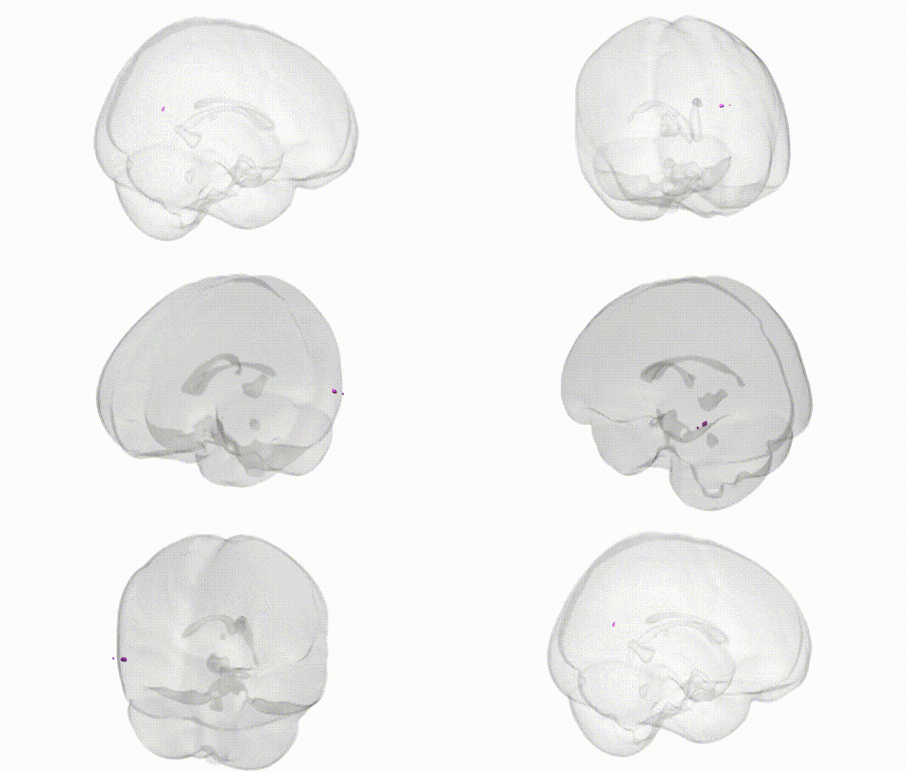
Full Quality Version: Download MP4
Wali Sidiqyar*, Gaurav Rudravaram*, Elyssa M. McMaster, Trent M. Schwartz, Adam M. Saunders, Kurt G. Schilling, Bennett A. Landman "Introducing SPINS: A Shared Public Visualization Library of Neuroanatomical Structures." Medical Imaging with Deep Learning- short paper
This resource is licensed under CC0 1.0 Universal (Public Domain).