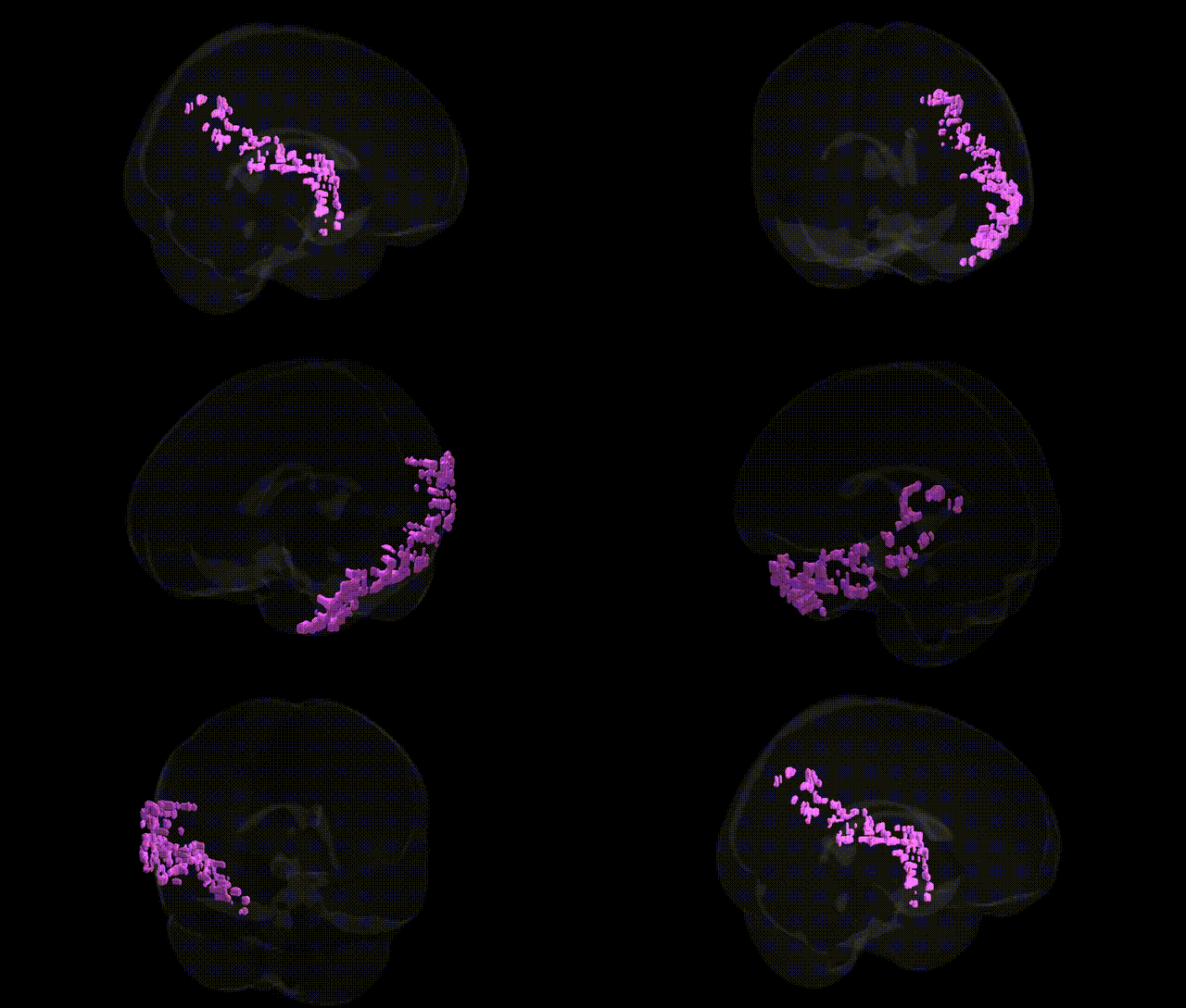
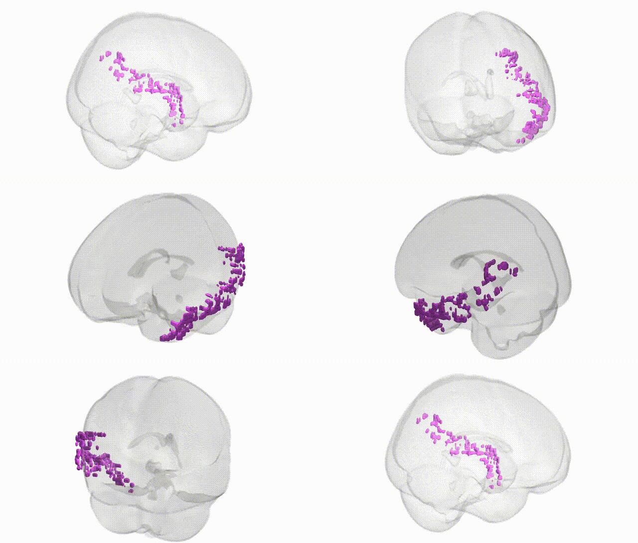
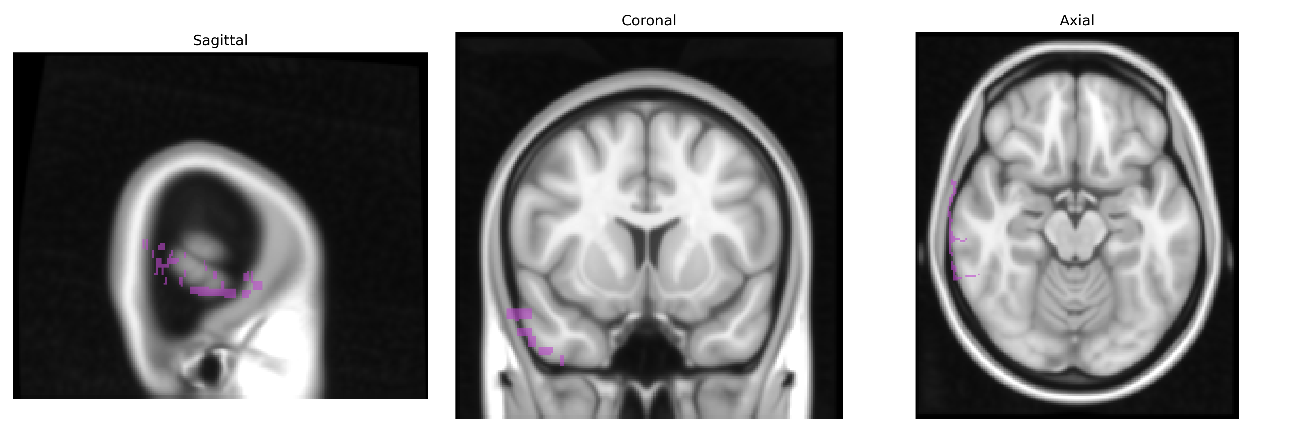
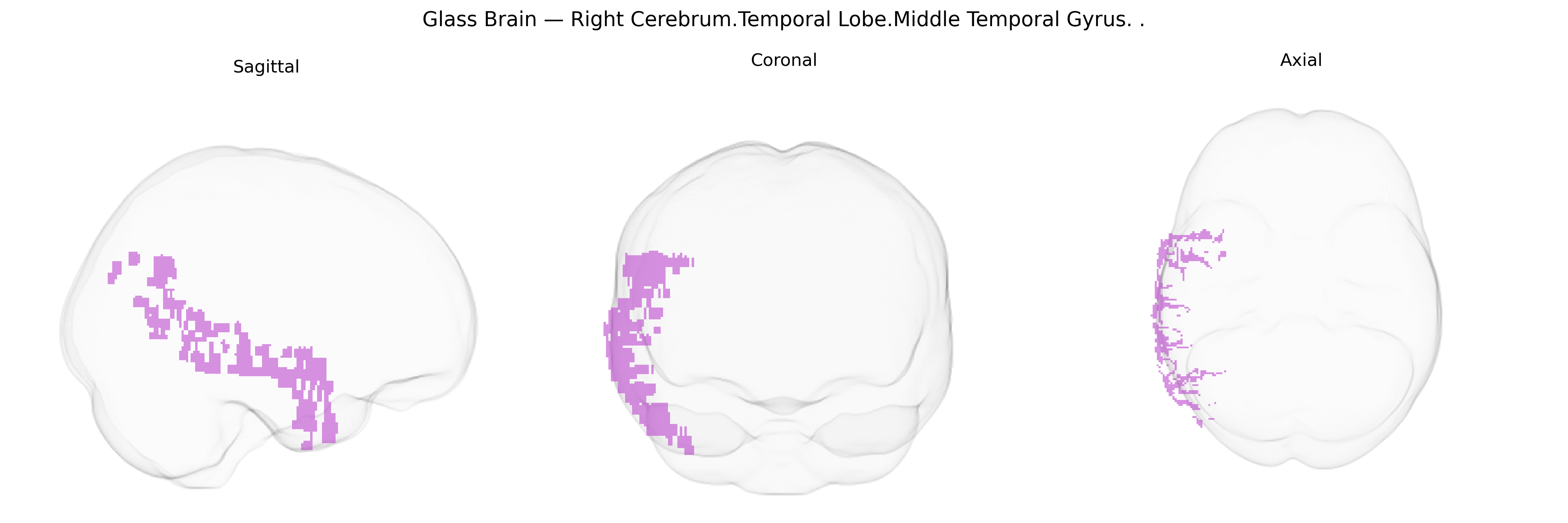
The bilateral right middle temporal gyrus (MTG) of the temporal lobe is a longitudinal cortical region located between the superior and inferior temporal gyri, extending from the temporal pole posteriorly toward the occipital junction along the lateral surface of the right cerebral hemisphere. Cytoarchitectonically, it encompasses portions of Brodmann areas such as 21 and 37, with extensive connectivity to auditory association cortex, visual association areas, inferior parietal regions, and prefrontal cortices via major white matter pathways (e.g., arcuate fasciculus, inferior longitudinal fasciculus). Functionally, the right MTG contributes to higher-order auditory and language-related processing (particularly prosody and aspects of semantic integration), visual and multimodal object recognition, social cognition (including processing of biological motion and social cues), and episodic memory retrieval. It also participates in large-scale networks related to semantic memory, default-mode activity, and integration of sensory information over time. Middle temporal gyrus
The bilateral right middle temporal gyrus (MTG) of the temporal lobe has been implicated in multiple genetic and genome-wide association studies, particularly in relation to language, social cognition, and neuropsychiatric and neurodegenerative disorders. Variants in FOXP2, CNTNAP2, and related language-associated genes have been linked to functional and structural differences in MTG, aligning with its role in semantic processing and speech perception. GWAS of cortical thickness and surface area (e.g., ENIGMA and UK Biobank consortia) have identified common variants in genes involved in neurodevelopment (such as MIR137, MSRA, and loci near WNT and Notch signaling genes) that influence temporal lobe and specifically middle temporal morphology, sometimes overlapping with loci associated with general cognitive ability and educational attainment. In schizophrenia and autism spectrum disorder, polygenic risk scores and imaging–genetic studies show that higher genetic liability is associated with altered MTG volume, connectivity, and activation patterns during social and language tasks. In Alzheimer’s disease and related dementias, risk variants in APOE (particularly ε4), CLU, and other GWAS-identified loci correlate with atrophy and metabolic changes in lateral temporal regions including the MTG, paralleling impairments in semantic memory. Additionally, GWAS of resting-state functional networks have linked common variants in synaptic and axonal guidance genes to connectivity within temporal language and default mode networks that prominently involve the right MTG, supporting a genetically mediated contribution to interindividual differences in higher-order temporal lobe function.
Overview generated by GPT-4o (2026).
Region ID: 30
Hemisphere: bilateral
Atlas: Talairach labels 1mm
Full Quality Version: Download MP4
Full Quality Version: Download MP4
Wali Sidiqyar*, Gaurav Rudravaram*, Elyssa M. McMaster, Trent M. Schwartz, Adam M. Saunders, Kurt G. Schilling, Bennett A. Landman "Introducing SPINS: A Shared Public Visualization Library of Neuroanatomical Structures." Medical Imaging with Deep Learning- short paper
This resource is licensed under CC0 1.0 Universal (Public Domain).