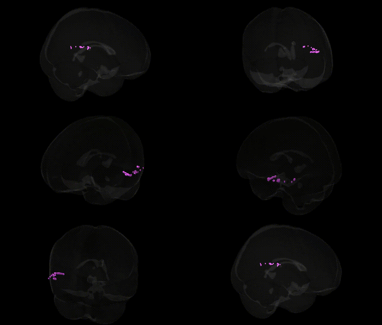
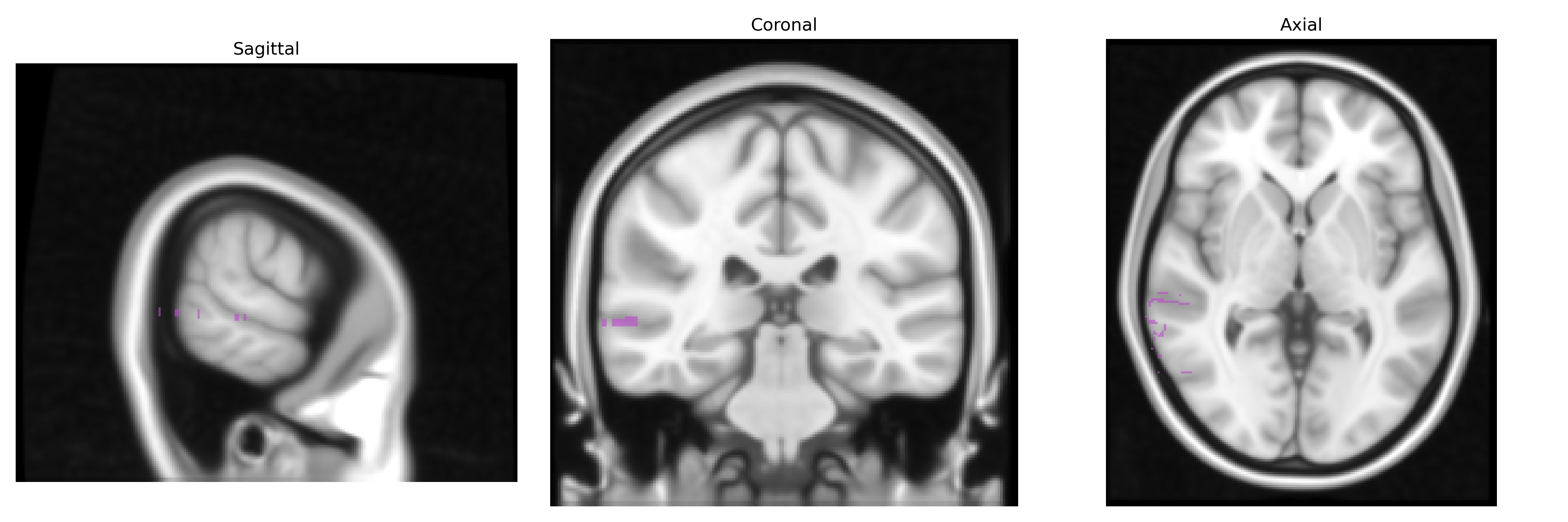
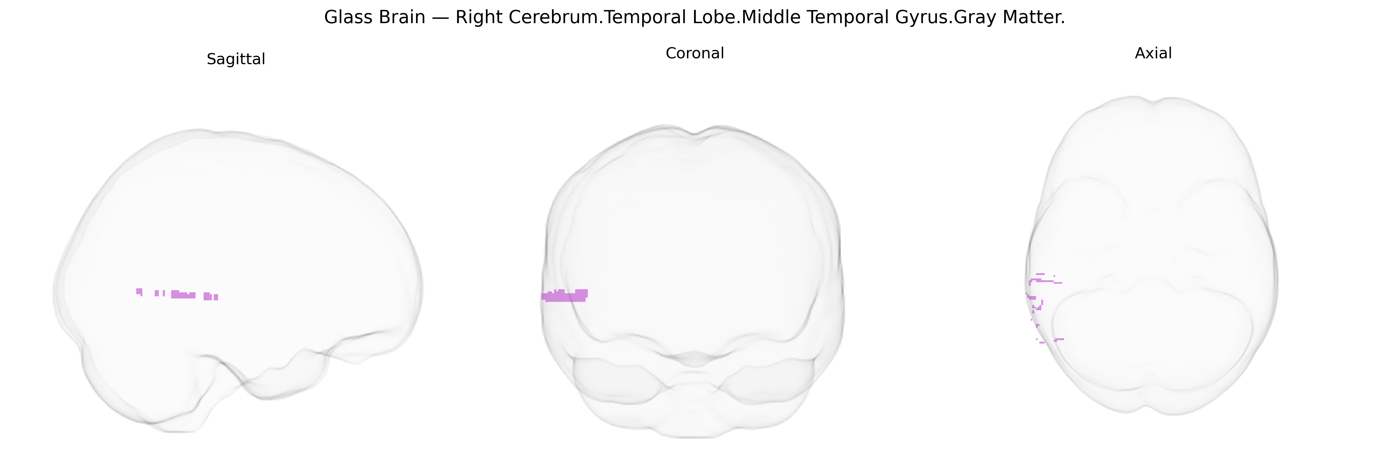
The bilateral right cerebrum temporal lobe middle temporal gyrus gray matter corresponds to cortical tissue located on the lateral surface of the temporal lobe, running between the superior and inferior temporal gyri and extending from the temporal pole posteriorly toward the angular and occipital regions. This gyral region participates in higher-order auditory and language processing, semantic memory, visual motion perception, and aspects of social cognition, integrating multimodal sensory inputs with stored conceptual knowledge. Functionally, it contributes to the comprehension of word meaning and sentence-level semantics, the interpretation of biological motion and faces, and broader networks involved in default-mode and semantic processing, with right-hemisphere emphasis often associated with nonverbal and social-emotional aspects of cognition. A related article is Middle temporal gyrus.
The bilateral right middle temporal gyrus gray matter, as defined in the Talairach 1 mm atlas, has been repeatedly implicated in imaging-genetics and GWAS studies of cortical structure and neuropsychiatric disease, with common variants influencing its thickness, surface area, and volume as well as functional properties such as language and social cognition. Large-scale ENIGMA and UK Biobank–based GWAS have identified associations between middle temporal gyrus morphology and loci near genes involved in neurodevelopment, synaptic signaling, and cell adhesion (for example, variants in or near genes such as GRIN2B, CNTNAP2, and FOXP2 have been linked to temporal lobe structure or function), and polygenic scores for schizophrenia, major depressive disorder, autism spectrum disorder, and Alzheimer’s disease show correlations with altered gray matter metrics in this region. The right middle temporal gyrus has also been implicated in GWAS of cognitive performance and educational attainment, where genetic variants affecting cortical development and plasticity contribute to interindividual differences in semantic processing and general intelligence. Furthermore, risk alleles for temporal lobe epilepsy and frontotemporal dementia have been associated with structural and functional abnormalities encompassing the right middle temporal gyrus, consistent with its role in language, memory, and social-emotional processing, although most such findings derive from broader temporal lobe or network-level analyses rather than this single parcel alone.
Overview generated by GPT-4o (2026).
Region ID: 555
Hemisphere: bilateral
Atlas: Talairach labels 1mm
Full Quality Version: Download MP4
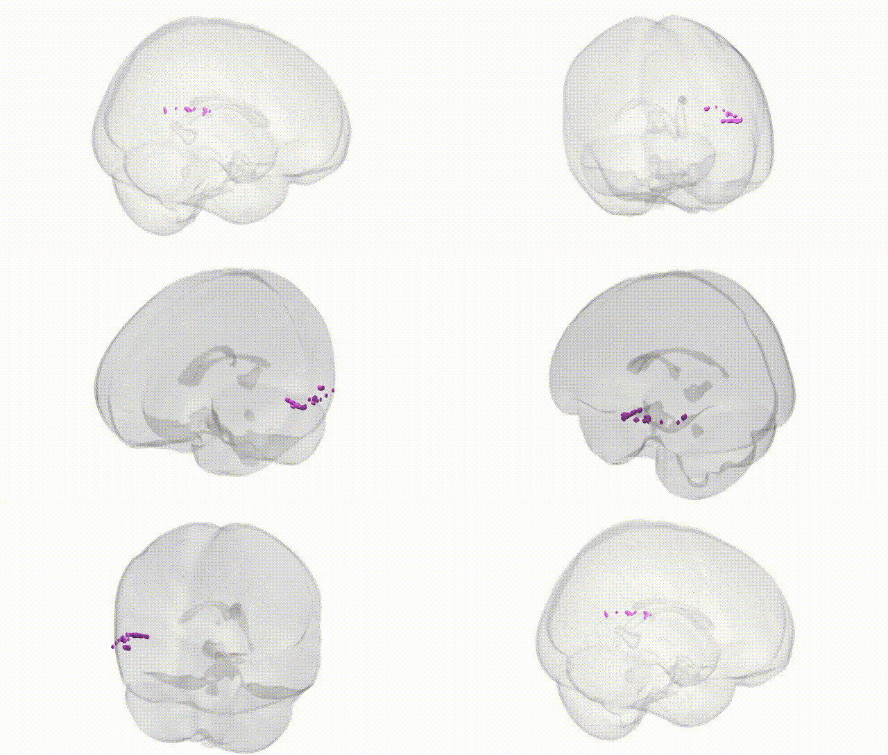
Full Quality Version: Download MP4
Wali Sidiqyar*, Gaurav Rudravaram*, Elyssa M. McMaster, Trent M. Schwartz, Adam M. Saunders, Kurt G. Schilling, Bennett A. Landman "Introducing SPINS: A Shared Public Visualization Library of Neuroanatomical Structures." Medical Imaging with Deep Learning- short paper
This resource is licensed under CC0 1.0 Universal (Public Domain).