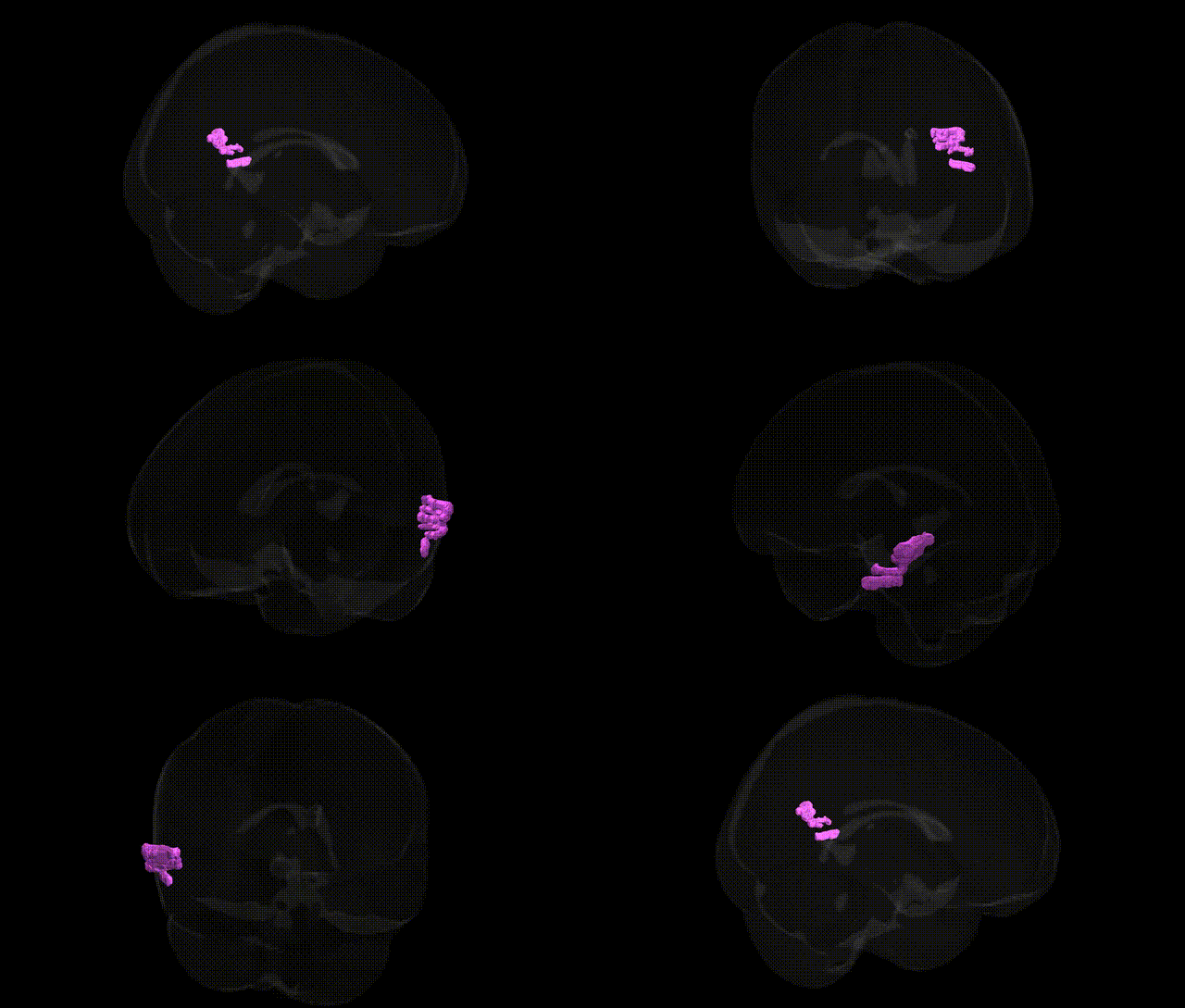
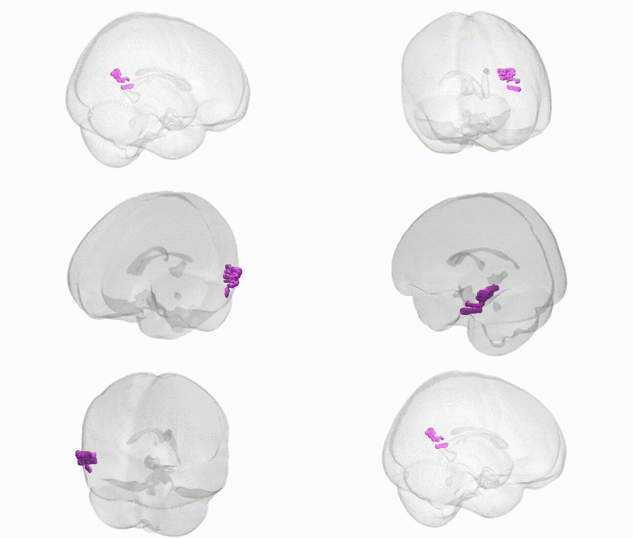
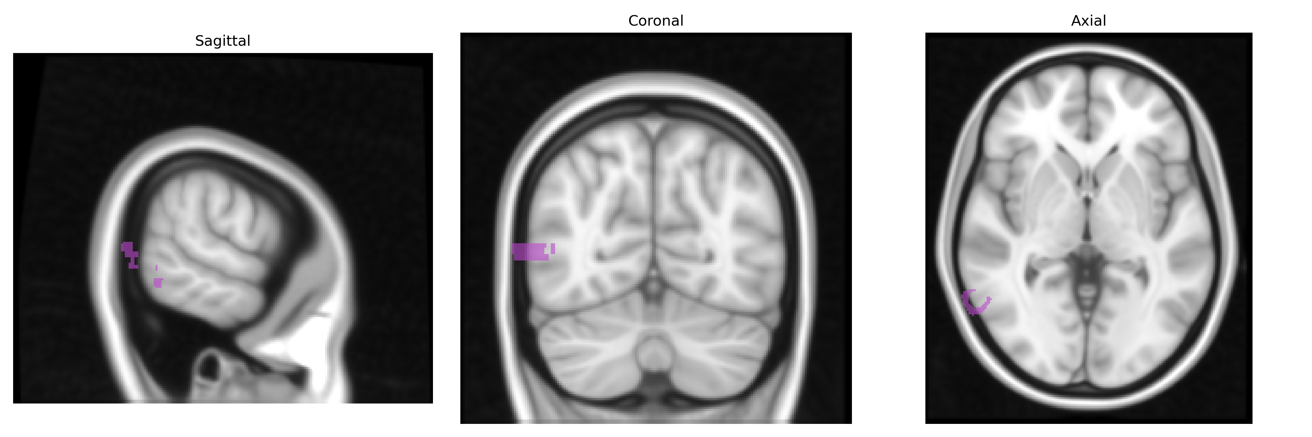
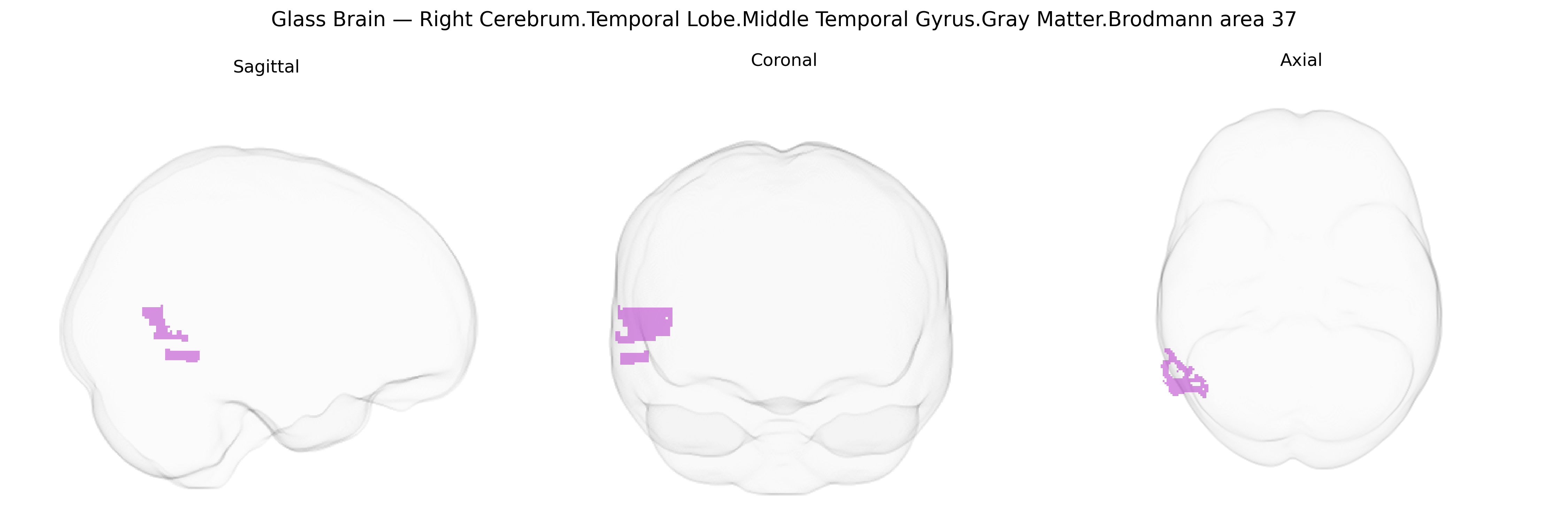
The bilateral Right Cerebrum.Temporal Lobe.Middle Temporal Gyrus.Gray Matter.Brodmann area 37 corresponds to a cytoarchitectonic territory located in the posterior portion of the middle and adjacent inferior temporal cortex, typically associated with visual object and word form processing. BA37 lies within the ventral visual stream and contributes to high-level perceptual functions such as face and object recognition, semantic integration of visual stimuli, and aspects of reading by interfacing visual input with stored lexical and conceptual representations. Functionally, this region is closely related to the fusiform gyrus and the so‑called fusiform face area, and lesions affecting BA37 can result in deficits such as visual agnosia or alexia, depending on the precise locus and extent of damage. There is no direct Wikipedia article specifically for “Brodmann area 37” as defined in the Talairach 1 mm Atlas; a closely related structure and functional territory is described under the Fusiform gyrus.
The bilateral right middle temporal gyrus (MTG) gray matter in Brodmann area 37, a ventral temporal region involved in semantic processing, visual word-form recognition, and face/object perception, has been implicated in several genetic and GWAS-based associations, although most findings are not uniquely specific to this exact Talairach-defined parcel. Imaging-genetics studies consistently link common variants in genes involved in synaptic plasticity and neurodevelopment—such as BDNF (e.g., Val66Met), DISC1, and NRG1—to structural and functional differences in temporal lobe cortices including the MTG; for example, BDNF polymorphisms have been associated with temporal lobe gray matter volume and memory performance. Large-scale GWAS of cortical thickness and surface area (e.g., ENIGMA, UK Biobank) have identified multiple loci (including variants near genes such as MIR124-3, WNT3, and microtubule- and axon-guidance–related genes) that influence temporal lobe morphology broadly, with some parcellation-based analyses reporting associations in lateral and inferior temporal regions encompassing or adjacent to BA37. Clinically, genetic risk for disorders with prominent temporal lobe involvement—such as schizophrenia, bipolar disorder, autism spectrum disorder, and temporal lobe epilepsy—has been associated with altered structure or activation of right MTG/BA37, with polygenic risk scores and specific risk variants (e.g., in CACNA1C, GRIN2A, and synaptic genes) correlating with temporal cortical changes in case–control and high-risk cohorts. GWAS of language, reading ability/dyslexia, and face recognition have also pointed to temporal-occipital regions overlapping BA37, linking variants in KIAA0319, DCDC2, and other neurodevelopmental genes to gray matter differences and connectivity patterns in ventral temporal cortex, though spatial resolution and parcellation differences mean that associations are usually reported for broader temporal lobe regions rather than the precise bilateral right BA37 label.
Overview generated by GPT-4o (2026).
Region ID: 328
Hemisphere: bilateral
Atlas: Talairach labels 1mm
Full Quality Version: Download MP4
Full Quality Version: Download MP4
Wali Sidiqyar*, Gaurav Rudravaram*, Elyssa M. McMaster, Trent M. Schwartz, Adam M. Saunders, Kurt G. Schilling, Bennett A. Landman "Introducing SPINS: A Shared Public Visualization Library of Neuroanatomical Structures." Medical Imaging with Deep Learning- short paper
This resource is licensed under CC0 1.0 Universal (Public Domain).