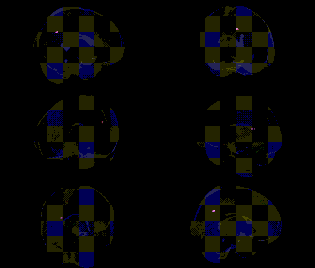
The bilateral Right Cerebrum.Temporal Lobe.Precuneus.White Matter label in the Talairach 1 mm atlas likely reflects white matter pathways situated near the interface of the temporal lobe and the medial parietal region encompassing the precuneus, rather than a discrete classical anatomical structure. This region contains association fibers that interconnect temporal cortical areas (involved in auditory processing, language, and memory) with parietal and medial parietal structures, including the precuneus, which is implicated in visuo-spatial integration, self-referential processing, and default-mode network activity. The white matter here contributes to large-scale cortical networks supporting episodic memory, spatial navigation, mental imagery, and aspects of consciousness by facilitating communication between temporal association cortices, posterior parietal areas, and medial cortical hubs. There is no direct Wikipedia article for this composite label; a related structure is the Precuneus.
Genetic associations involving the bilateral right cerebrum temporal lobe precuneus white matter—approximated here by studies targeting the precuneus and adjacent temporo‑parietal white matter—arise largely from imaging‑genetics and GWAS of brain structure and connectivity rather than region‑specific Talairach-based analyses. Heritability studies show that white‑matter microstructure (e.g., fractional anisotropy) in posterior cingulate–precuneus pathways is highly heritable and influenced by polygenic variation affecting axonal development, myelination, and oligodendrocyte function, with notable contributions from genes in neurodevelopmental and cell-adhesion pathways (e.g., neuregulin/NRG–ErbB signaling, LINGO1, and contactin/NCAM family members) identified in large diffusion MRI GWAS consortia such as ENIGMA and UK Biobank. Variants in APOE, CLU, PICALM, and other Alzheimer’s disease risk loci have been linked to altered structural connectivity and white‑matter integrity in default mode network hubs including the precuneus, consistent with this region’s early involvement in amyloid and tau pathology. Imaging‑genetics work in schizophrenia, autism spectrum disorder, and major depressive disorder implicates polygenic risk scores in modulating temporal–parietal/precuneus white‑matter properties, with effects reported for genes involved in synaptic plasticity and glutamatergic signaling (e.g., GRIN2B, CACNA1C) that may mediate disrupted connectivity in these disorders. In addition, GWAS of cognitive traits (general intelligence, episodic memory, and self-referential/mentalizing abilities) show that polygenic influences on these functions partially converge on structural and functional variation in the precuneus and its white‑matter tracts, suggesting that complex, highly polygenic architectures underlie individual differences in this region’s connectivity and related neurocognitive and neuropsychiatric phenotypes.
Overview generated by GPT-4o (2026).
Region ID: 866
Hemisphere: bilateral
Atlas: Talairach labels 1mm
Full Quality Version: Download MP4
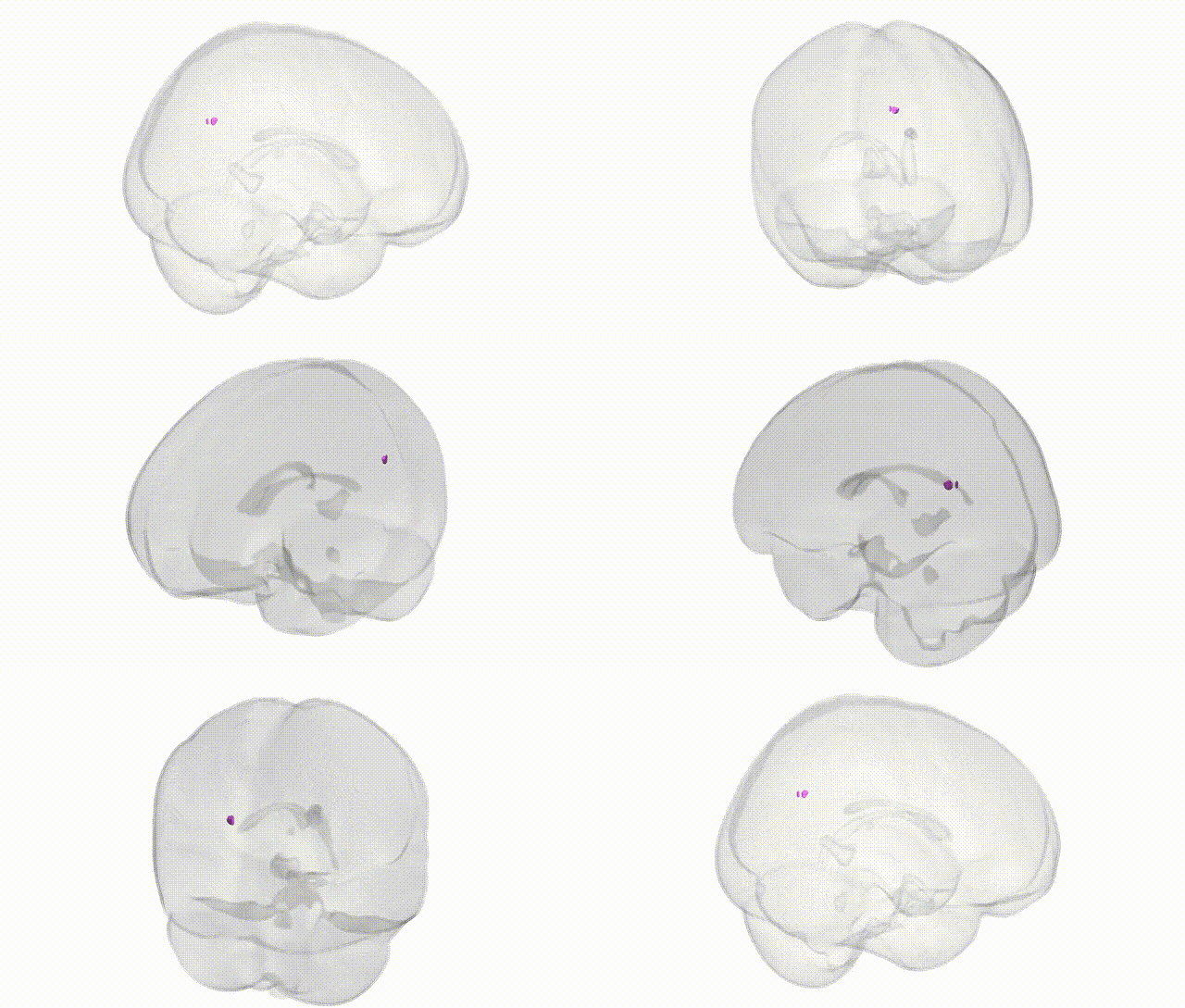
Full Quality Version: Download MP4
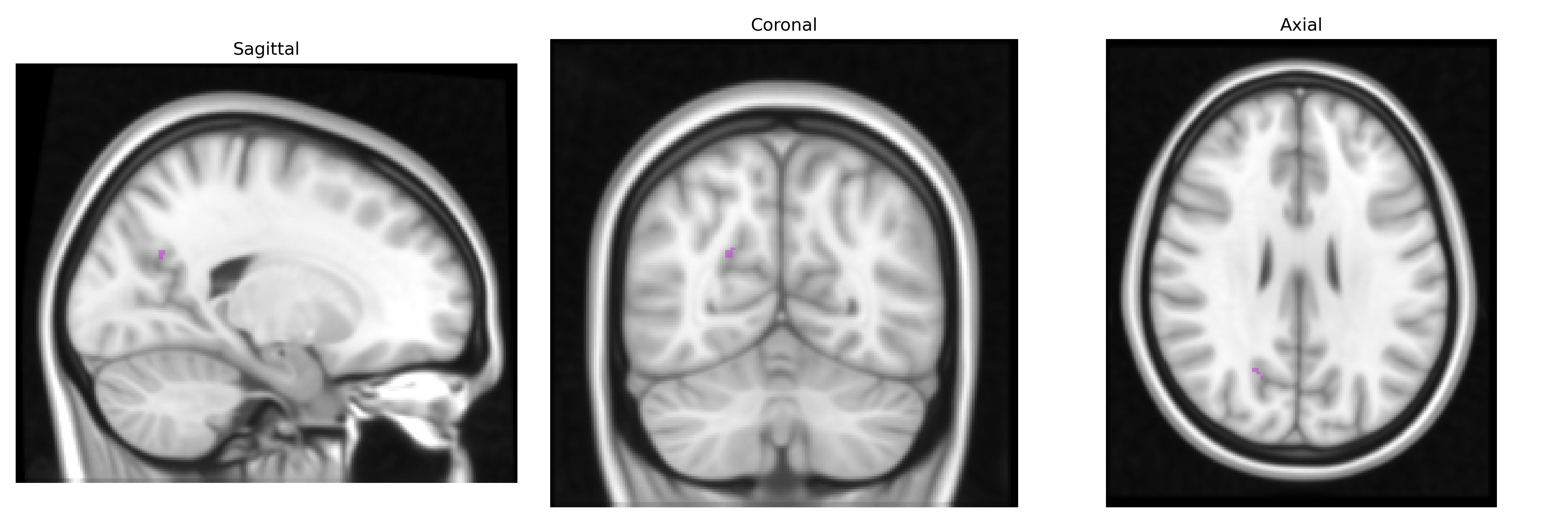
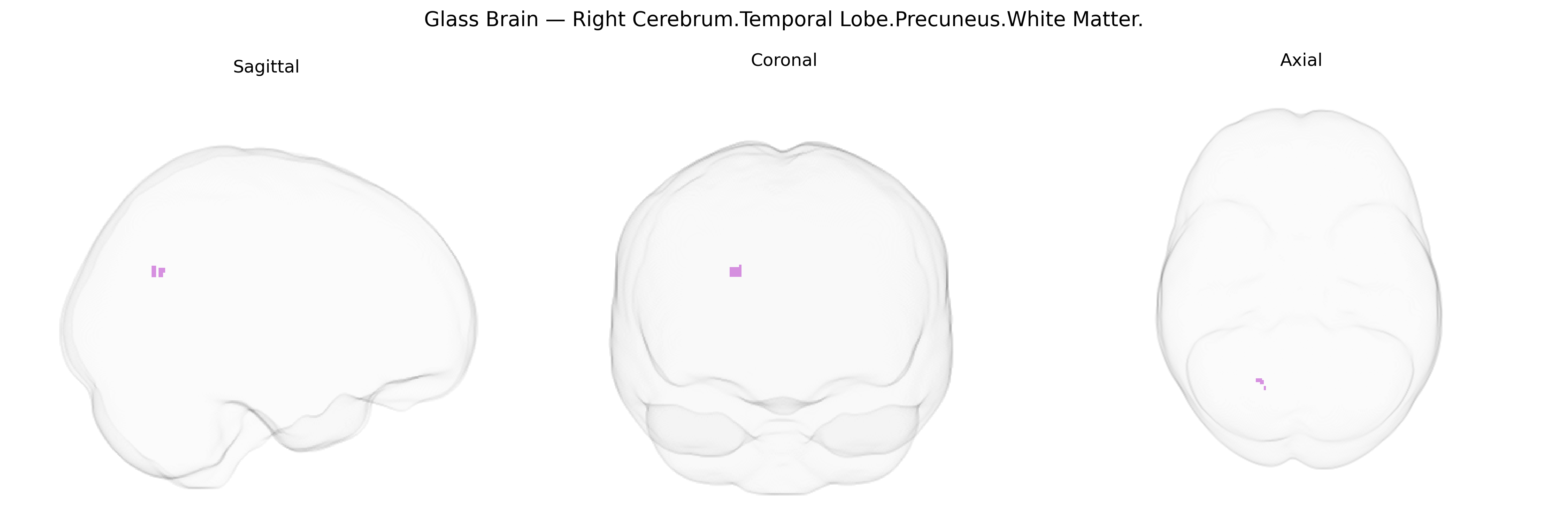
Wali Sidiqyar*, Gaurav Rudravaram*, Elyssa M. McMaster, Trent M. Schwartz, Adam M. Saunders, Kurt G. Schilling, Bennett A. Landman "Introducing SPINS: A Shared Public Visualization Library of Neuroanatomical Structures." Medical Imaging with Deep Learning- short paper
This resource is licensed under CC0 1.0 Universal (Public Domain).