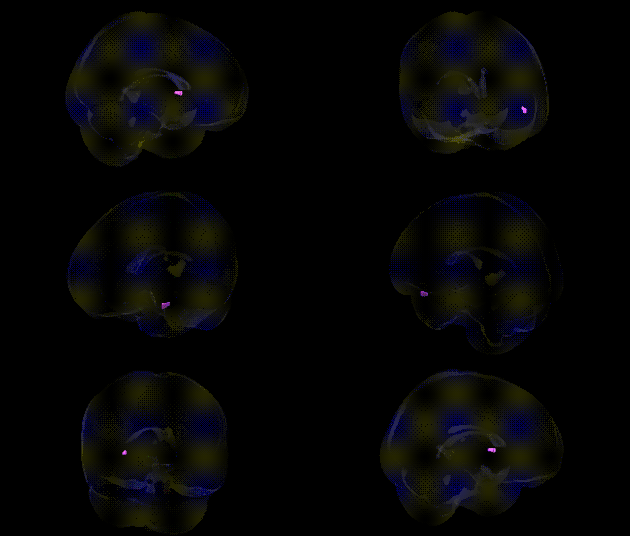
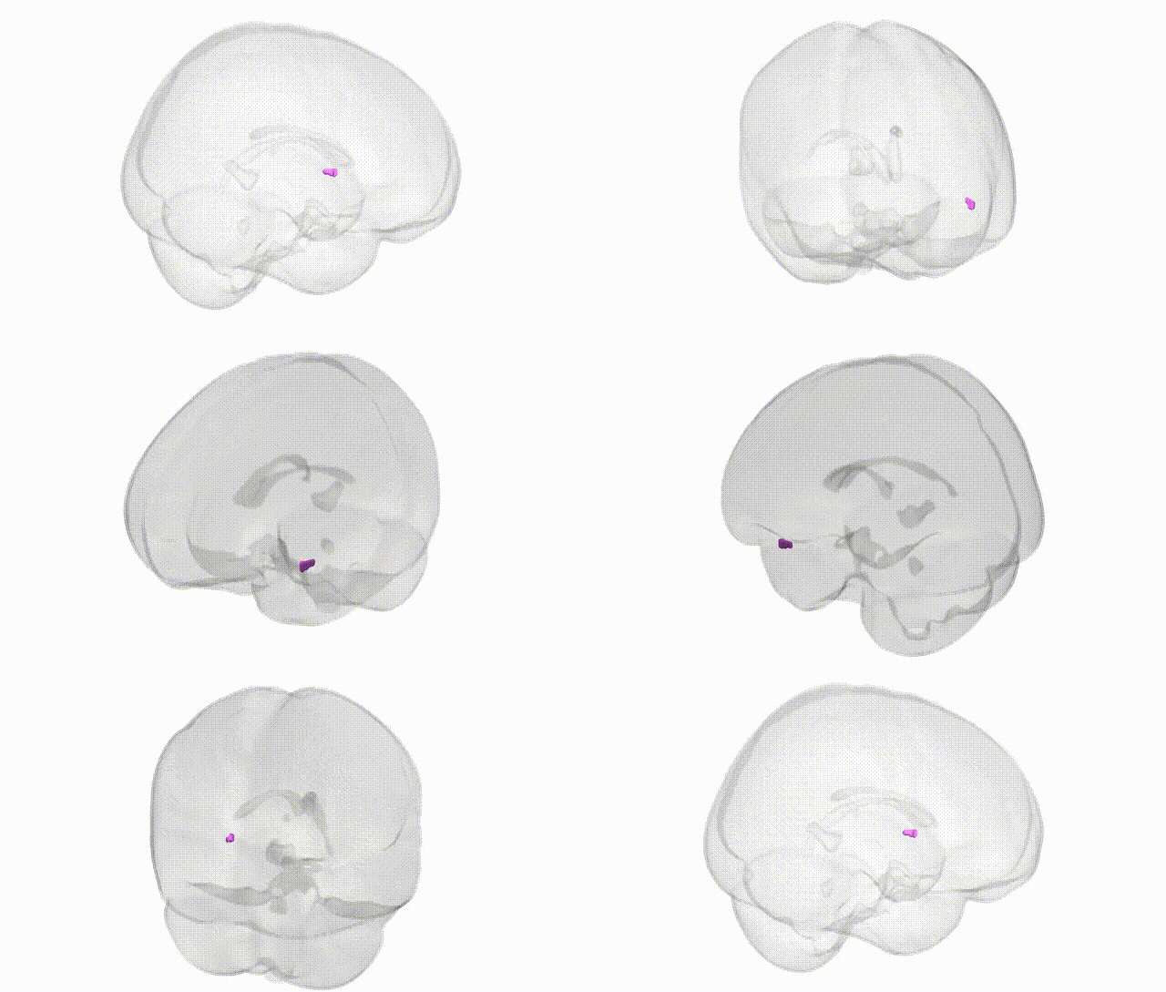
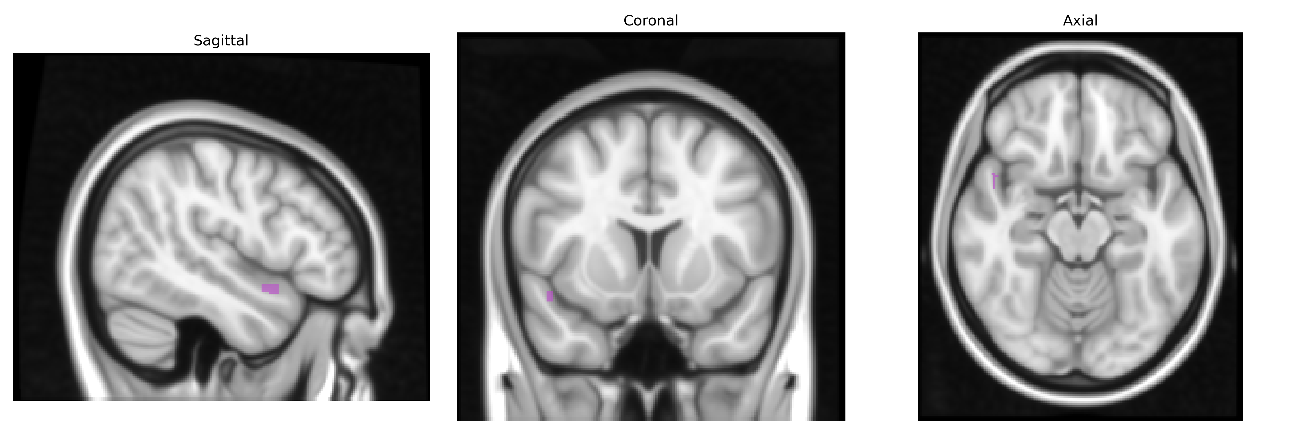
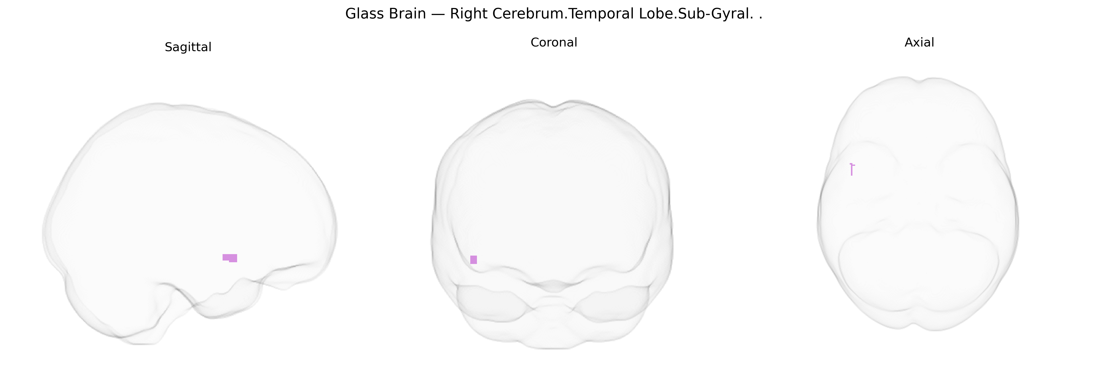
The bilateral Right Cerebrum.Temporal Lobe.Sub-Gyral region, as defined in the Talairach 1 mm atlas, corresponds to subcortical white matter located beneath the cortical gyri of the right temporal lobe. This area contains association and projection fibers that interconnect temporal cortical regions with each other and with distant structures such as the frontal lobe, parietal lobe, limbic system, and subcortical nuclei. Functionally, these sub-gyral fibers support higher-order auditory processing, language functions (particularly aspects of speech perception and semantic processing), memory integration via connections with the hippocampal formation, and multimodal sensory association involving visual and auditory inputs. Damage or disruption in this region can affect temporal lobe network communication, potentially contributing to language impairments, memory deficits, or temporal lobe epilepsy. There is no direct Wikipedia article for this exact sub-gyral label; a related structure is the Temporal lobe.
The bilateral right temporal sub-gyral region (encompassing deep portions of the superior, middle, and inferior temporal gyri and adjacent white matter) has been implicated in several genetic and genome-wide association findings, largely via imaging-genetics and neuropsychiatric GWAS. Variants in genes involved in synaptic function and neurodevelopment—such as DISC1, NRG1, GRM3, CACNA1C, and CNTNAP2—have been associated with gray matter volume, cortical thickness, or connectivity changes in right temporal lobe subcortical and sub-gyral areas, particularly in schizophrenia, bipolar disorder, and autism spectrum conditions. Large-scale ENIGMA consortia studies have identified common variants (for example near HMGA2, FRMD4B, and other loci) associated with temporal lobe surface area and thickness, with some effects lateralized to the right hemisphere. Right temporal sub-gyral structures are also highlighted in GWAS of language and reading-related traits, where genes such as FOXP2, DCDC2, and KIAA0319 show associations with temporal white matter microstructure and functional activation supporting phonological processing. In neurodegenerative conditions, APOE ε4 and other Alzheimer’s disease risk loci (e.g., CLU, PICALM) correlate with atrophy and hypometabolism in right temporal sub-gyral and adjacent limbic regions. Additionally, GWAS of brain connectivity and resting-state networks have linked common variants in genes regulating axon guidance and myelination (e.g., NRXN1, LINGO1) to diffusion MRI measures in deep temporal white matter, including sub-gyral right temporal pathways connecting auditory, language, and limbic networks. Overall, while most findings are not specific to the Talairach “Right Cerebrum.Temporal Lobe.Sub-Gyral” label in isolation, converging genetic evidence ties this region to the heritable architecture of language, psychosis, affective disorders, autism spectrum traits, and neurodegeneration through effects on temporal lobe structure and connectivity.
Overview generated by GPT-4o (2026).
Region ID: 384
Hemisphere: bilateral
Atlas: Talairach labels 1mm
Full Quality Version: Download MP4
Full Quality Version: Download MP4
Wali Sidiqyar*, Gaurav Rudravaram*, Elyssa M. McMaster, Trent M. Schwartz, Adam M. Saunders, Kurt G. Schilling, Bennett A. Landman "Introducing SPINS: A Shared Public Visualization Library of Neuroanatomical Structures." Medical Imaging with Deep Learning- short paper
This resource is licensed under CC0 1.0 Universal (Public Domain).