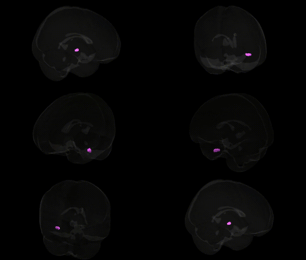
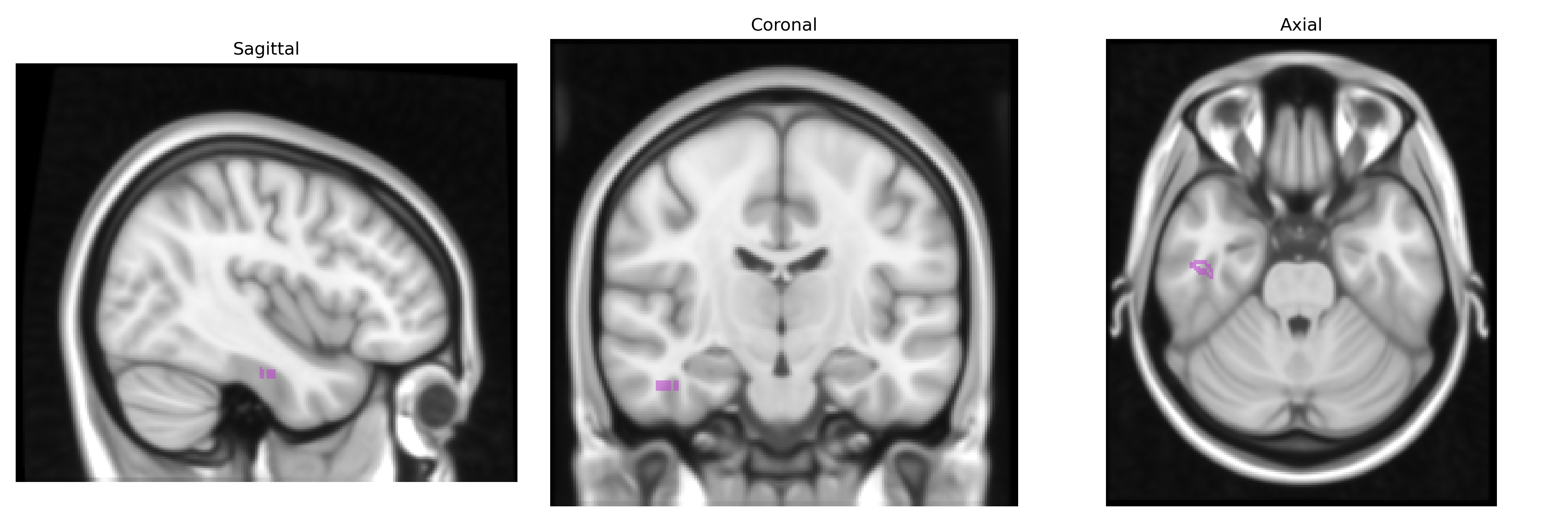
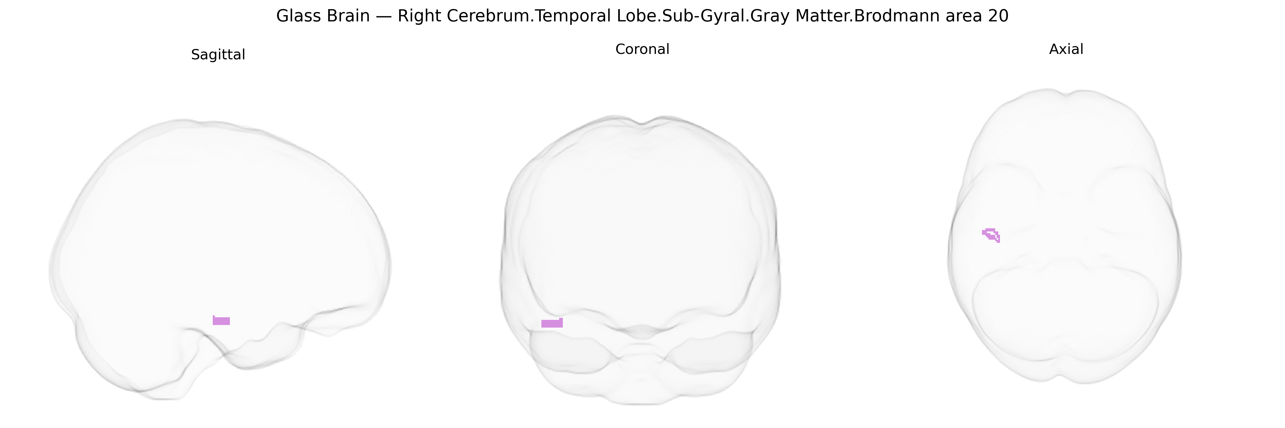
Bilateral Right Cerebrum.Temporal Lobe.Sub-Gyral.Gray Matter.Brodmann area 20 corresponds to inferior temporal association cortex located on the ventral surface of the temporal lobe, deep to the cortical gyri. Brodmann area 20 is involved in high-level visual processing, including object recognition, complex form analysis, and aspects of semantic integration of visual information, contributing to the ventral “what” pathway. Neuronal populations in this region receive convergent input from earlier visual areas and project to other temporal and limbic structures, supporting visual memory and the integration of visual stimuli with stored representations. Functionally, this area is implicated in the recognition of faces and objects and in linking visual patterns with meaning, and its disruption can contribute to visual agnosias and impairments in semantic processing. There is no direct link for Brodmann area 20; a closely related structure is the Inferior temporal gyrus.
The bilateral right temporal lobe sub-gyral gray matter of Brodmann area 20 (inferior temporal cortex, including parts of the fusiform and inferotemporal regions) has been implicated in several genetic and genome-wide association findings through its roles in high-level visual processing, semantic memory, and face/object recognition. GWAS and imaging-genetics studies have linked variation in genes involved in synaptic plasticity, neurodevelopment, and glutamatergic signaling (for example, BDNF, CNTNAP2, NRGN, GRIN2A, and genes within major neurodevelopmental CNV loci) to structural measures such as cortical thickness and surface area in the inferior and fusiform temporal cortex overlapping BA20, as well as to functional activation during language and visual recognition tasks. Risk loci for schizophrenia, autism spectrum disorder, and major depressive disorder—many identified by large consortia such as the Psychiatric Genomics Consortium and ENIGMA—show downstream effects on temporal lobe morphology and connectivity, including right inferior temporal regions that encompass BA20, and altered structure or function in this area has been repeatedly reported in these conditions. Additional GWAS of brain morphometry have identified polygenic contributions to interindividual differences in inferior temporal cortex volume and thickness, with enrichment of neurodevelopmental pathways and transcriptional profiles expressed in temporal association cortices, supporting a broadly polygenic, pleiotropic architecture rather than single region-specific risk genes.
Overview generated by GPT-4o (2026).
Region ID: 172
Hemisphere: bilateral
Atlas: Talairach labels 1mm
Full Quality Version: Download MP4
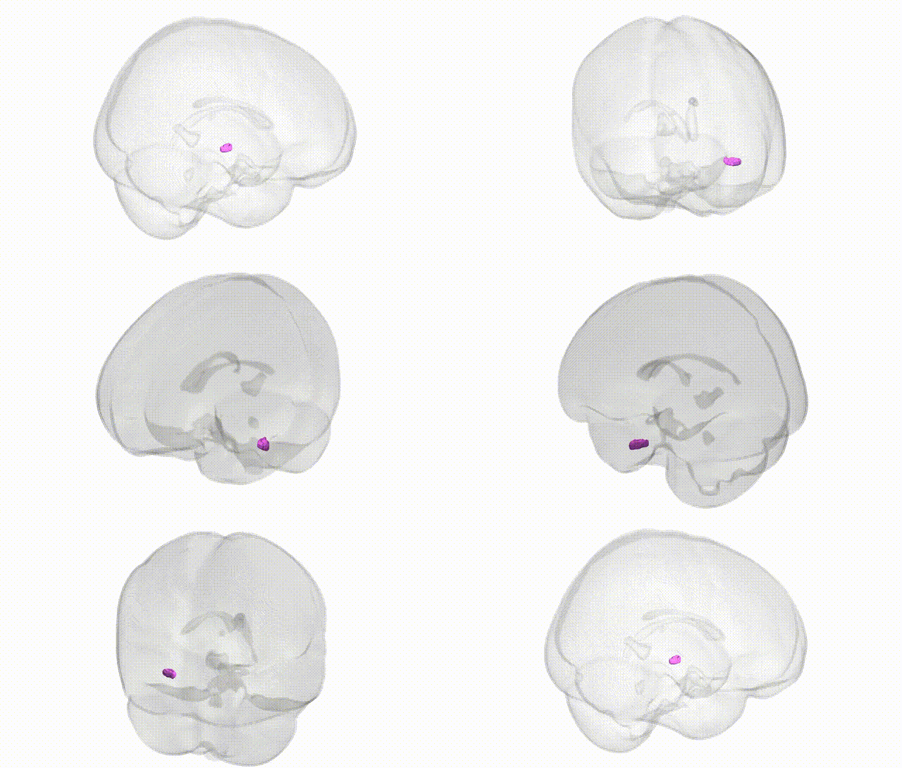
Full Quality Version: Download MP4
Wali Sidiqyar*, Gaurav Rudravaram*, Elyssa M. McMaster, Trent M. Schwartz, Adam M. Saunders, Kurt G. Schilling, Bennett A. Landman "Introducing SPINS: A Shared Public Visualization Library of Neuroanatomical Structures." Medical Imaging with Deep Learning- short paper
This resource is licensed under CC0 1.0 Universal (Public Domain).