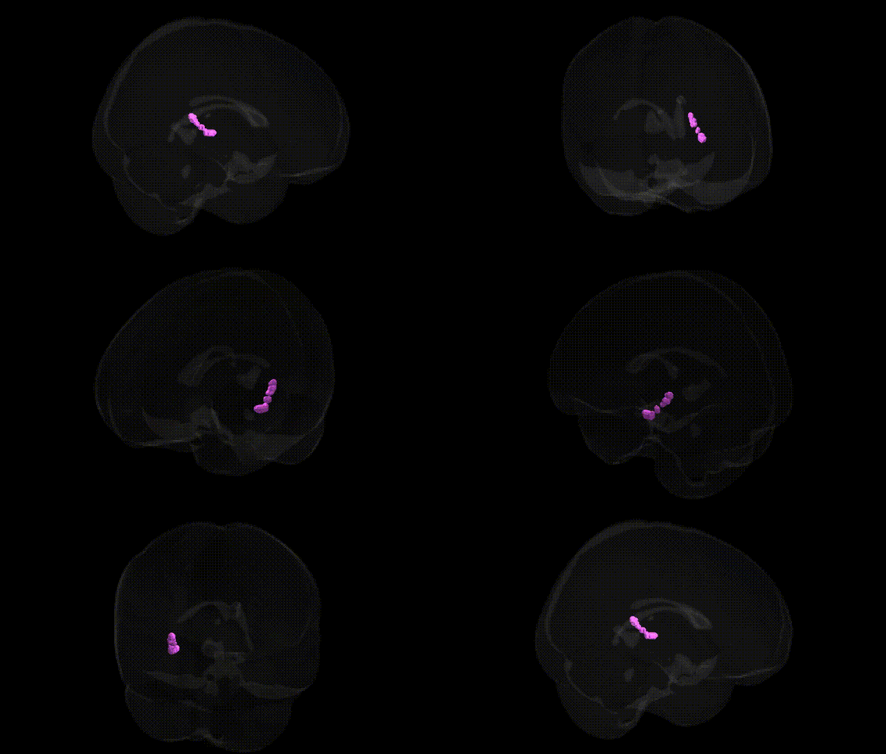
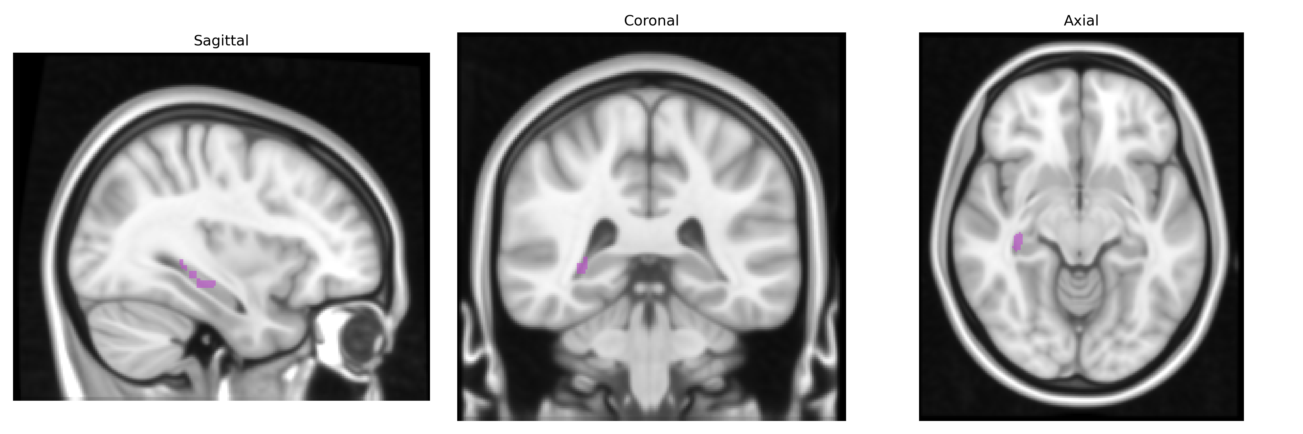
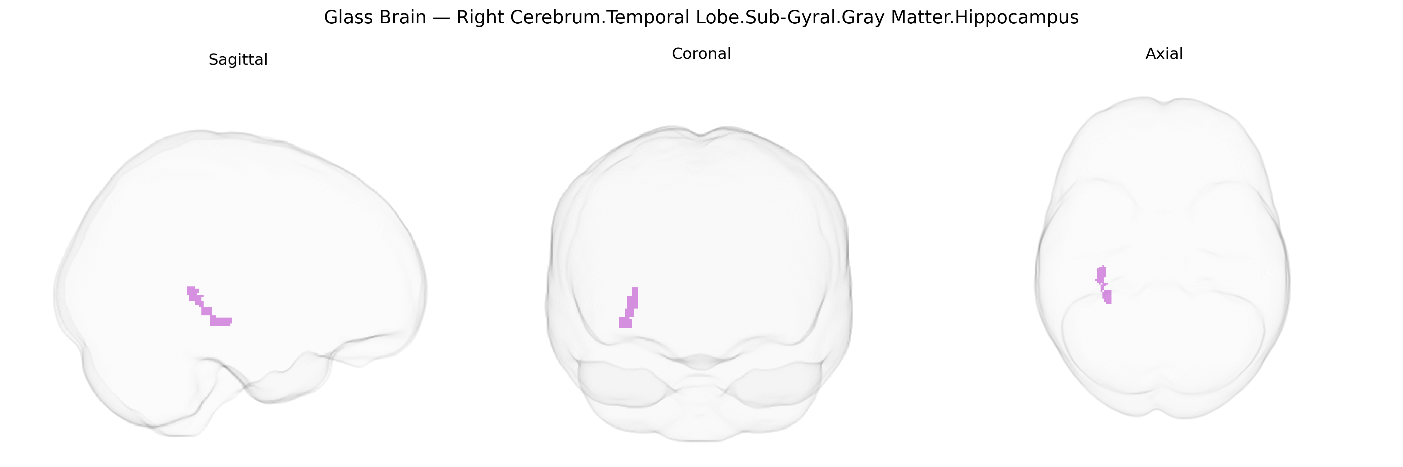
The bilateral Right Cerebrum.Temporal Lobe.Sub-Gyral.Gray Matter.Hippocampus corresponds to the hippocampal formation located deep within the medial temporal lobes, comprising primarily gray matter structures involved in learning, memory consolidation, spatial navigation, and contextual processing of experiences. This region receives multimodal cortical input and projects extensively to limbic and cortical association areas, supporting the encoding of episodic and declarative memories and playing a critical role in the formation of long-term memories from short-term traces. It is also highly plastic, exhibits a characteristic layered cytoarchitecture (notably in the cornu ammonis and dentate gyrus), and is vulnerable to hypoxia, neurodegenerative processes, and temporal lobe epilepsy. Hippocampus
The hippocampal gray matter in the bilateral right temporal sub-gyral region, as defined in the Talairach 1 mm Atlas, is strongly implicated in genetic studies of memory, neurodegeneration, and psychiatric illness: GWAS of hippocampal volume and shape (e.g., ENIGMA and UK Biobank consortia) have identified common variants in genes such as APOE (notably ε4), SORL1, CLU, PICALM, and BIN1 that influence hippocampal atrophy and Alzheimer’s disease risk; additional loci (e.g., in MS4A, CR1, and ABCA7) have been associated with hippocampal structure and amyloid-related pathology. Polygenic risk scores for Alzheimer’s disease, schizophrenia, and major depressive disorder correlate with reduced hippocampal volume or altered microstructure, suggesting shared genetic architectures affecting this region across disorders. Variants in BDNF (particularly Val66Met), COMT, and genes involved in glutamatergic signaling and synaptic plasticity have been linked to individual differences in hippocampal-dependent episodic memory, stress reactivity, and hippocampal volume or connectivity. Large-scale imaging-genetics analyses also implicate genes involved in neurodevelopment and axon guidance (e.g., DCC, ROBO pathways) in shaping hippocampal morphology, and common risk alleles for temporal lobe epilepsy and mood disorders show convergent effects on this region, reinforcing its role as a genetically modulated hub for cognition, emotional regulation, and vulnerability to neuropsychiatric disease.
Overview generated by GPT-4o (2026).
Region ID: 338
Hemisphere: bilateral
Atlas: Talairach labels 1mm
Full Quality Version: Download MP4
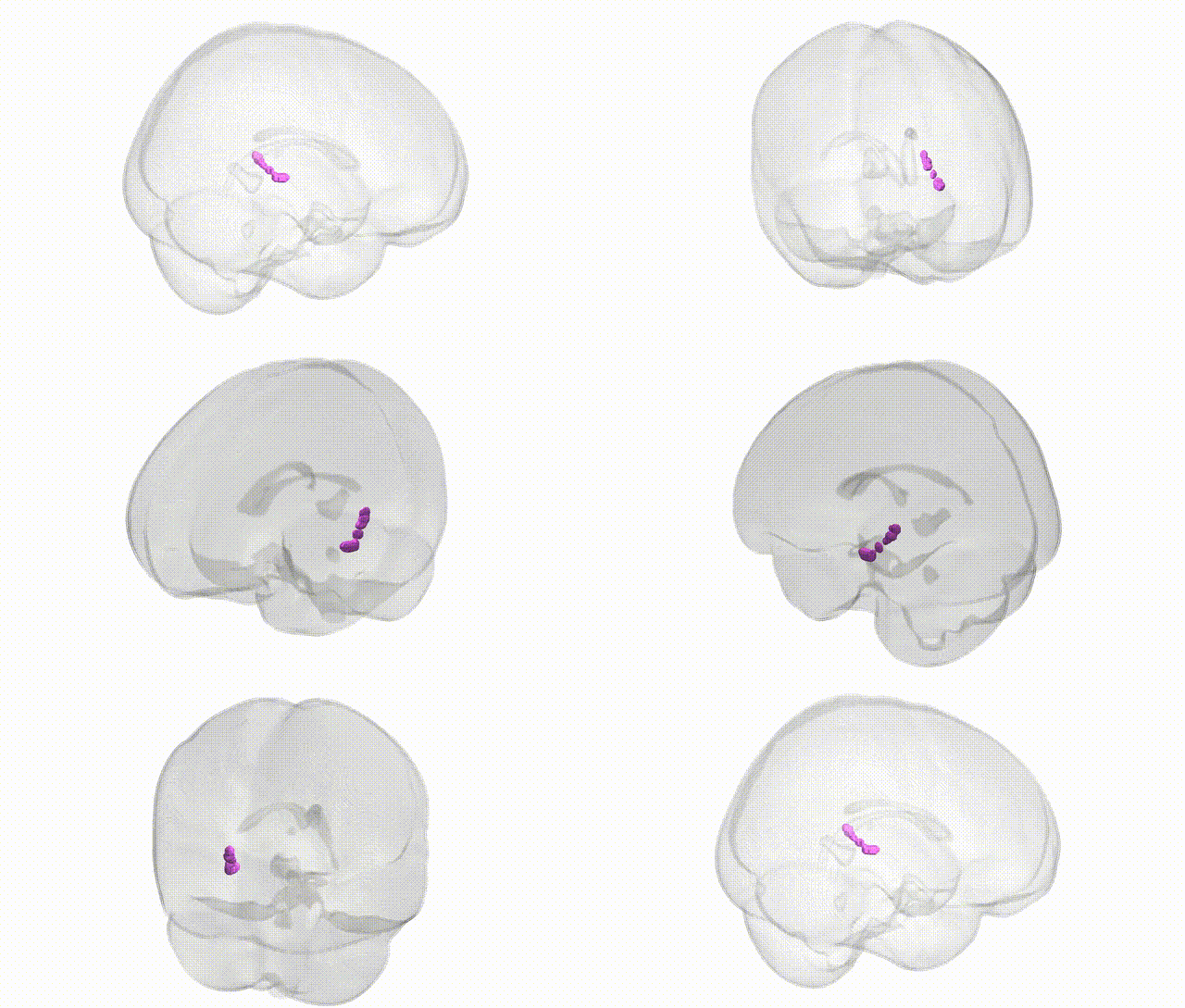
Full Quality Version: Download MP4
Wali Sidiqyar*, Gaurav Rudravaram*, Elyssa M. McMaster, Trent M. Schwartz, Adam M. Saunders, Kurt G. Schilling, Bennett A. Landman "Introducing SPINS: A Shared Public Visualization Library of Neuroanatomical Structures." Medical Imaging with Deep Learning- short paper
This resource is licensed under CC0 1.0 Universal (Public Domain).