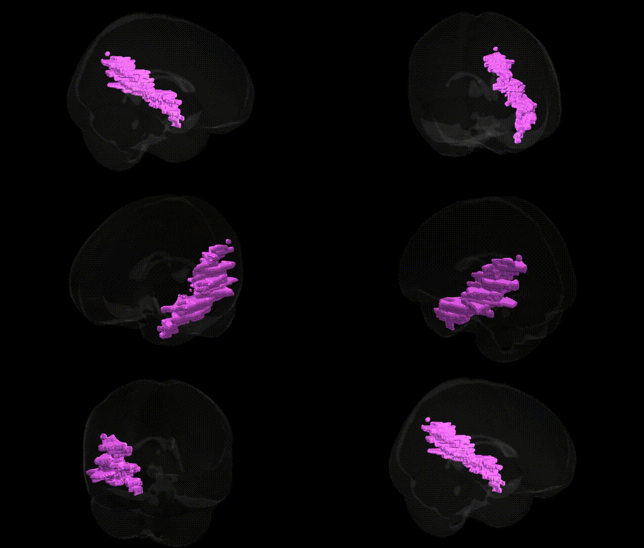
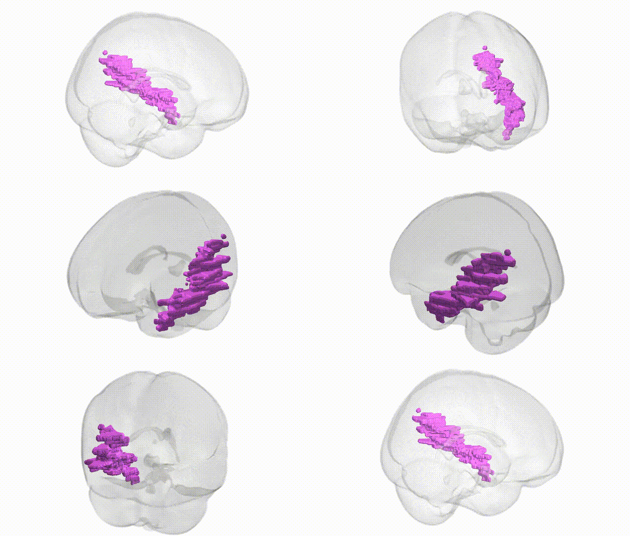
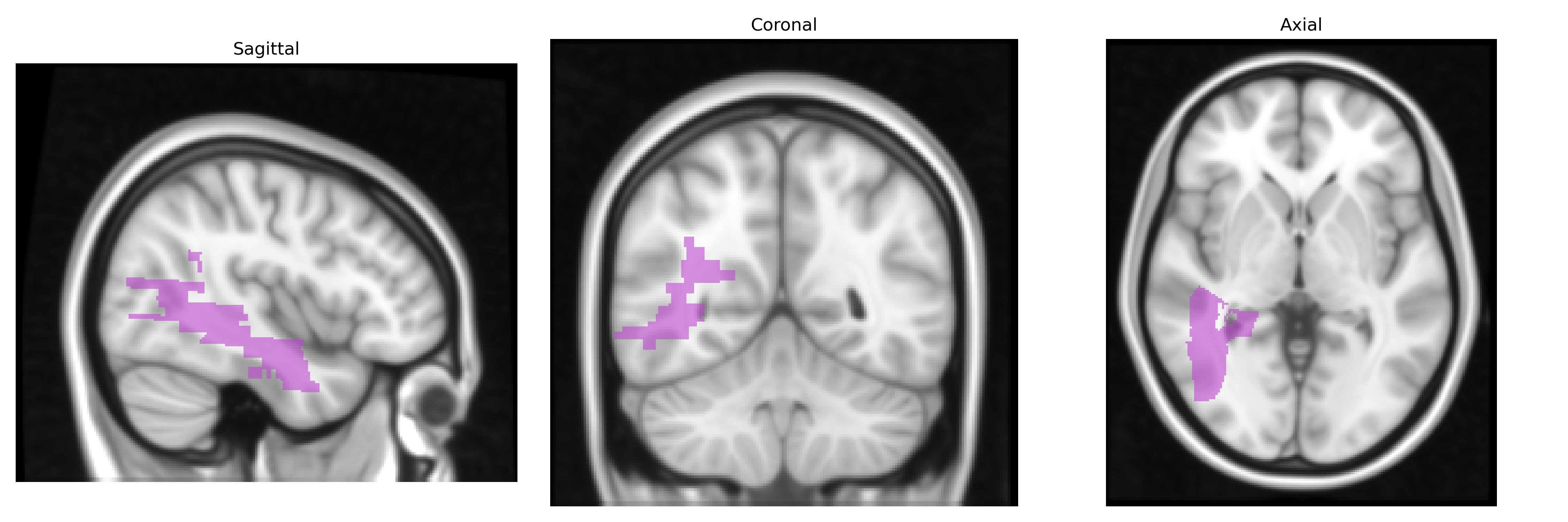
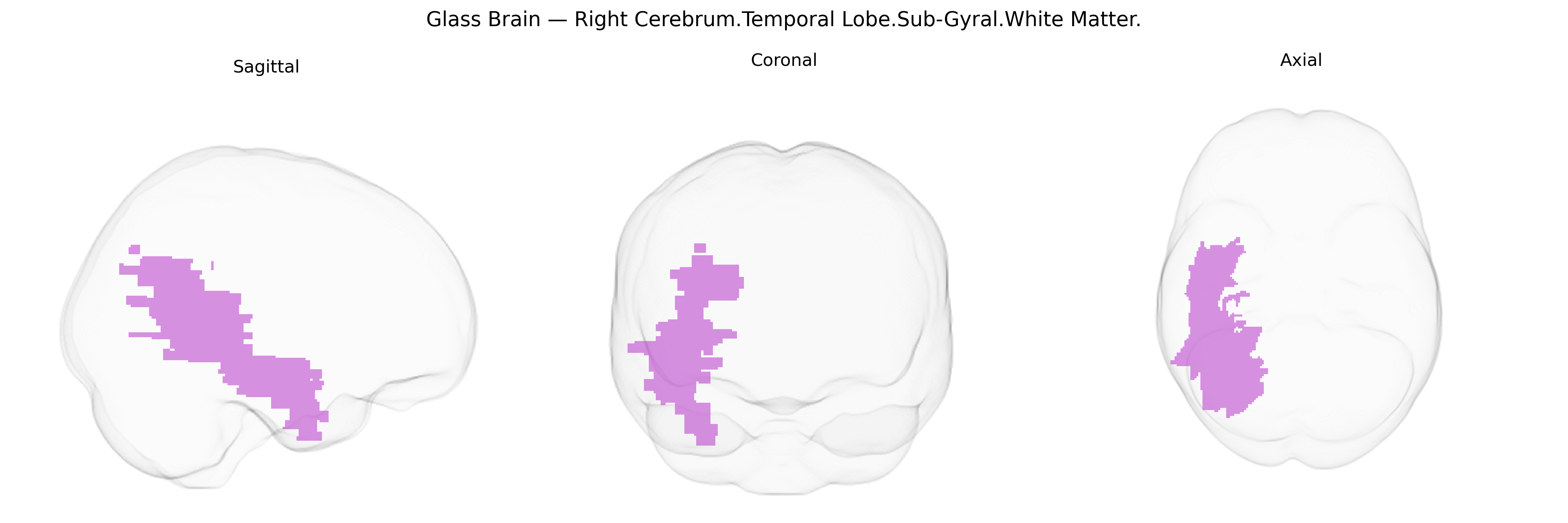
The bilateral right cerebrum temporal lobe sub-gyral white matter comprises the deep myelinated fiber tracts located beneath the cortical gyri of the right temporal lobe, including association, commissural, and projection pathways that interconnect temporal cortical areas with each other and with distant regions such as frontal, parietal, limbic, and subcortical structures. These fibers support integration of auditory processing, language (particularly prosody and aspects of speech perception), semantic memory, object and face recognition, and components of socio-emotional and mnemonic functions attributed to the temporal lobe. The sub-gyral white matter includes portions of major tracts such as the inferior longitudinal fasciculus, inferior fronto-occipital fasciculus, and arcuate/uncinate systems, and serves as a critical conduit for information flow between superior, middle, and inferior temporal gyri and deeper limbic and thalamic circuits. There is no direct link; related structure: Temporal lobe.
Genetic associations involving the bilateral right temporal sub-gyral white matter (as approximated by Talairach 1 mm atlas labels) derive largely from genome-wide and imaging-genetics studies of temporal lobe white-matter microstructure, temporal lobe connectivity, and related neuropsychiatric and neurodegenerative conditions rather than this specific parcel alone. Variants in genes affecting myelination and axonal integrity (such as those in the MAG, MBP, CNTNAP2, and NRG1 pathways) have been linked to diffusion MRI measures (e.g., fractional anisotropy) in temporal white matter, while large consortia (ENIGMA and UK Biobank) have identified polygenic influences on temporal lobe volume and white-matter tracts coursing through sub-gyral regions, including parts of the inferior and middle temporal and adjacent association fibers. GWAS of schizophrenia, bipolar disorder, autism spectrum disorder, and major depression implicate temporal white-matter alterations and highlight common risk loci in synaptic and neurodevelopmental genes (e.g., CACNA1C, DISC1, GRIN2A) that show downstream effects on temporal lobe connectivity. In Alzheimer’s disease and other dementias, APOE ε4 and additional GWAS loci (e.g., CLU, PICALM) associate with temporal lobe white-matter degeneration and altered diffusion metrics, while temporal white-matter changes in carriers of mutations in presenilin genes and MAPT reinforce a genetic contribution to vulnerability in this region. Overall, genetic findings converge on pathways involved in synaptic plasticity, neurodevelopment, and myelin biology that modulate the structure and integrity of temporal sub-gyral white matter and, by extension, risk for cognitive decline and major psychiatric illnesses, even though explicit, parcel-level GWAS for the “Right Cerebrum.Temporal Lobe.Sub-Gyral.White Matter” label remain limited.
Overview generated by GPT-4o (2026).
Region ID: 52
Hemisphere: bilateral
Atlas: Talairach labels 1mm
Full Quality Version: Download MP4
Full Quality Version: Download MP4
Wali Sidiqyar*, Gaurav Rudravaram*, Elyssa M. McMaster, Trent M. Schwartz, Adam M. Saunders, Kurt G. Schilling, Bennett A. Landman "Introducing SPINS: A Shared Public Visualization Library of Neuroanatomical Structures." Medical Imaging with Deep Learning- short paper
This resource is licensed under CC0 1.0 Universal (Public Domain).