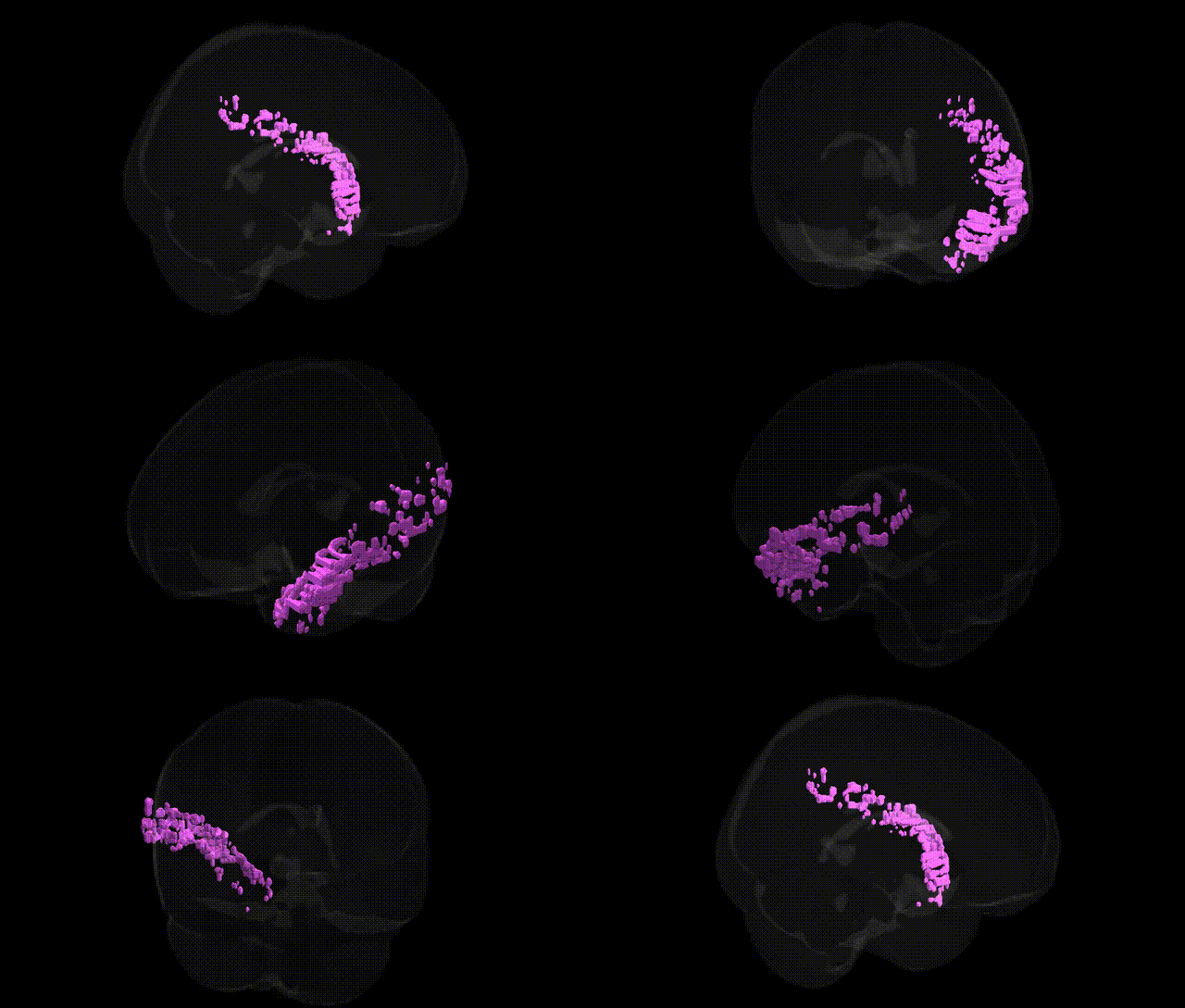
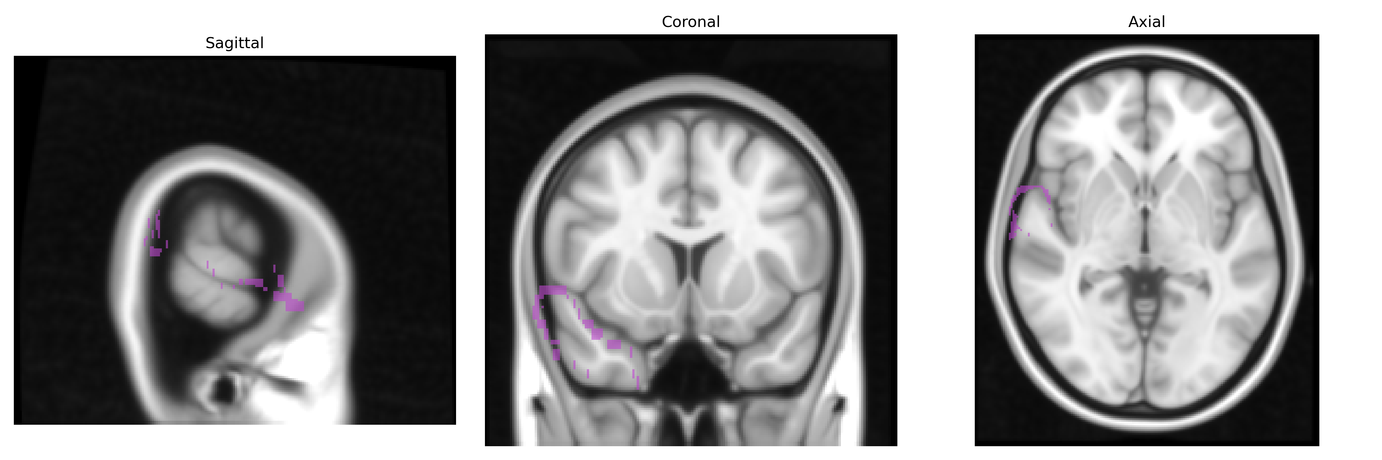
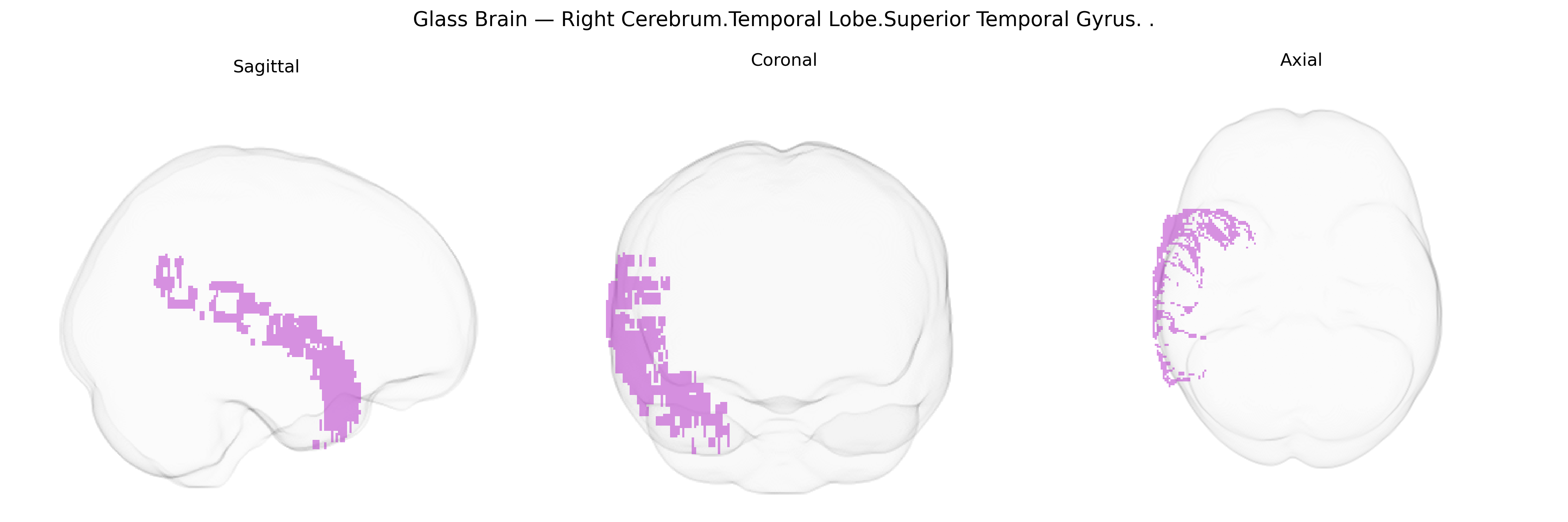
The bilateral Right Cerebrum.Temporal Lobe.Superior Temporal Gyrus corresponds to the right superior temporal gyrus (STG), a key neocortical region of the temporal lobe situated between the lateral sulcus (Sylvian fissure) and the superior temporal sulcus. It encompasses primary and secondary auditory cortices (including Heschl’s gyrus on the transverse temporal plane) and participates in processing complex acoustic features, speech perception, phonological decoding, and aspects of language comprehension, as well as integration of auditory with multisensory information. Functionally, the right STG is particularly associated with prosody, music and pitch processing, and the perception of voice and environmental sounds, and it is implicated in social cognition and aspects of emotional processing of auditory stimuli. Lesions or dysfunction in this region can lead to deficits in auditory perception, impairments in understanding speech intonation and emotional tone, and disturbances in higher-order auditory–language functions. Superior temporal gyrus
The bilateral right superior temporal gyrus (STG) of the temporal lobe has been repeatedly implicated in genetic studies of language, social cognition, and psychiatric and neurodevelopmental disorders. Twin and family studies indicate high heritability of STG volume and cortical thickness, with GWAS identifying common variants in genes involved in neurodevelopment, synaptic structure, and axon guidance (for example, loci near DCDC2, CNTNAP2, and FOXP2 in language and reading-related cohorts, and microdeletion/duplication regions such as 16p11.2 and 22q11.2 for broader temporal lobe phenotypes). Imaging–genetics and large-scale consortia (e.g., ENIGMA) have linked temporal lobe structural variation, including in the superior temporal gyrus, to schizophrenia, bipolar disorder, major depression, and autism spectrum disorder, often implicating genes in glutamatergic and GABAergic signaling, neuronal adhesion (e.g., NRXN1, NRG1), and calcium-channel function. GWAS of auditory hallucinations, psychosis-proneness, and social communication traits have associated risk loci with altered right STG volume or functional activation, while language and reading GWAS highlight temporal cortex–related variants contributing to dyslexia and specific language impairment. Additionally, polygenic risk scores for schizophrenia, autism, and Alzheimer’s disease show associations with temporal cortical morphology, suggesting that genetic liability for these conditions partly converges on right STG structure and connectivity, although most findings reflect distributed networks rather than a region-specific genetic signature.
Overview generated by GPT-4o (2026).
Region ID: 43
Hemisphere: bilateral
Atlas: Talairach labels 1mm
Full Quality Version: Download MP4
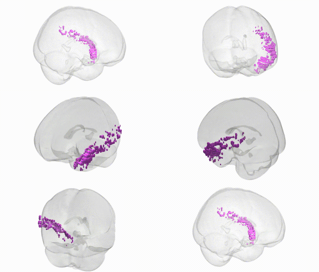
Full Quality Version: Download MP4
Wali Sidiqyar*, Gaurav Rudravaram*, Elyssa M. McMaster, Trent M. Schwartz, Adam M. Saunders, Kurt G. Schilling, Bennett A. Landman "Introducing SPINS: A Shared Public Visualization Library of Neuroanatomical Structures." Medical Imaging with Deep Learning- short paper
This resource is licensed under CC0 1.0 Universal (Public Domain).