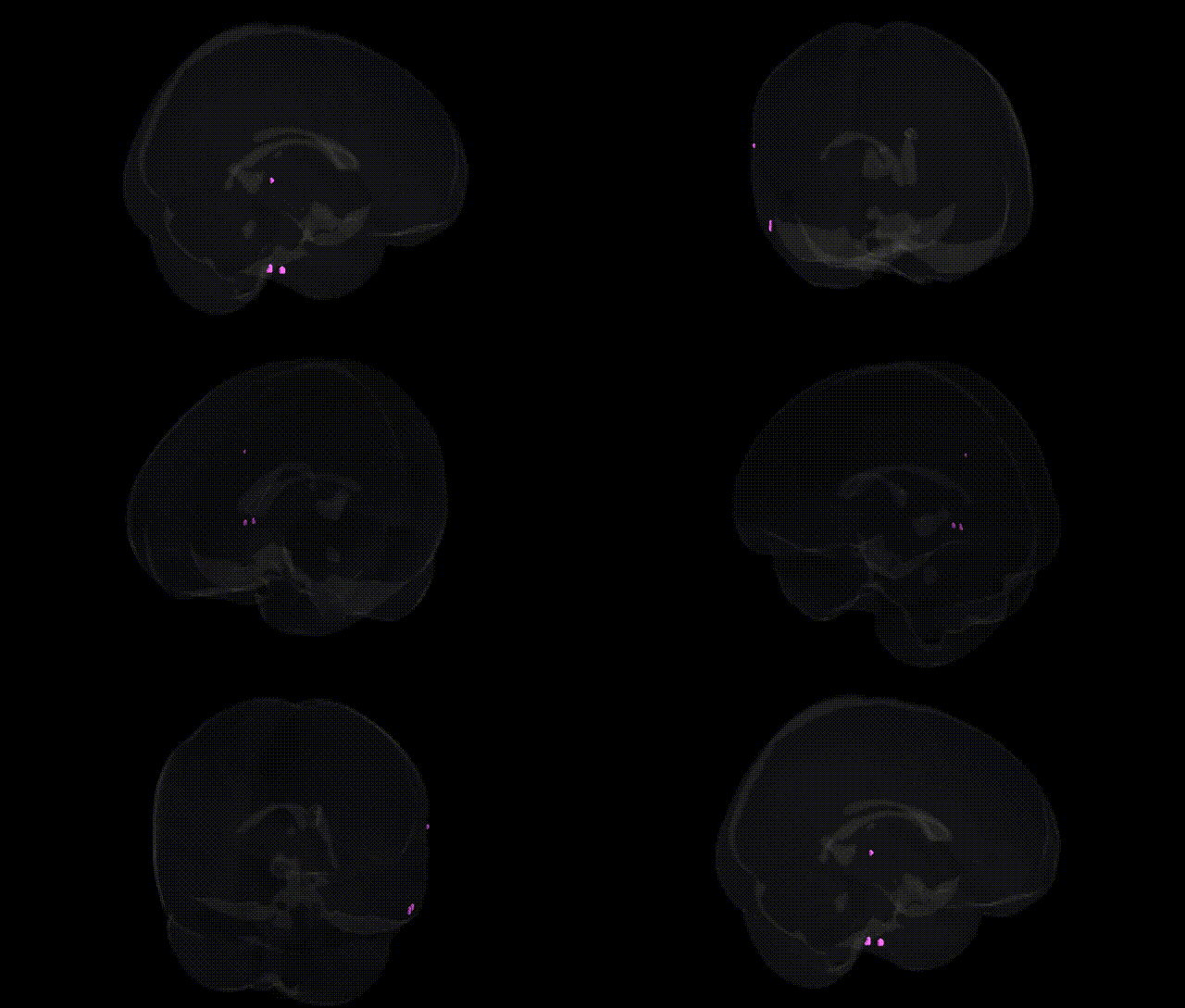
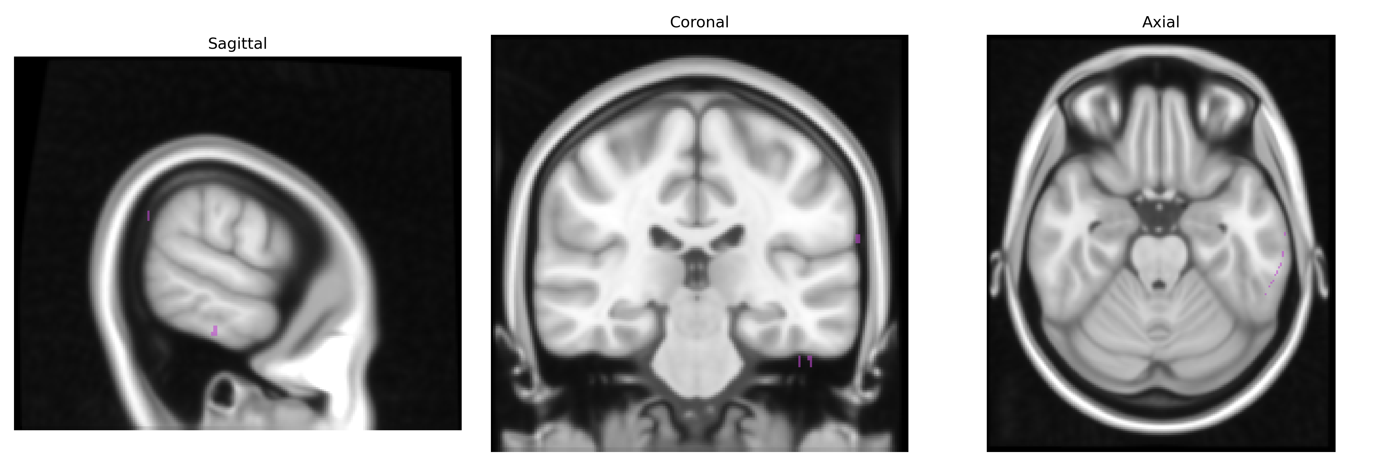
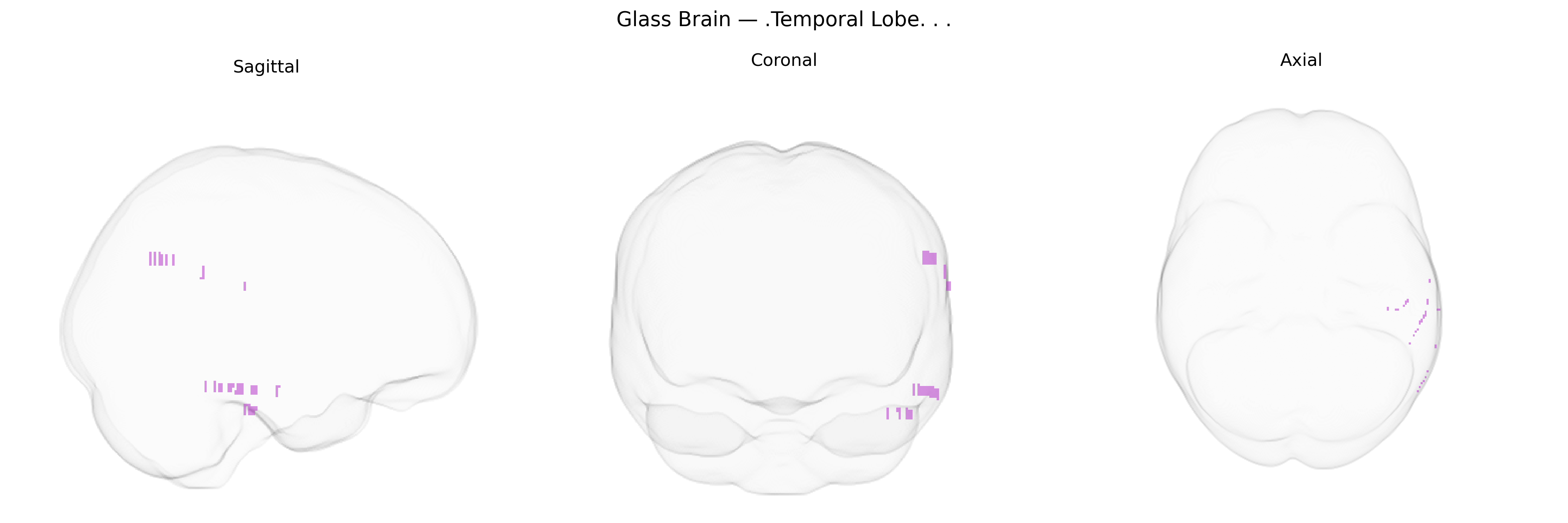
The bilateral temporal lobe is a paired cerebral cortical region located inferior to the lateral (Sylvian) fissure and lateral to the inferior horn of the lateral ventricles, extending from the temporal pole posteriorly toward the occipital lobe. It comprises lateral neocortical areas (e.g., superior, middle, and inferior temporal gyri) as well as medial temporal structures (including parts of the hippocampal formation and parahippocampal region), although the precise extent depends on the specific atlas definition, such as the Talairach 1 mm Atlas. Functionally, the temporal lobe subserves auditory processing (primary and secondary auditory cortex), language comprehension (typically dominant hemisphere posterior superior temporal gyrus), high-level visual and object recognition, as well as memory encoding and retrieval via the medial temporal lobe circuitry. It also contributes to emotional processing and multimodal integration through extensive reciprocal connections with frontal, parietal, limbic, and subcortical structures. Temporal lobe
Genetic associations with the bilateral temporal lobe, as defined in the Talairach 1 mm atlas, have been implicated across neurodevelopmental, neuropsychiatric, and neurodegenerative conditions, as well as structural and functional brain phenotypes identified in GWAS of imaging traits. Variants in genes such as DISC1, NRG1, RELN, CNTNAP2, and those in the 22q11.2 deletion region have been repeatedly linked to temporal lobe structure and function in schizophrenia and related psychotic disorders, often through reduced temporal gray matter volume or altered activation during language and auditory tasks. APOE ε4 and other Alzheimer’s disease risk loci (e.g., in CLU, PICALM, CR1) show robust associations with medial and lateral temporal atrophy, particularly in hippocampal and parahippocampal regions, and with progression of temporal-lobe–predominant neurodegeneration. Large-scale imaging genetics consortia (e.g., ENIGMA, UK Biobank) have identified common variants in genes related to neuronal development and synaptic function, including HMGA2, DCC, and others near loci on chromosomes 3, 5, and 12, that correlate with temporal lobe volume, cortical thickness, and surface area. Temporal lobe–linked genetic effects are also observed in autism spectrum disorder (e.g., SHANK3, NRXN1, and other synaptic genes), dyslexia and language disorders (e.g., KIAA0319, DCDC2, FOXP2 network genes), and epilepsy, where loci such as LGI1 and GRIN2A, as well as polygenic burden for excitatory–inhibitory imbalance, have been associated with temporal lobe epilepsy and related structural alterations. Overall, GWAS and candidate gene studies converge on the temporal lobe as a key anatomical substrate through which diverse risk variants for cognition, language, psychiatric illness, and neurodegeneration exert measurable effects on brain structure and function.
Overview generated by GPT-4o (2026).
Region ID: 90
Hemisphere: bilateral
Atlas: Talairach labels 1mm
Full Quality Version: Download MP4
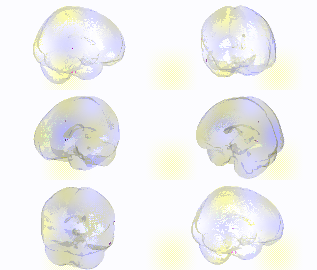
Full Quality Version: Download MP4
Wali Sidiqyar*, Gaurav Rudravaram*, Elyssa M. McMaster, Trent M. Schwartz, Adam M. Saunders, Kurt G. Schilling, Bennett A. Landman "Introducing SPINS: A Shared Public Visualization Library of Neuroanatomical Structures." Medical Imaging with Deep Learning- short paper
This resource is licensed under CC0 1.0 Universal (Public Domain).