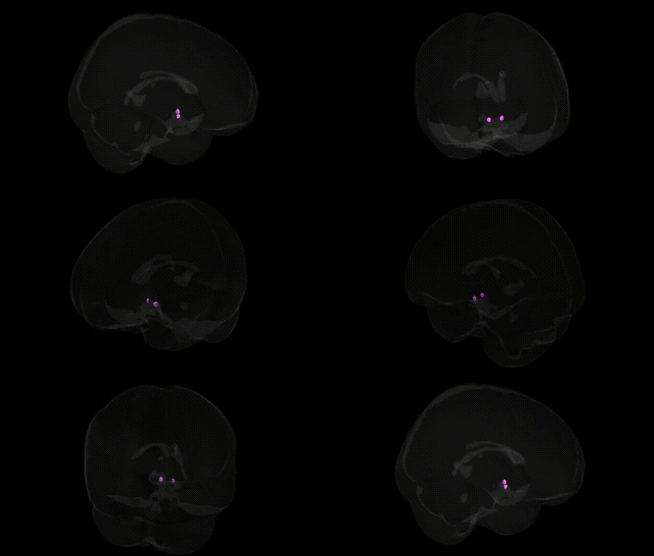
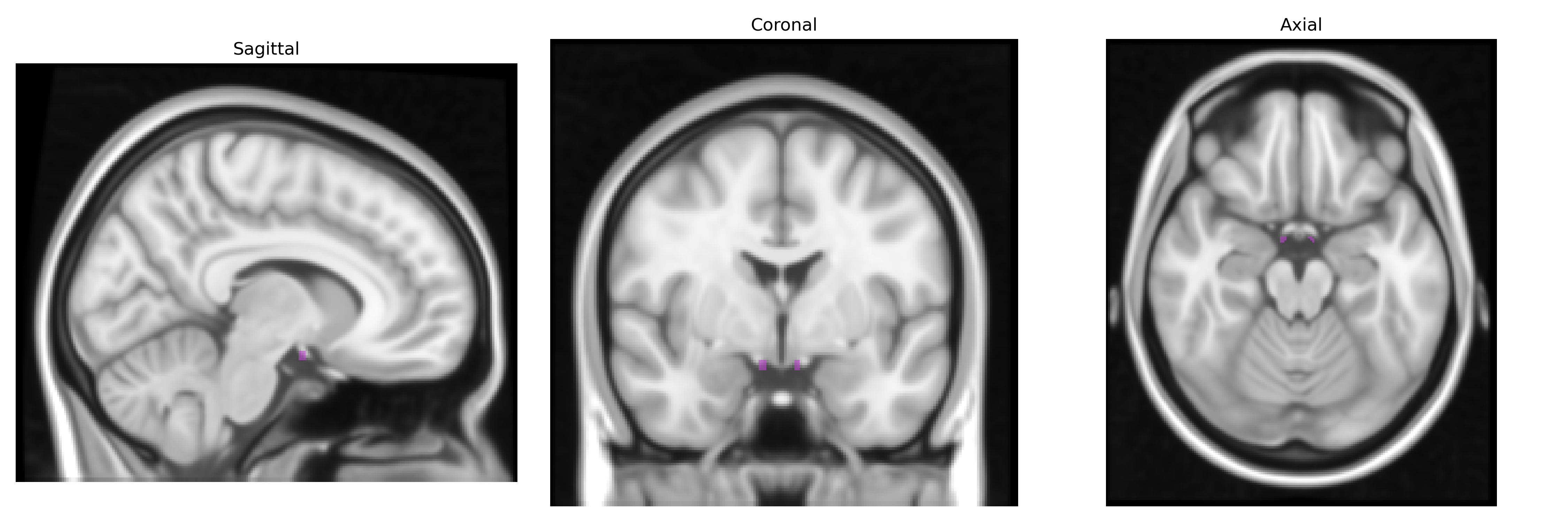
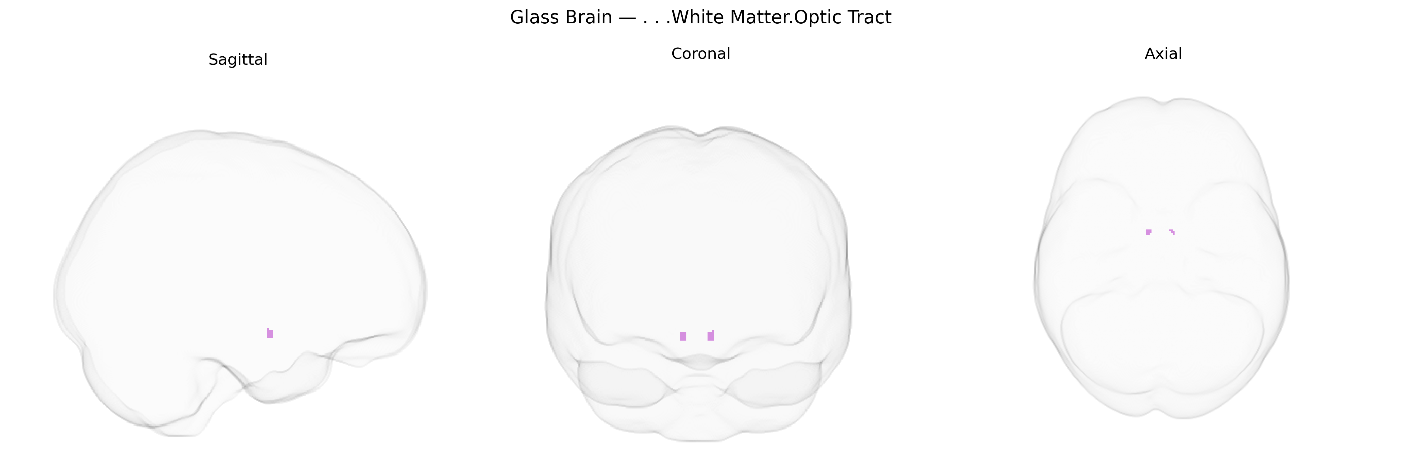
The bilateral White Matter Optic Tract comprises paired bundles of myelinated axons that carry visual information from the optic chiasm to the lateral geniculate nucleus of the thalamus, as well as to other visual relay centers such as the pretectal area and superior colliculus. These tracts represent post-chiasmatic continuation of retinal ganglion cell fibers, transmitting signals that have already undergone partial decussation, and thus encode information from contralateral visual hemifields. Structurally, the optic tracts course posterolaterally around the cerebral peduncles within the white matter, maintaining a retinotopic organization critical for subsequent visual processing in thalamic and cortical regions. There is no direct link for “White Matter Optic Tract,” but it is part of the Optic tract.
The bilateral White Matter Optic Tract, which carries retinal ganglion cell axons to the lateral geniculate nucleus, is not typically isolated as a specific GWAS target but falls within broader genetic findings on optic nerve, visual pathway, and white matter integrity. Variants in genes involved in myelination and axonal structure (e.g., MBP, MAG, PLP1), oligodendrocyte function (e.g., MOG, SOX10), and general neurodevelopment (e.g., L1CAM, ROBO3, ATOH7) have been implicated in disorders where optic tract/optic radiation involvement is common, such as multiple sclerosis, leukodystrophies, congenital optic nerve hypoplasia, and hereditary spastic paraplegias, often via demyelination or axonal degeneration observed on MRI. Large imaging-genetics studies (e.g., UK Biobank) have linked common variants in genes regulating axon guidance, synaptic signaling, and white matter microstructure (including loci near EPHA, NTRK, and cell-adhesion genes) to diffusion MRI measures in visual pathway tracts, including optic radiations and adjacent white matter, though optic tract–specific associations are typically reported at the level of broader visual white matter networks. In glaucoma and other optic neuropathies, risk variants in genes such as MYOC, OPTN, and TBK1 contribute to retinal ganglion cell loss that secondarily affects the optic nerve and tract, while mitochondrial genes (e.g., MT-ND family in Leber’s hereditary optic neuropathy) have been linked to degeneration of the anterior visual pathway with downstream white matter changes. Overall, genetic associations for this Talairach-defined region are inferred largely from disorders and quantitative imaging traits that impact the visual white matter pathway rather than from region-specific GWAS signals.
Overview generated by GPT-4o (2026).
Region ID: 273
Hemisphere: bilateral
Atlas: Talairach labels 1mm
Full Quality Version: Download MP4
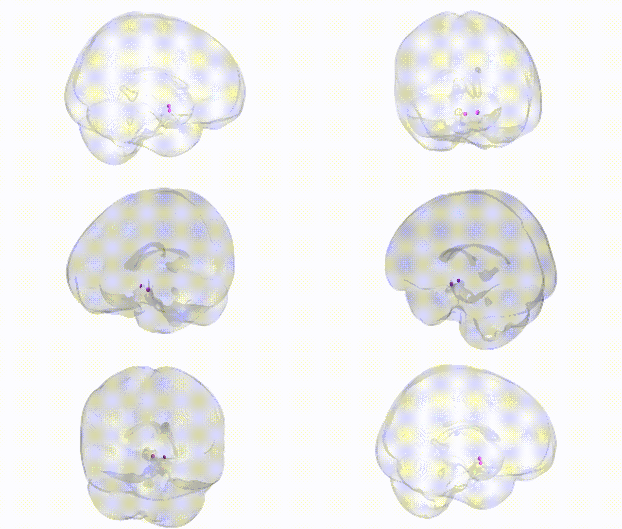
Full Quality Version: Download MP4
Wali Sidiqyar*, Gaurav Rudravaram*, Elyssa M. McMaster, Trent M. Schwartz, Adam M. Saunders, Kurt G. Schilling, Bennett A. Landman "Introducing SPINS: A Shared Public Visualization Library of Neuroanatomical Structures." Medical Imaging with Deep Learning- short paper
This resource is licensed under CC0 1.0 Universal (Public Domain).