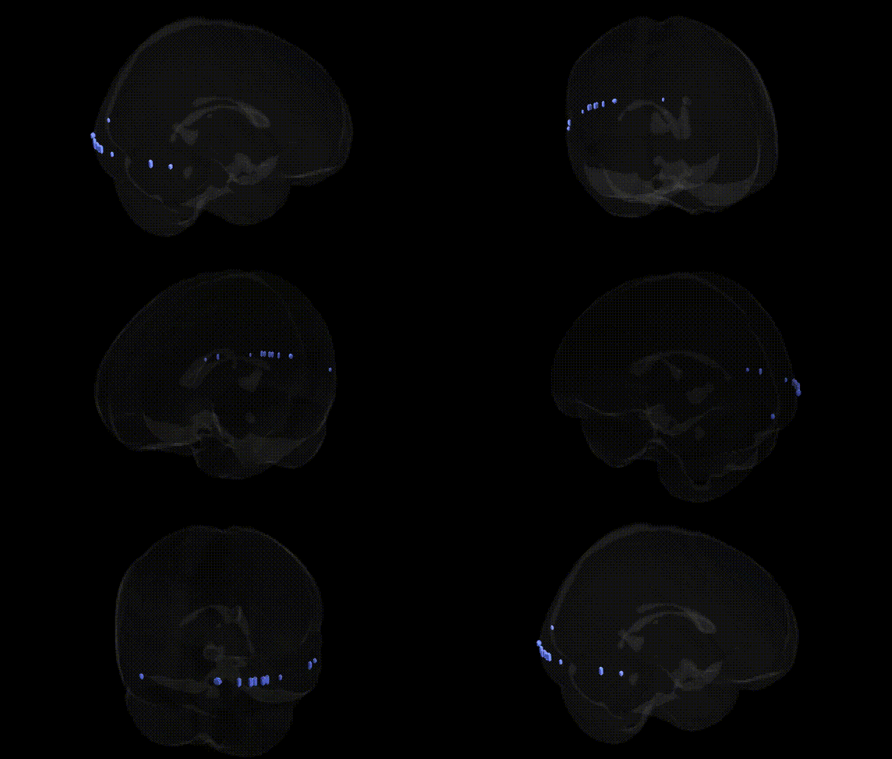
The bilateral gray matter comprises the neuronal cell bodies, dendrites, unmyelinated axons, synapses, and glial cells that form the core processing substrate of the central nervous system. In the brain, gray matter is organized into cortical layers and subcortical nuclei that support perception, motor control, memory, emotion, and executive functions, with bilateral distribution across both hemispheres. It is highly vascularized and metabolically active, serving as the primary site for synaptic integration and neural computation, in contrast to the predominantly myelinated fiber tracts of white matter that mediate long-range signal transmission. Gray matter volume and integrity are critical biomarkers in neurodevelopment, aging, and various neurological and psychiatric conditions. There is no direct link for “Gray Matter” as a specific Talairach region; a closely related article is Grey matter.
The bilateral gray matter, as defined in the Talairach 2 mm atlas, encompasses distributed cortical and subcortical tissue rather than a single discrete functional region; accordingly, genetic associations arise from studies of total or regional gray matter volume and cortical thickness rather than this specific atlas label. Large neuroimaging GWAS efforts, such as ENIGMA and UK Biobank–based studies, have identified numerous common variants associated with global and regional gray matter measures, including loci near genes involved in neurodevelopment (e.g., HMGA2, MEF2C, BDNF, DCC, CNTNAP2) and synaptic function (e.g., GRIN2B, CACNA1C). Polygenic risk scores for schizophrenia, bipolar disorder, major depressive disorder, ADHD, and autism spectrum disorder are consistently associated with reductions or alterations in gray matter volume and cortical thickness across widespread bilateral regions, particularly frontal and temporal cortices and hippocampus, suggesting shared genetic architectures between psychiatric risk and gray matter morphology. Similarly, genetic liability for Alzheimer’s disease and other neurodegenerative disorders (e.g., APOE, BIN1, CLU variants) is linked to reduced gray matter in medial temporal and parietal areas, while genes influencing brain growth and intracranial volume (e.g., variants near HMGA2 and IGF1-related pathways) contribute to overall gray matter differences. Together, these findings indicate that the bilateral gray matter compartment is strongly shaped by highly polygenic influences spanning neurodevelopmental, synaptic, and neurodegenerative pathways, with many of the same variants implicated in cognitive traits, psychiatric disorders, and age-related brain atrophy.
Overview generated by GPT-4o (2026).
Region ID: 518
Hemisphere: bilateral
Atlas: Talairach labels 2mm
Full Quality Version: Download MP4
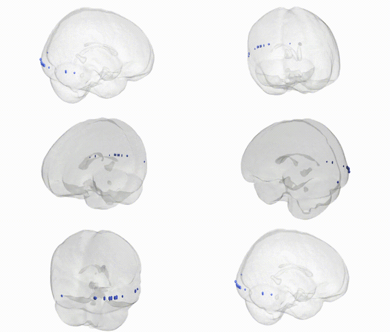
Full Quality Version: Download MP4
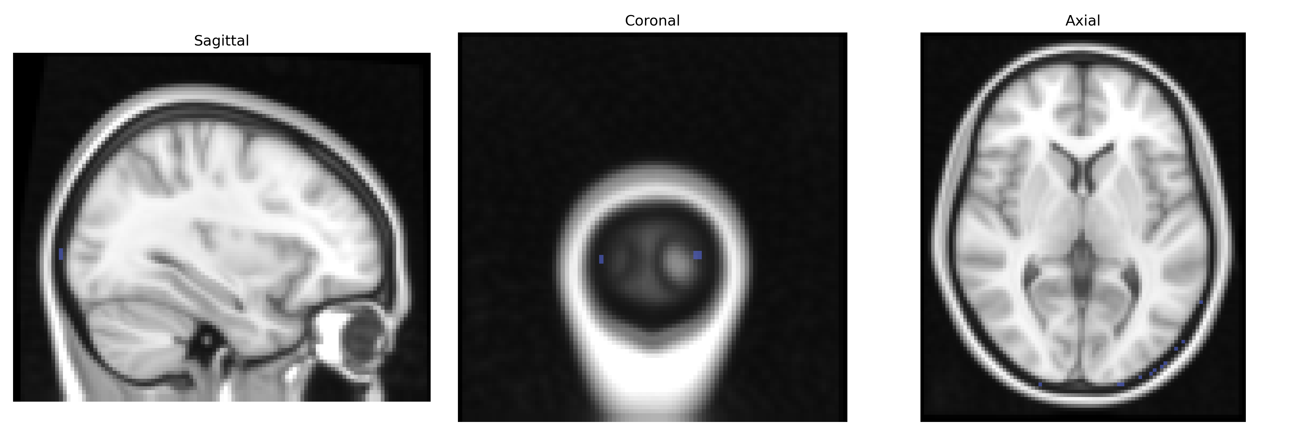
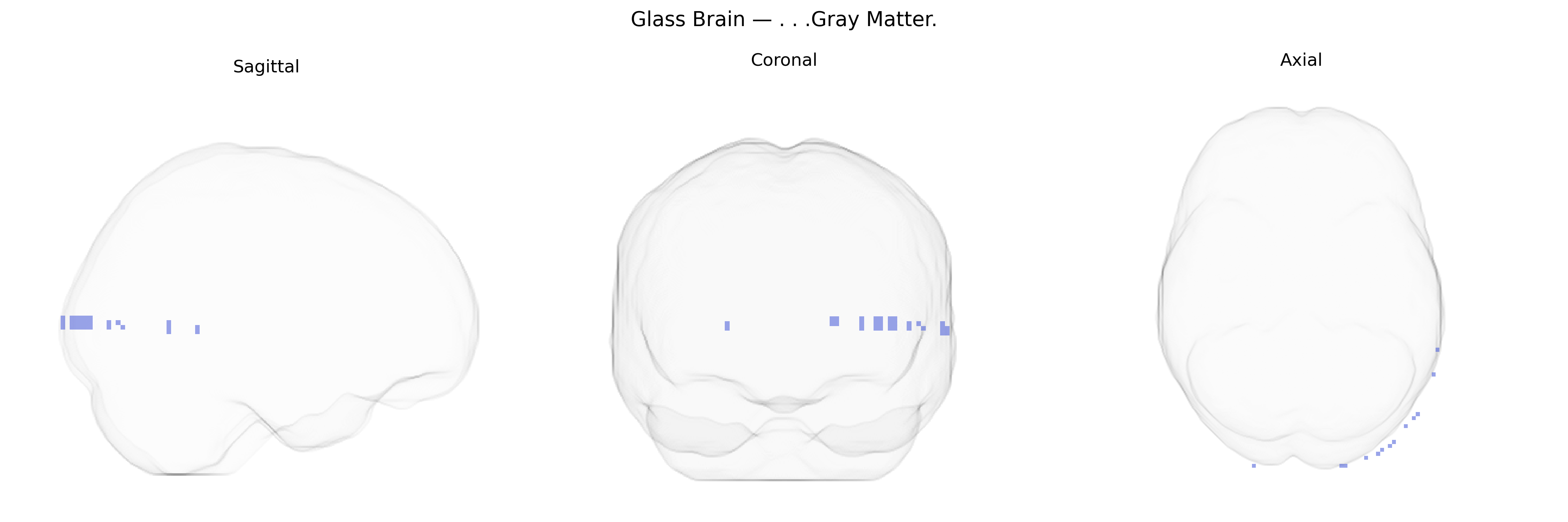
Wali Sidiqyar*, Gaurav Rudravaram*, Elyssa M. McMaster, Trent M. Schwartz, Adam M. Saunders, Kurt G. Schilling, Bennett A. Landman "Introducing SPINS: A Shared Public Visualization Library of Neuroanatomical Structures." Medical Imaging with Deep Learning- short paper
This resource is licensed under CC0 1.0 Universal (Public Domain).