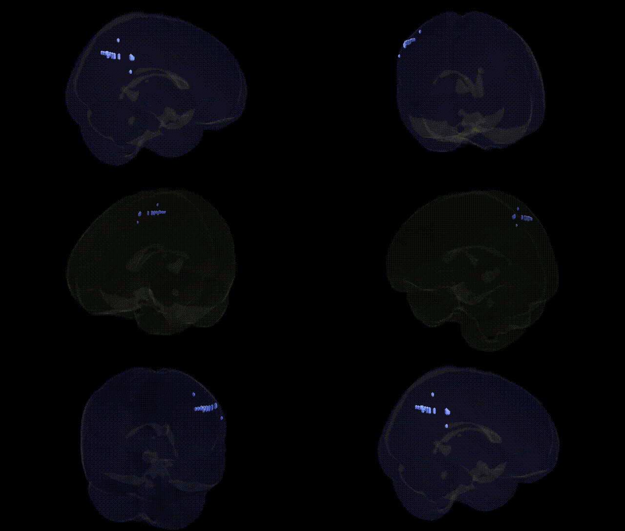
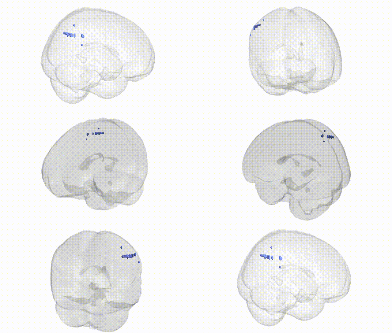
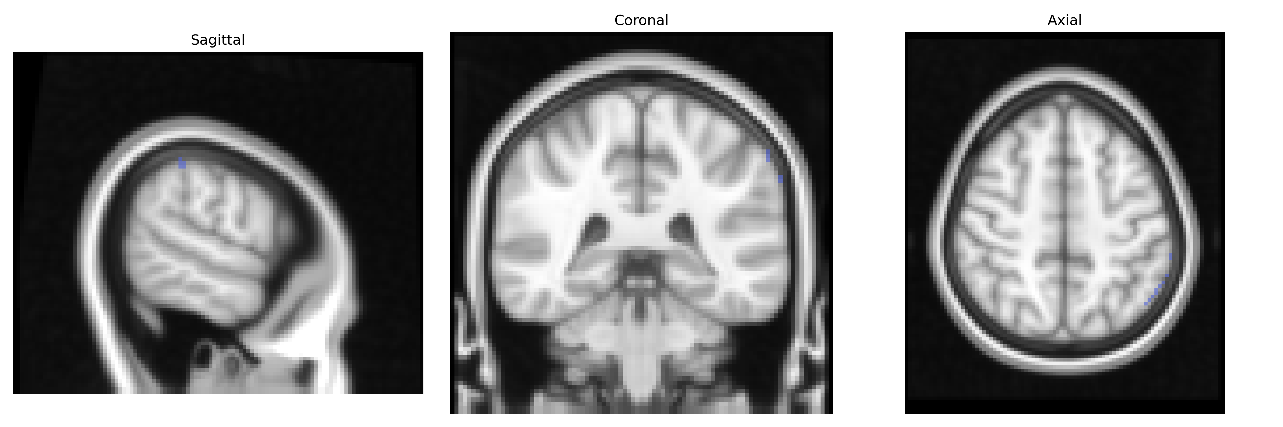
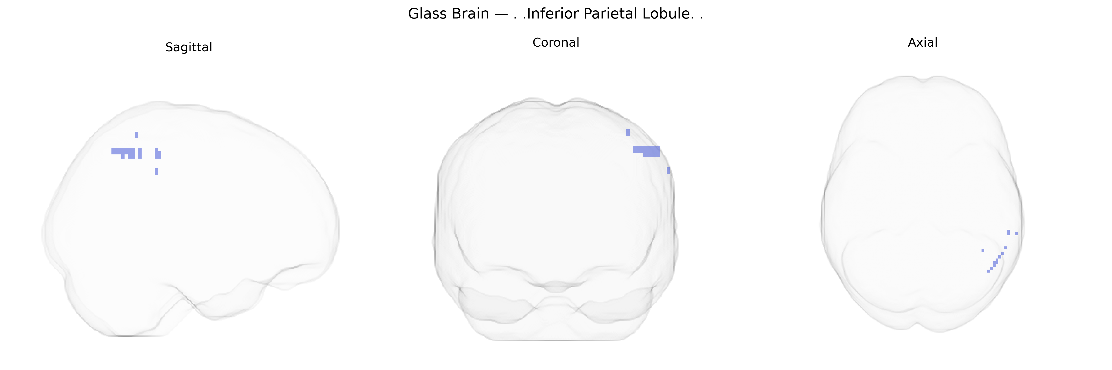
The bilateral Inferior Parietal Lobule (IPL) is a heteromodal associative cortex located in the posterior parietal lobe, bordering the superior temporal and occipital regions, and comprising mainly the angular gyrus and supramarginal gyrus. It integrates multimodal sensory information (visual, auditory, somatosensory) to support higher-order functions including spatial attention, body representation, visuomotor coordination, language processing (particularly semantic integration and phonological processing), numerical cognition, and aspects of social cognition such as theory of mind. The IPL is strongly lateralized functionally, with the left IPL more involved in language and symbolic processing and the right IPL more engaged in spatial attention and awareness. It forms key nodes in fronto-parietal and default mode networks, with extensive connectivity to prefrontal, temporal, and occipital cortices, and is frequently implicated in disorders of attention, neglect, apraxia, and language.
Inferior parietal lobule
The bilateral inferior parietal lobule (IPL), as defined in the Talairach 2 mm atlas, has been implicated in multiple genetic and genome-wide association studies through its roles in attention, language, social cognition, and higher-order integration. GWAS and imaging-genetics work have linked IPL structure and function to variants in genes associated with neurodevelopment and synaptic plasticity, including FOXP2, CNTNAP2, and DCDC2 for language and reading-related traits, and genes such as DRD2, COMT, and BDNF that modulate dopaminergic signaling and cortical plasticity in attentional and working memory networks. SNPs in CACNA1C, ZNF804A, and other major psychiatric risk loci have been associated with altered IPL activation and connectivity in schizophrenia and bipolar disorder, while APOE ε4 and additional Alzheimer’s disease risk variants (e.g., in CLU, CR1) relate to IPL atrophy, hypometabolism, and disrupted default mode network connectivity in aging and dementia. IPL abnormalities have also been tied to autism spectrum conditions and social cognition via genes affecting synapse formation and excitation–inhibition balance (e.g., SHANK3, NRXN1/3), and to structural and functional differences in attention-deficit/hyperactivity disorder involving dopaminergic and glutamatergic pathway genes. Across large neuroimaging GWAS, polygenic influences on cortical surface area and thickness highlight the IPL as a heritable association cortex region where common variants contribute to interindividual differences in cognition, educational attainment, and risk for multiple neuropsychiatric disorders.
Overview generated by GPT-4o (2026).
Region ID: 933
Hemisphere: bilateral
Atlas: Talairach labels 2mm
Full Quality Version: Download MP4
Full Quality Version: Download MP4
Wali Sidiqyar*, Gaurav Rudravaram*, Elyssa M. McMaster, Trent M. Schwartz, Adam M. Saunders, Kurt G. Schilling, Bennett A. Landman "Introducing SPINS: A Shared Public Visualization Library of Neuroanatomical Structures." Medical Imaging with Deep Learning- short paper
This resource is licensed under CC0 1.0 Universal (Public Domain).