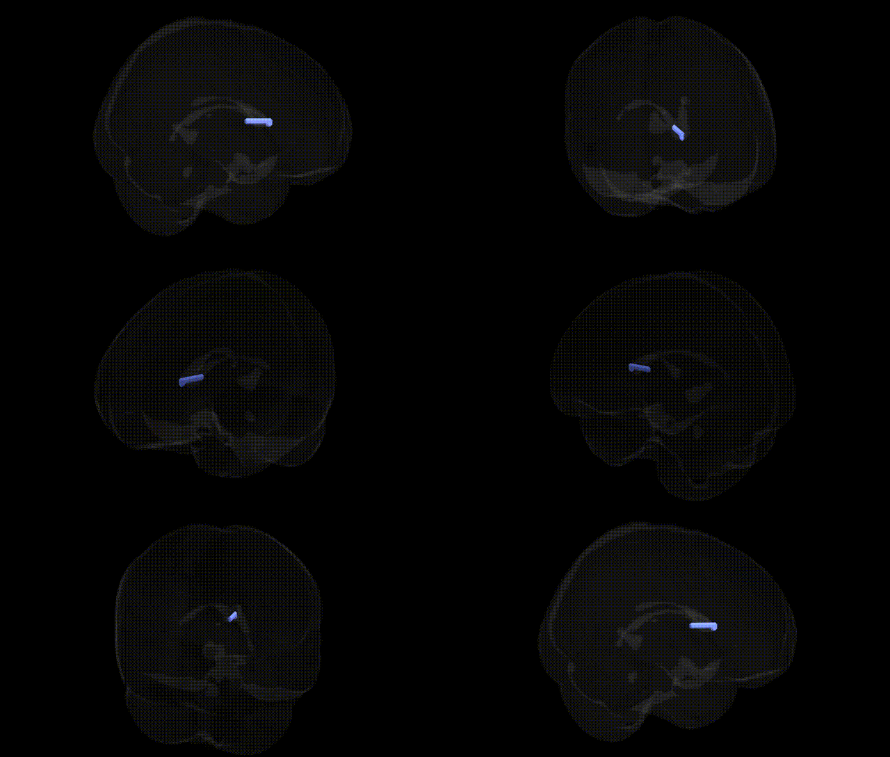
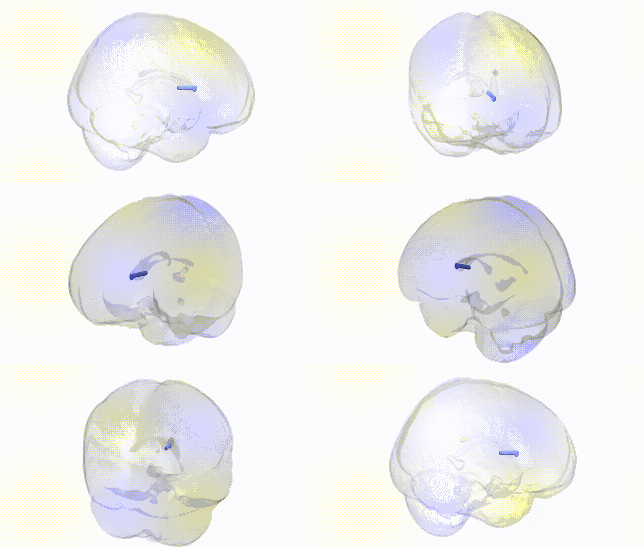
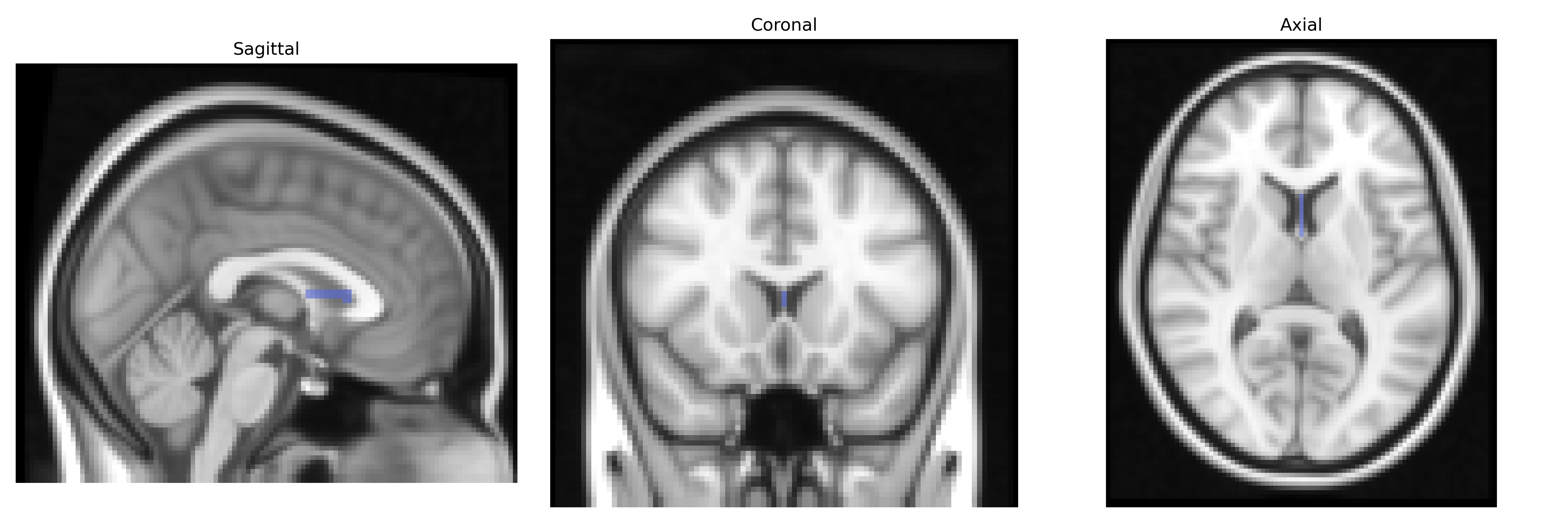
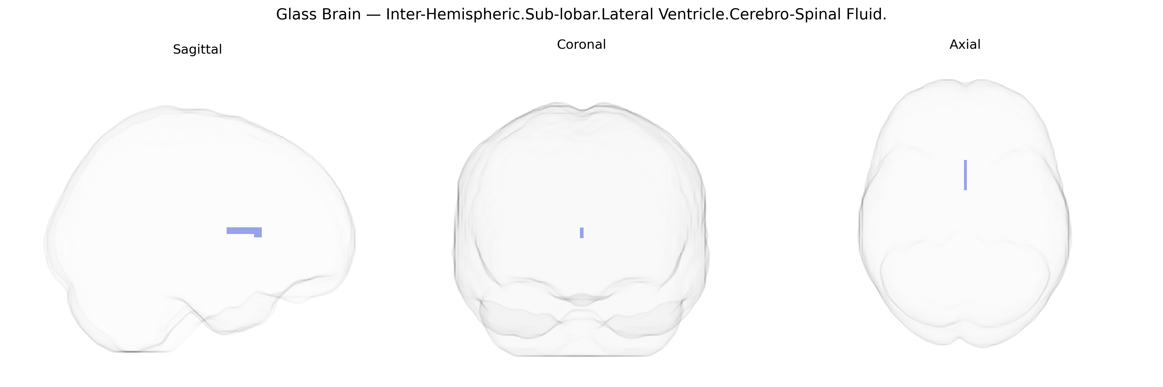
The bilateral Inter-Hemispheric Sub-lobar Lateral Ventricle Cerebro-Spinal Fluid region corresponds to the cerebrospinal fluid (CSF) occupying the lateral ventricles within the sub-lobar, deep compartment of both cerebral hemispheres, situated near the midline between them. The lateral ventricles are paired, C-shaped cavities extending through the frontal, parietal, occipital, and temporal lobes, and they communicate with the third ventricle via the interventricular foramina (foramina of Monro). CSF in these spaces is produced primarily by the choroid plexus and circulates through the ventricular system and subarachnoid space, providing mechanical protection, buoyancy, and a regulated chemical environment for the brain, while also contributing to waste clearance. Although the label emphasizes inter-hemispheric and sub-lobar positioning, it functionally denotes the ventricular CSF compartment rather than neural parenchyma. There is no direct link for “Inter-Hemispheric Sub-lobar Lateral Ventricle CSF”; a related structure is the Lateral ventricle.
Genetic associations with bilateral Inter-Hemispheric Sub-lobar Lateral Ventricle cerebrospinal fluid (CSF) volumes, as defined in the Talairach 2 mm atlas, have largely emerged from neuroimaging GWAS that treat ventricular/CSF space as a structural endophenotype of brain aging and neurodegeneration. Multiple large-scale studies (e.g., ENIGMA, UK Biobank) have identified loci where common variants influence lateral ventricular volume, including regions near or within genes such as APOE, BIN1, CLU, PICALM, and CR1 that are also implicated in Alzheimer’s disease risk, supporting a shared genetic architecture between ventricular enlargement and neurodegenerative processes. Other GWAS have linked ventricular and CSF volume variation to polygenic risk for schizophrenia, bipolar disorder, and major depressive disorder, as well as to general cognitive performance and educational attainment, suggesting that genes influencing neurodevelopmental trajectories and brain atrophy also modulate the size of these CSF spaces. In addition, loci associated with intracranial volume and global brain morphology (e.g., variants near HMGA2 and other growth-related genes) show correlated effects on lateral ventricle CSF volumes, consistent with a genetic contribution to overall brain size and its age-related structural reconfiguration. Overall, ventricular CSF measures are repeatedly used as quantitative traits in GWAS, and their genetic correlates cluster strongly with pathways involved in neurodevelopment, synaptic function, and neurodegeneration.
Overview generated by GPT-4o (2026).
Region ID: 744
Hemisphere: bilateral
Atlas: Talairach labels 2mm
Full Quality Version: Download MP4
Full Quality Version: Download MP4
Wali Sidiqyar*, Gaurav Rudravaram*, Elyssa M. McMaster, Trent M. Schwartz, Adam M. Saunders, Kurt G. Schilling, Bennett A. Landman "Introducing SPINS: A Shared Public Visualization Library of Neuroanatomical Structures." Medical Imaging with Deep Learning- short paper
This resource is licensed under CC0 1.0 Universal (Public Domain).