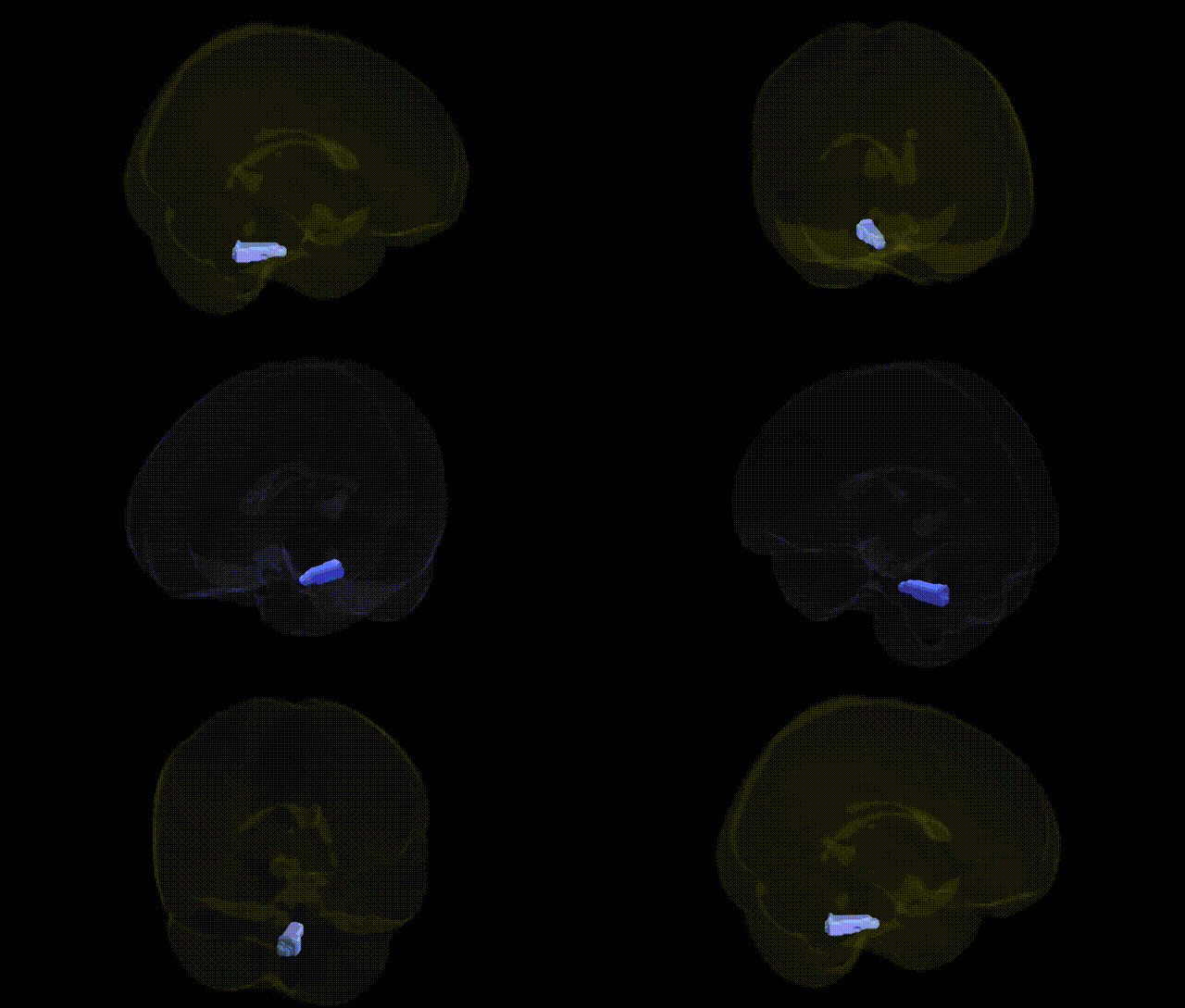
The bilateral Left Brainstem.Medulla, as defined in the Talairach 2 mm Atlas, corresponds to the left half of the medulla oblongata segment of the brainstem, a caudal structure continuous with the spinal cord and lying inferior to the pons. The medulla contains essential autonomic and relay nuclei, including centers for cardiovascular and respiratory regulation, as well as nuclei for cranial nerves IX–XII involved in swallowing, vocalization, tongue movement, and visceral sensory processing. Major ascending and descending tracts, such as the corticospinal pathways and dorsal column–medial lemniscus system, traverse the medulla, with important decussations (e.g., pyramidal decussation) occurring in this region. Functionally, the left medullary portion participates in control of vital homeostatic functions, somatosensory and motor integration, and coordination of reflexes that maintain posture, breathing, and heart rate.
The bilateral left brainstem medulla, as defined in the Talairach 2 mm atlas, has relatively sparse region-specific genetic association data, since most GWAS and imaging-genetics studies treat the brainstem as a composite structure rather than isolating the medulla or its left-sided subdivisions. Nonetheless, genetic influences on medullary structure and function are implicated indirectly through variants associated with autonomic regulation (e.g., blood pressure, heart rate variability), respiratory control, and sudden infant death syndrome, where brainstem abnormalities are frequently observed. Genes involved in serotonergic signaling (such as SLC6A4 and HTR1A/2A), neurodevelopmental pathways, and ionic channel function have been linked to disorders that prominently involve medullary circuits, including congenital central hypoventilation syndrome (PHOX2B), familial dysautonomia (IKBKAP/ELP1), and some forms of epilepsy and sudden unexplained death in epilepsy, which show neuropathological changes in medullary cardiorespiratory nuclei. Large-scale neuroimaging GWAS, including UK Biobank-based studies, have identified polygenic effects on brainstem volume and microstructure, but these typically do not resolve associations specifically to the left medulla, and current evidence connects genetic variants more to global brainstem morphology and broader autonomic, motor, and arousal systems than to a discrete Talairach-defined left medullary region.
Overview generated by GPT-4o (2026).
Region ID: 5
Hemisphere: bilateral
Atlas: Talairach labels 2mm
Full Quality Version: Download MP4
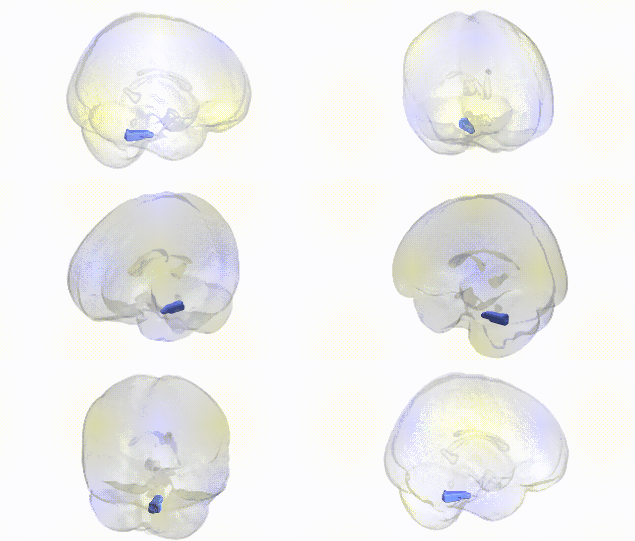
Full Quality Version: Download MP4
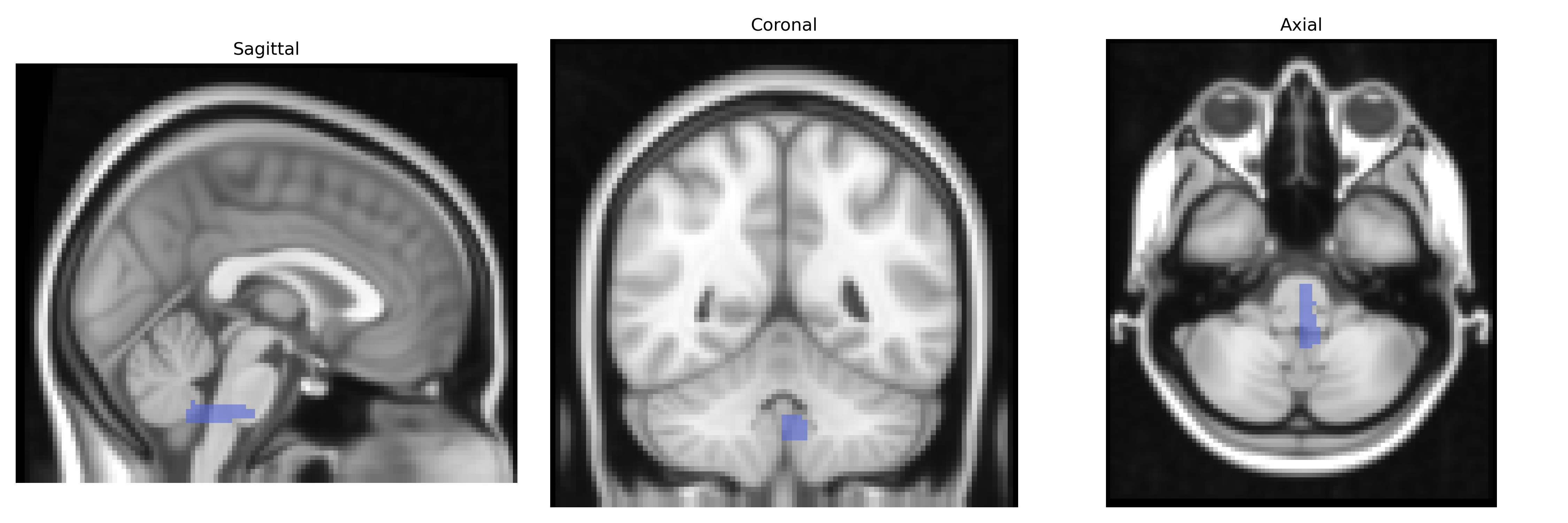
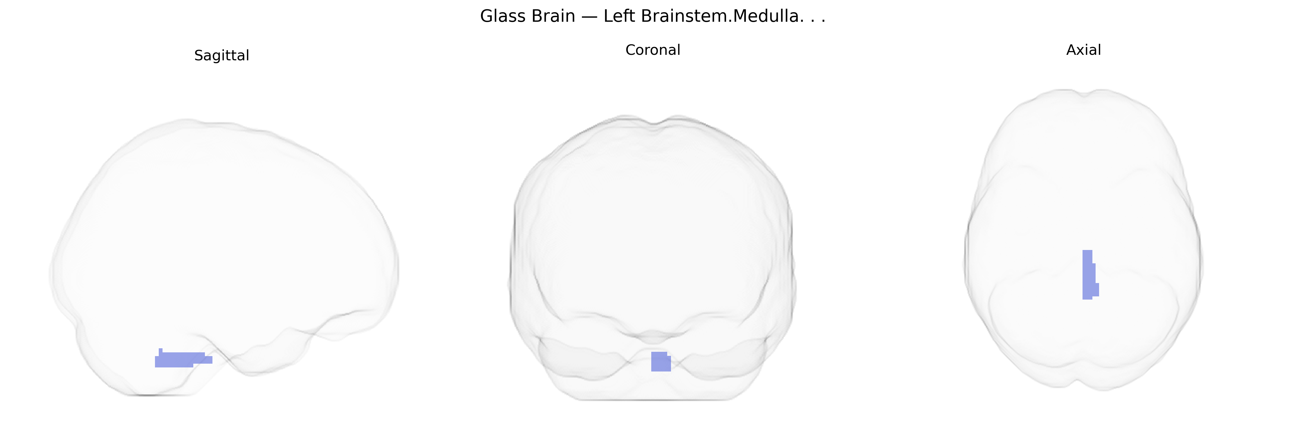
Wali Sidiqyar*, Gaurav Rudravaram*, Elyssa M. McMaster, Trent M. Schwartz, Adam M. Saunders, Kurt G. Schilling, Bennett A. Landman "Introducing SPINS: A Shared Public Visualization Library of Neuroanatomical Structures." Medical Imaging with Deep Learning- short paper
This resource is licensed under CC0 1.0 Universal (Public Domain).