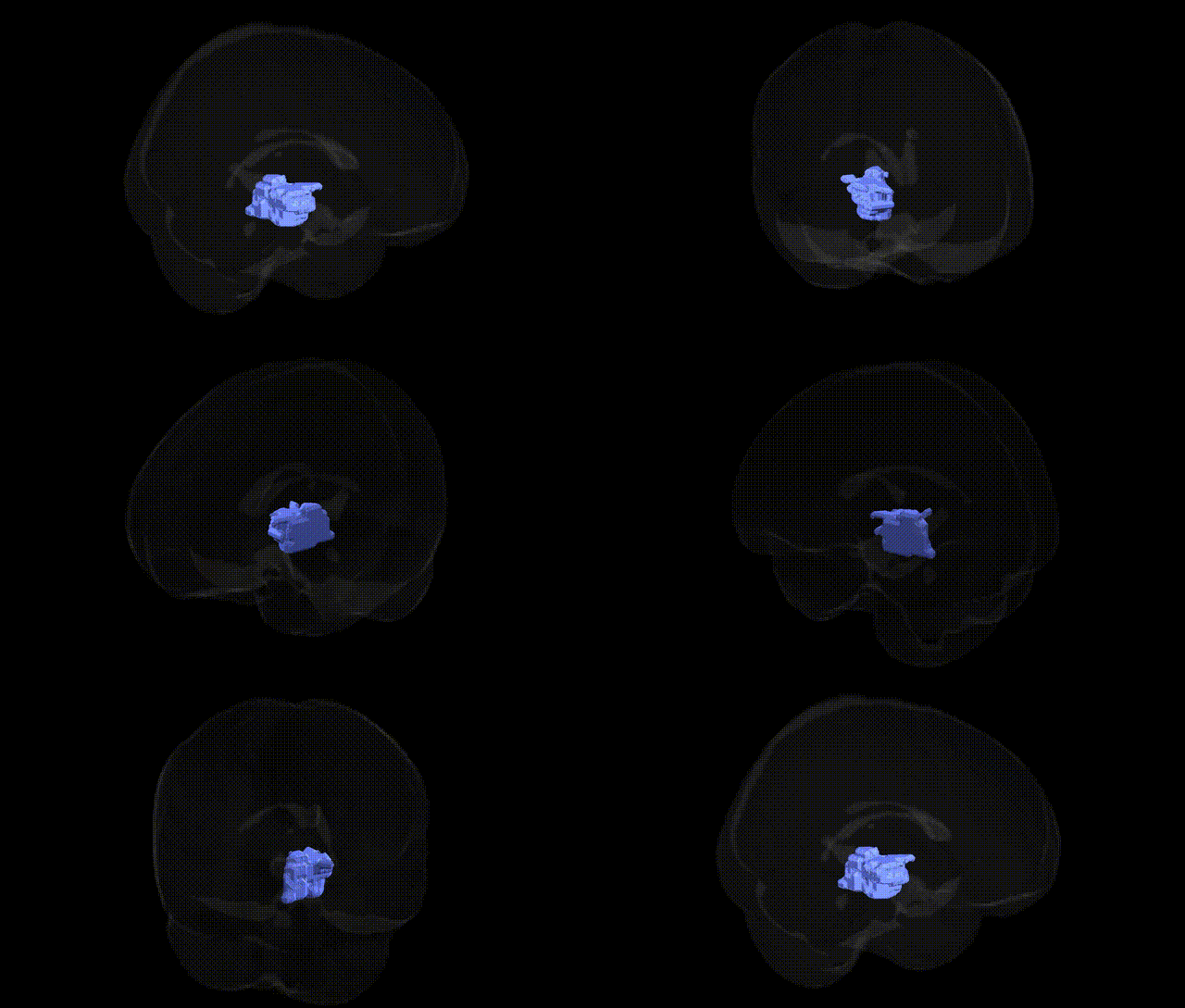
The bilateral left brainstem midbrain region in the Talairach 2 mm Atlas corresponds to the mesencephalic portion of the brainstem, situated between the pons caudally and the diencephalon rostrally. It contains critical ascending and descending pathways, including components of the corticospinal, corticobulbar, and sensory tracts, as well as nuclei such as the substantia nigra, red nucleus, and oculomotor and trochlear nerve nuclei. Functionally, this region is central to motor control (particularly movement initiation and coordination), modulation of muscle tone, eye movement regulation, auditory and visual reflex integration via the colliculi, and aspects of arousal and attention through connections with the reticular formation and higher cortical structures. There is no single direct Wikipedia article for “Left Brainstem Midbrain” as a Talairach label; a closely related structure is the Midbrain.
The bilateral left brainstem midbrain, as defined in the Talairach 2 mm atlas, is a relatively small and heterogeneous region, and current genetic association literature rarely targets it as an isolated structure; instead, midbrain-related genetic findings typically arise from studies of dopaminergic pathways, motor and oculomotor circuits, pain modulation, and neuropsychiatric or neurodegenerative disorders. GWAS and candidate-gene studies implicate genes affecting dopamine synthesis, transport, and receptor signaling—such as COMT, DRD2, DRD3, DAT1/SLC6A3, and MAOA—in traits and disorders involving midbrain nuclei (including substantia nigra and ventral tegmental area), with relevance to Parkinson’s disease, schizophrenia, substance use, impulsivity, and cognitive control. Parkinson’s disease GWAS highlight loci including SNCA, LRRK2, GBA, and others that influence nigrostriatal pathways originating in the midbrain, while psychiatric GWAS (e.g., for major depression, ADHD, and schizophrenia) identify risk variants in genes regulating synaptic transmission and neurodevelopment (such as CACNA1C, GRIN2A, BDNF, and multiple polygenic loci) that correlate with functional and structural changes in midbrain circuits observed via imaging genetics. Additional associations link midbrain-related pain and autonomic control to variants in opioid-system genes (e.g., OPRM1) and serotonergic genes (SLC6A4, HTR1A), although these findings generally reference functional networks rather than a specific Talairach-defined “Left Brainstem.Midbrain” parcel. Overall, the genetic architecture of traits and diseases involving this region is highly polygenic and network-based, and existing GWAS map risk loci to broader dopamino-serotonergic and motor circuits in which the midbrain is a central hub, rather than to this atlas-defined region alone.
Overview generated by GPT-4o (2026).
Region ID: 215
Hemisphere: bilateral
Atlas: Talairach labels 2mm
Full Quality Version: Download MP4
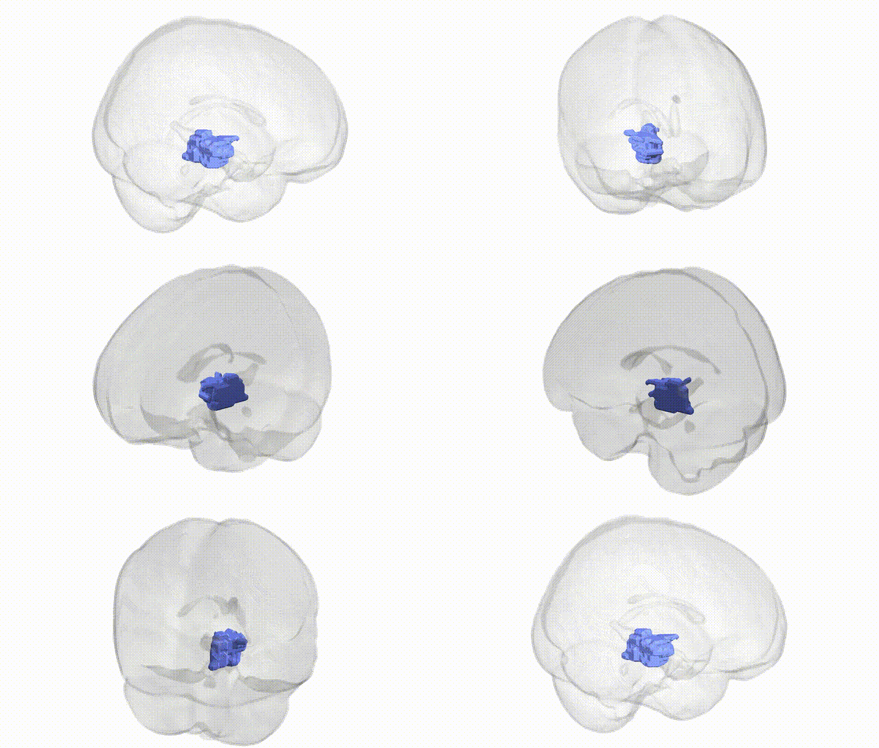
Full Quality Version: Download MP4
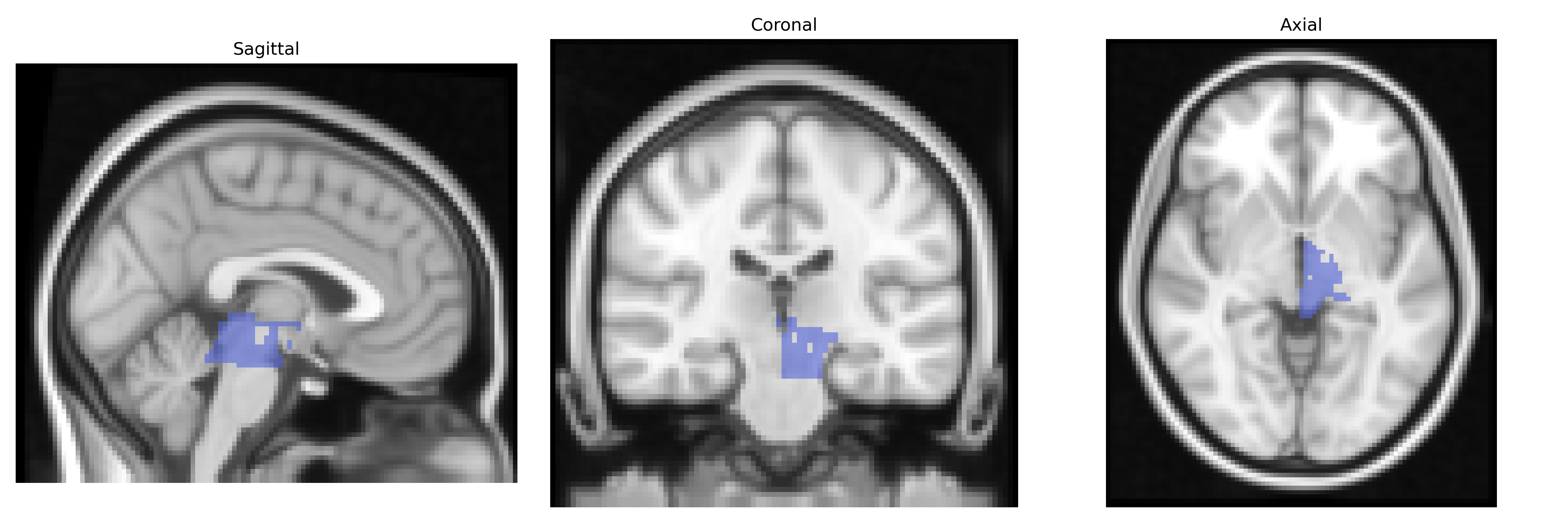
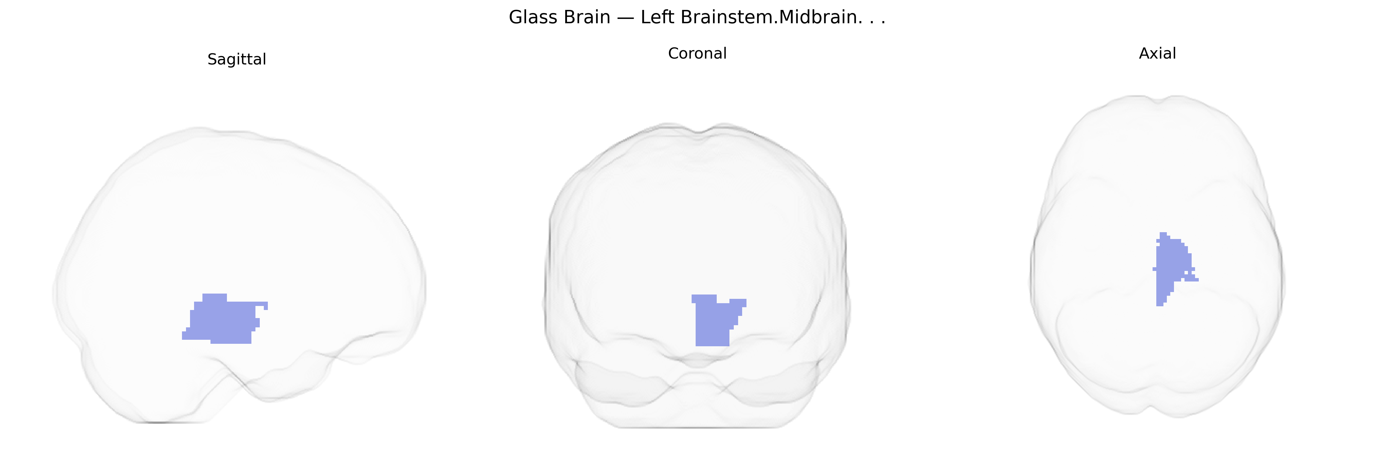
Wali Sidiqyar*, Gaurav Rudravaram*, Elyssa M. McMaster, Trent M. Schwartz, Adam M. Saunders, Kurt G. Schilling, Bennett A. Landman "Introducing SPINS: A Shared Public Visualization Library of Neuroanatomical Structures." Medical Imaging with Deep Learning- short paper
This resource is licensed under CC0 1.0 Universal (Public Domain).