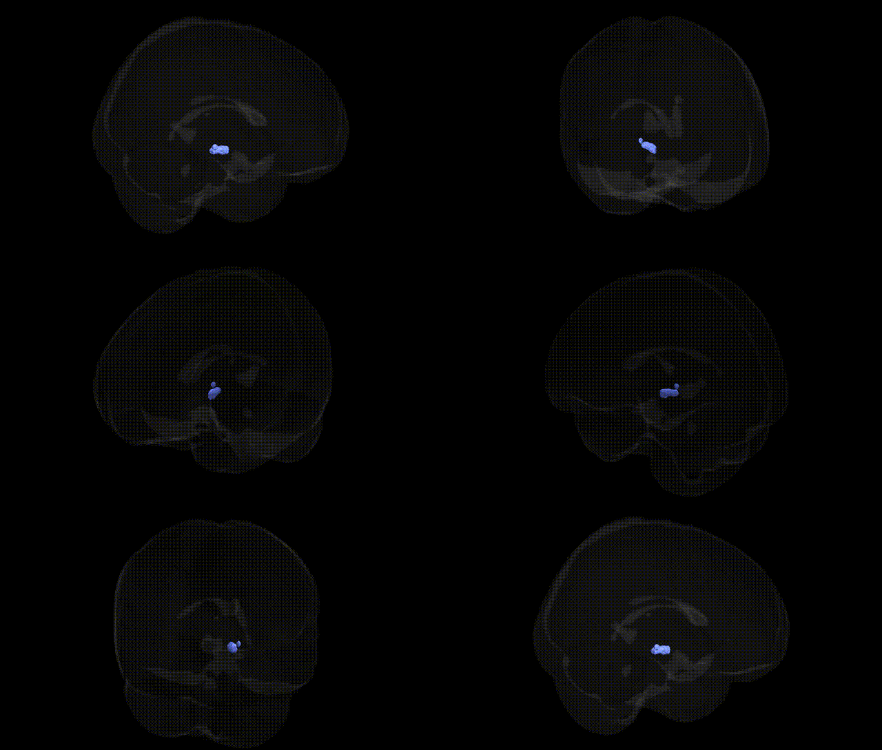
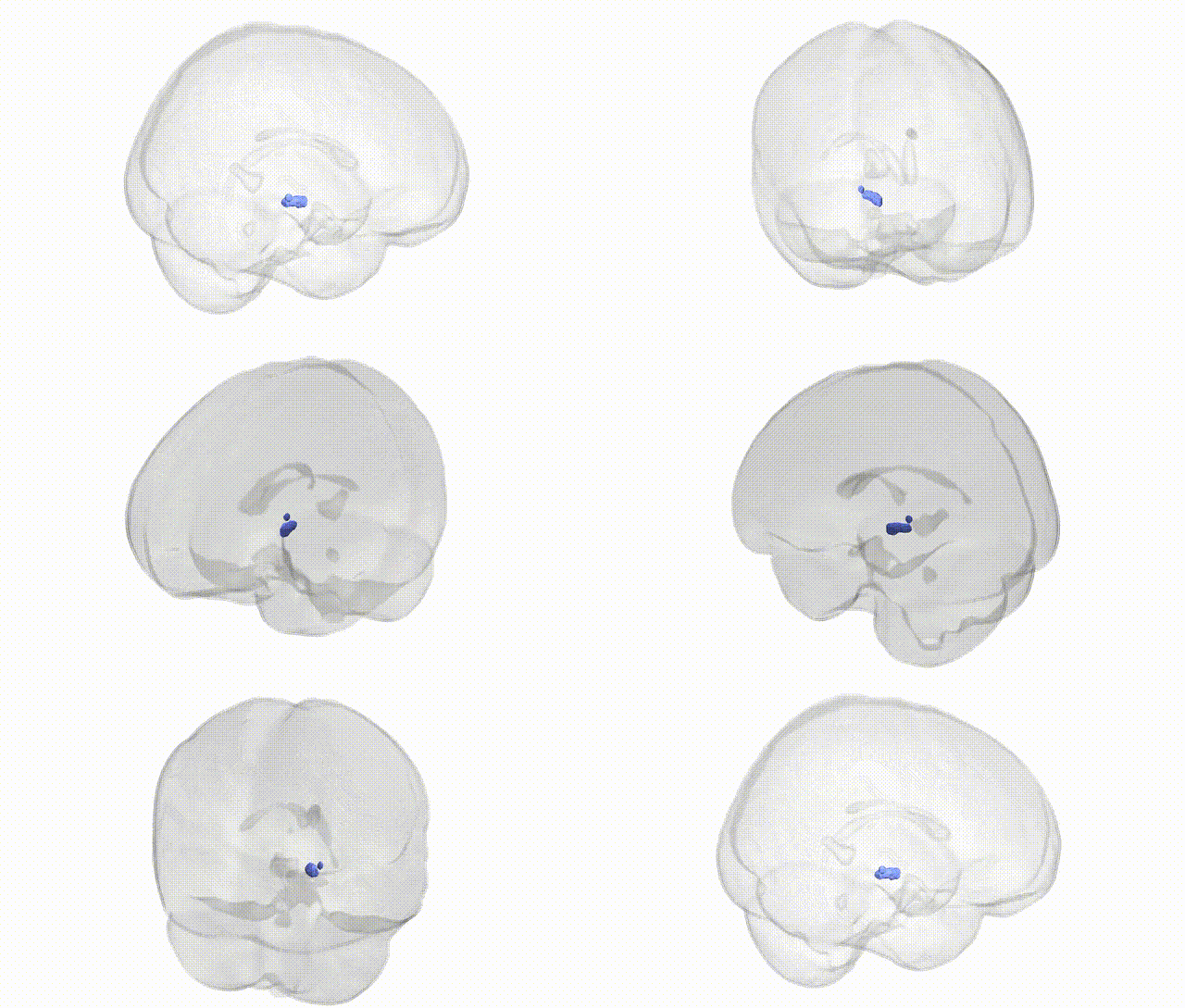
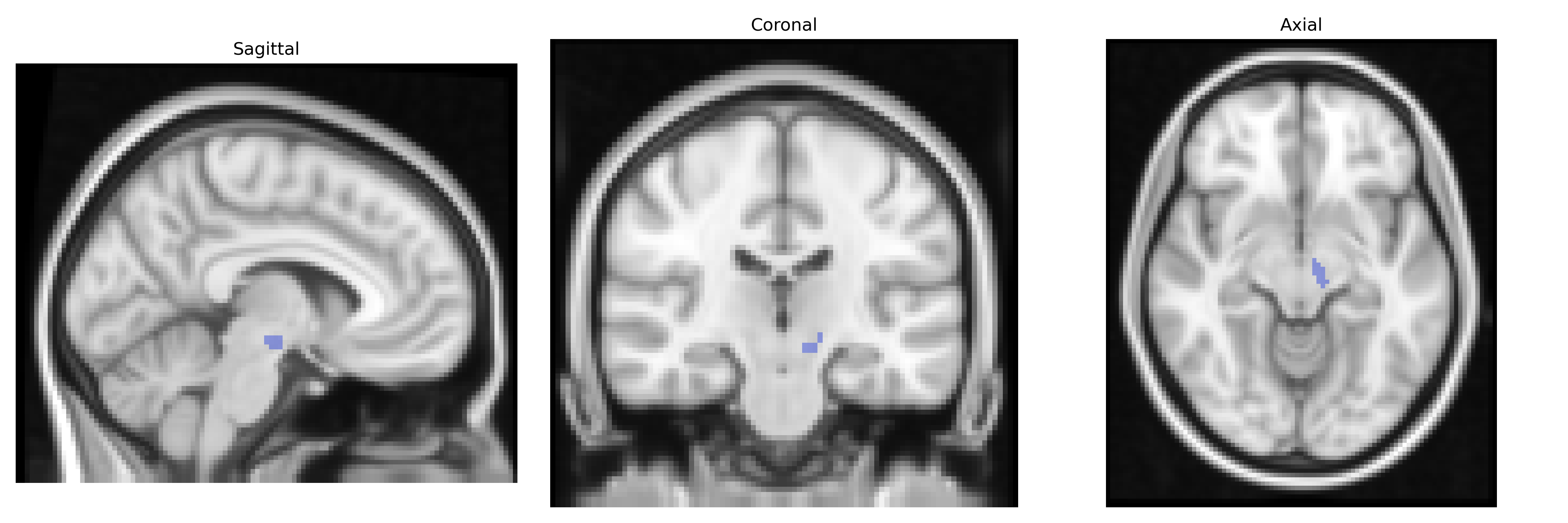
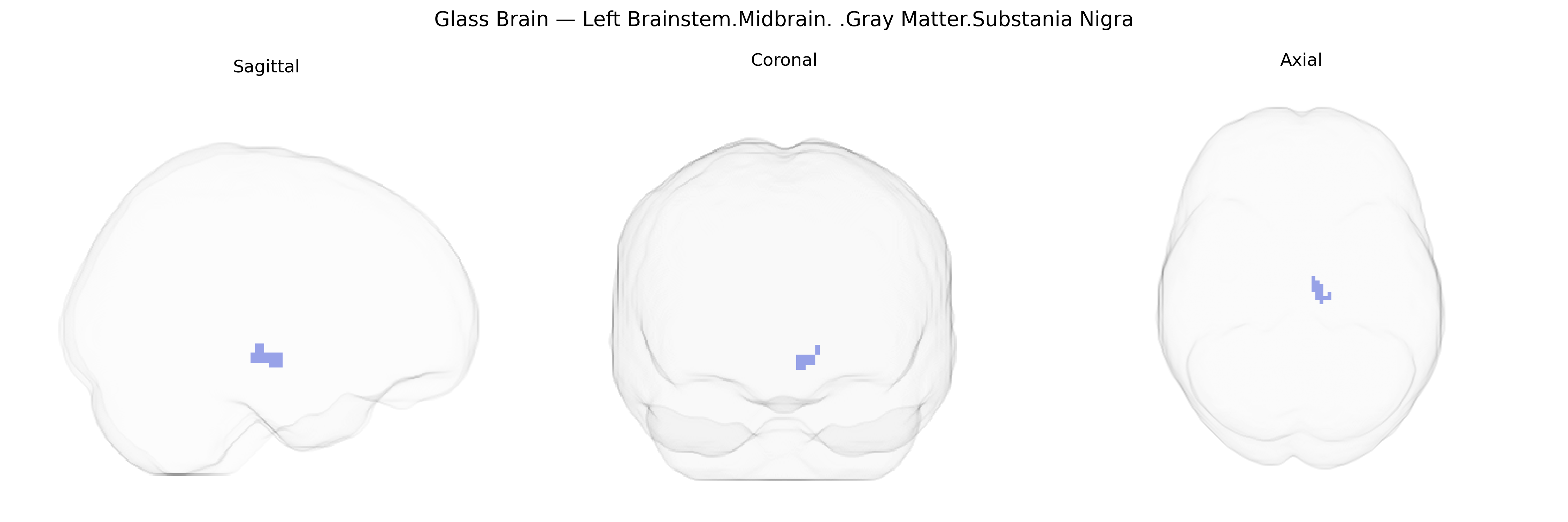
The bilateral Left Brainstem.Midbrain.Gray Matter.Substantia Nigra corresponds to paired nuclei in the ventral midbrain that play a key role in motor control, reward processing, and neuromodulation via dopaminergic signaling. Structurally, the substantia nigra is divided into pars compacta, rich in dopaminergic neurons projecting primarily to the striatum, and pars reticulata, containing GABAergic output neurons that influence thalamocortical and brainstem motor pathways. Functionally, this region is critical for the initiation and modulation of voluntary movement, reinforcement learning, and habit formation; degeneration of its dopaminergic neurons is a hallmark of Parkinson’s disease and contributes to multiple movement disorders. As part of the basal ganglia circuitry, the substantia nigra integrates cortical, striatal, and subcortical inputs to regulate the balance between facilitation and inhibition of motor programs and associated cognitive and affective processes. Substantia nigra
The bilateral left brainstem midbrain gray matter region corresponding to the Substantia Nigra (Talairach 2 mm atlas) is strongly implicated in genetic studies of movement disorders, especially Parkinson’s disease (PD), where GWAS have identified multiple risk loci (e.g., SNCA, LRRK2, GBA, MAPT, BST1, HLA region, and others) that influence dopaminergic neuron integrity and α‑synuclein aggregation, contributing to nigral degeneration and reduced dopamine signaling. Variants in SNCA and LRRK2 are among the most robustly replicated, with some alleles linked to earlier onset and more rapid decline in nigral function, while GBA mutations confer substantial PD risk and more severe phenotypes. Beyond PD, genetic associations involving nigral dysfunction or dopaminergic signaling have been reported in restless legs syndrome (MEIS1 and BTBD9 loci), essential tremor, and susceptibility to antipsychotic‑induced parkinsonism, as well as in neuropsychiatric traits such as addiction-related behaviors and reward processing through genes affecting dopamine pathways (e.g., DRD2, COMT) that modulate midbrain dopaminergic activity. Imaging‑genetics studies also link common variants in dopaminergic and synaptic genes to structural and functional MRI measures of midbrain and Substantia Nigra, indicating that polygenic risk for PD and related traits can manifest as altered nigral volume, iron content, or connectivity in healthy and clinical populations.
Overview generated by GPT-4o (2026).
Region ID: 341
Hemisphere: bilateral
Atlas: Talairach labels 2mm
Full Quality Version: Download MP4
Full Quality Version: Download MP4
Wali Sidiqyar*, Gaurav Rudravaram*, Elyssa M. McMaster, Trent M. Schwartz, Adam M. Saunders, Kurt G. Schilling, Bennett A. Landman "Introducing SPINS: A Shared Public Visualization Library of Neuroanatomical Structures." Medical Imaging with Deep Learning- short paper
This resource is licensed under CC0 1.0 Universal (Public Domain).