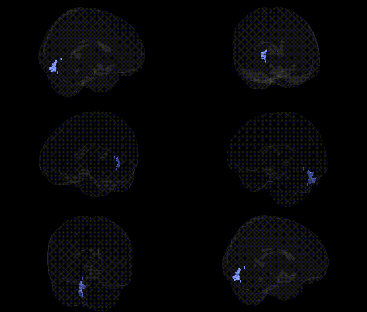
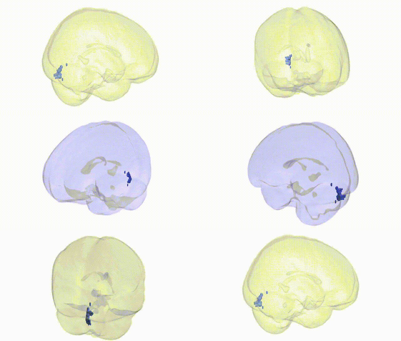
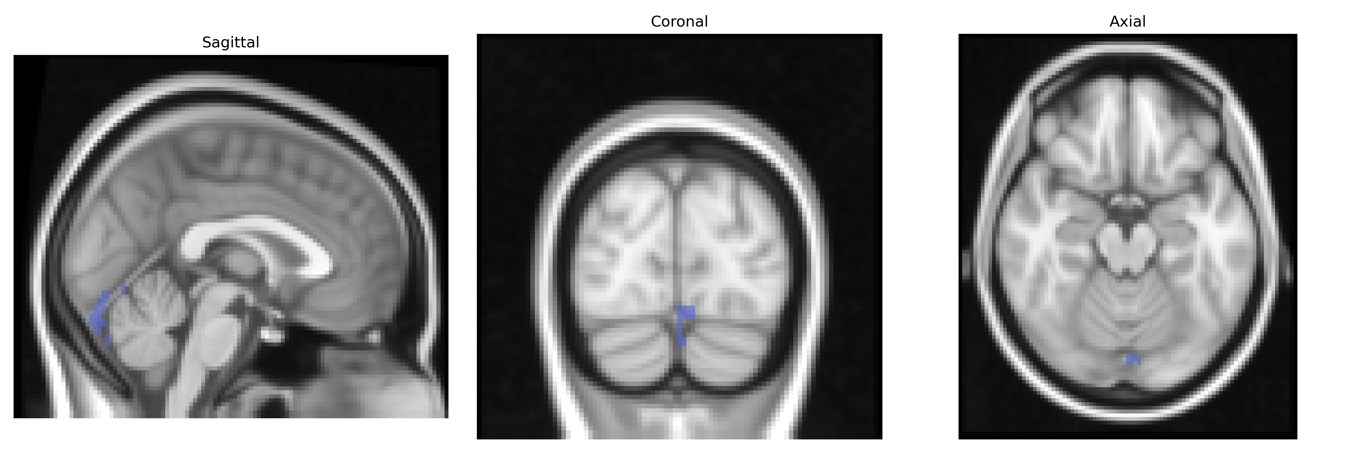
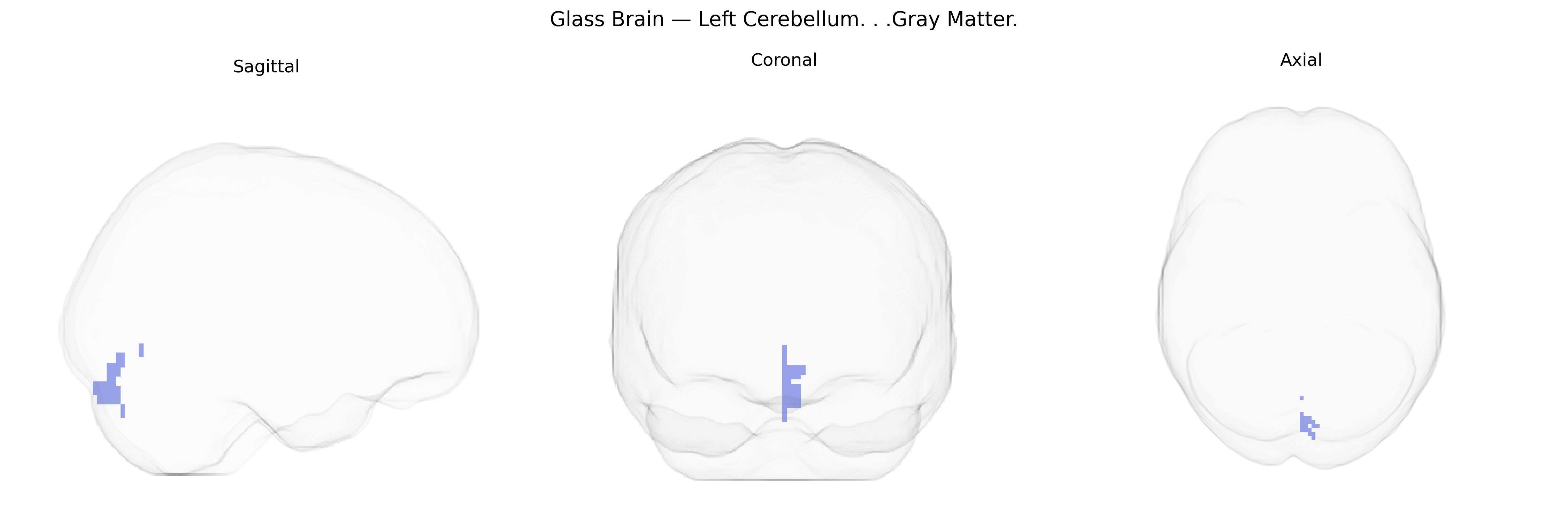
The bilateral Left Cerebellum gray matter, as defined in the Talairach 2 mm atlas, comprises the cortical regions of the left cerebellar hemisphere, including the layered cerebellar cortex (molecular, Purkinje cell, and granular layers) that overlay the deep cerebellar nuclei. This tissue is primarily involved in the coordination, timing, and precision of voluntary movements, as well as in motor learning and adaptation through the integration of proprioceptive, vestibular, and cortical inputs. Functionally, the left cerebellar hemisphere participates in the modulation of ipsilateral body movements and, via crossed cerebro-cerebellar pathways, contributes to the fine-tuning of contralateral cerebral motor and cognitive processes. There is no direct Wikipedia article specifically titled “Left Cerebellum Gray Matter”; a related and encompassing structure is the cerebellum: Cerebellum.
The left cerebellar gray matter has emerged in imaging genetics and GWAS studies as a region whose volume and functional variation are moderately heritable and associated with multiple polygenic influences, rather than single, large-effect loci. Large-scale brain morphology GWAS (e.g., ENIGMA and UK Biobank analyses) have identified common variants near genes involved in neurodevelopment, synaptic function, and axonal guidance—such as KIAA0586, TBR1, and genes in the WNT and FGF pathways—that contribute to cerebellar volume, although these signals are typically bilateral and not strictly confined to the left side. Cerebellar gray matter alterations have been repeatedly implicated in neurodevelopmental and psychiatric disorders, including autism spectrum disorder, schizophrenia, bipolar disorder, and major depressive disorder, where polygenic risk scores for these conditions correlate with structural and functional measures in cerebellar regions, sometimes overlapping the left cerebellum labels in the Talairach atlas. Genetic risk for cognitive traits (general intelligence, educational attainment, processing speed) and motor coordination also shows associations with cerebellar morphology, supporting the cerebellum’s role in both cognitive and motor networks. In addition, variants in genes linked to neurodegeneration (e.g., ATXN family in spinocerebellar ataxias, C9orf72 and other ALS-related loci) contribute to cerebellar atrophy patterns that can involve bilateral cerebellar gray matter, including the left hemisphere. Overall, current evidence indicates that the bilateral left cerebellum gray matter region is influenced by widespread polygenic architecture related to brain development, cognition, and psychiatric risk, with no single genetic variant uniquely specific to this Talairach-defined region, but consistent involvement in distributed genotype–phenotype relationships across the cerebellum.
Overview generated by GPT-4o (2026).
Region ID: 66
Hemisphere: bilateral
Atlas: Talairach labels 2mm
Full Quality Version: Download MP4
Full Quality Version: Download MP4
Wali Sidiqyar*, Gaurav Rudravaram*, Elyssa M. McMaster, Trent M. Schwartz, Adam M. Saunders, Kurt G. Schilling, Bennett A. Landman "Introducing SPINS: A Shared Public Visualization Library of Neuroanatomical Structures." Medical Imaging with Deep Learning- short paper
This resource is licensed under CC0 1.0 Universal (Public Domain).