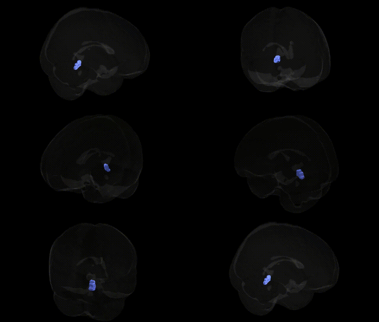
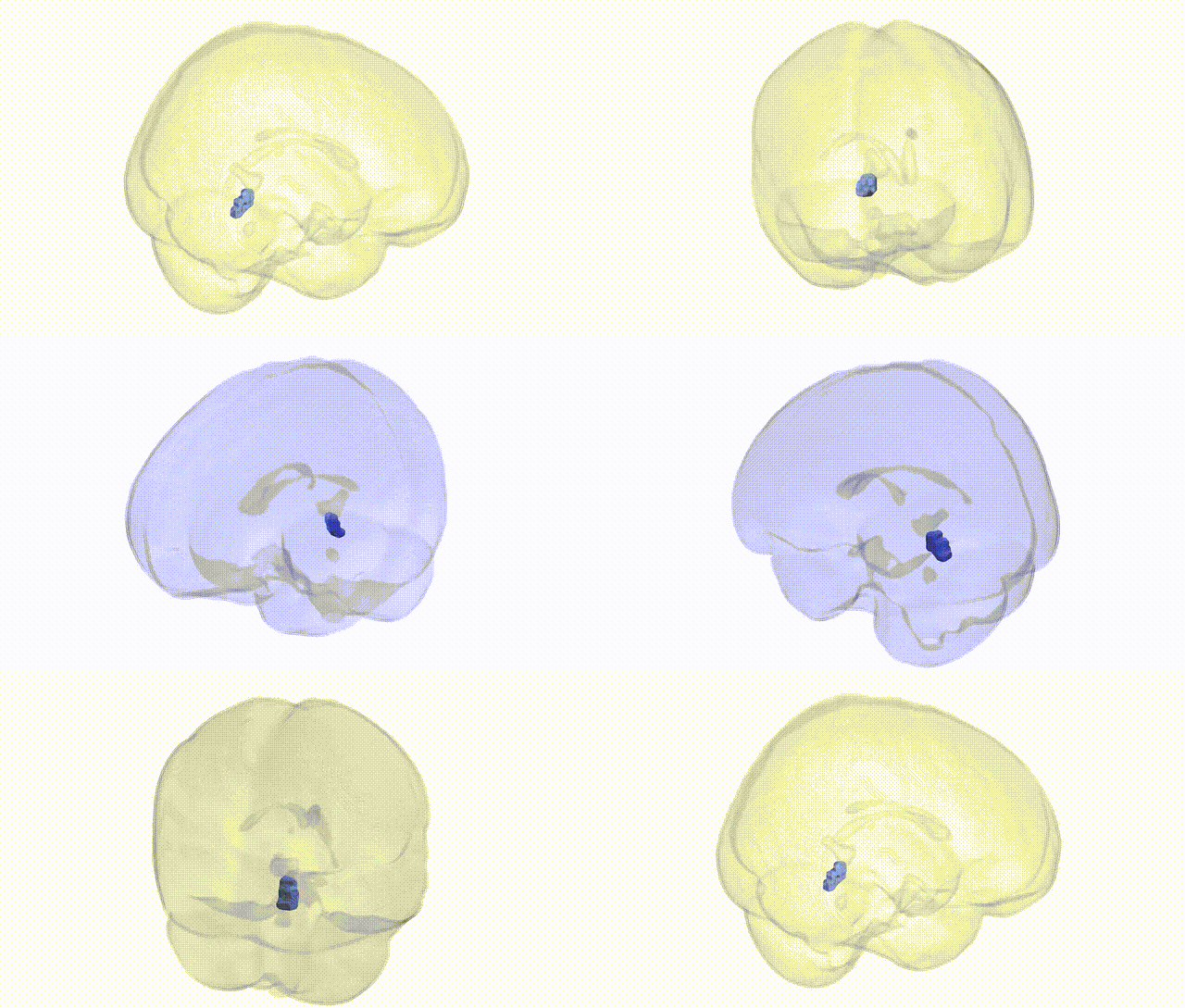
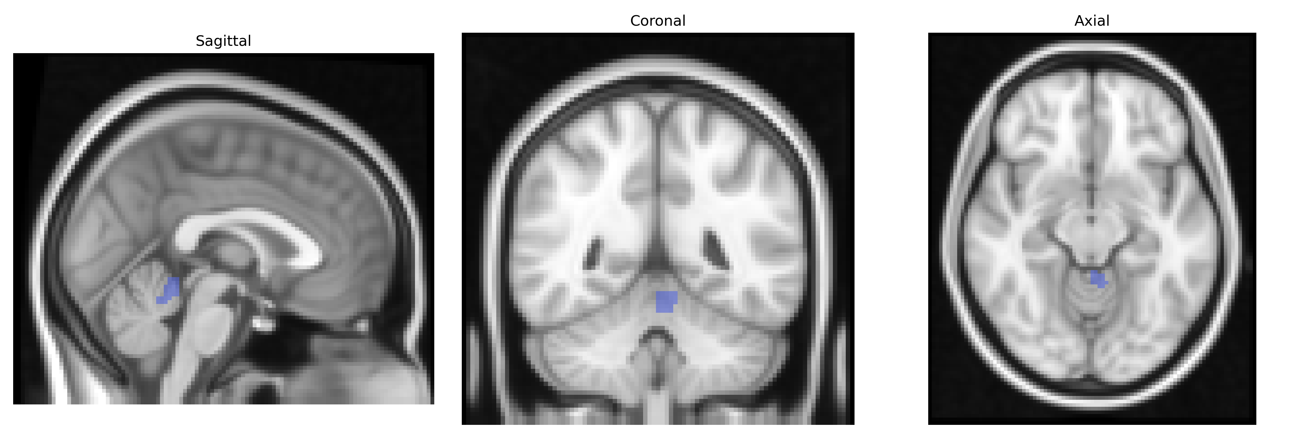
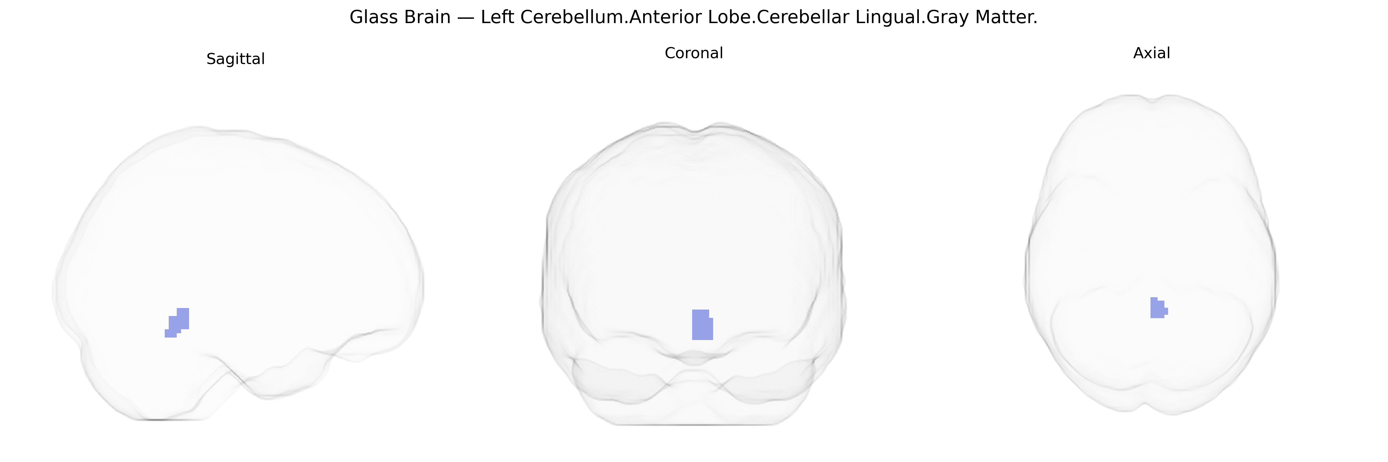
The bilateral Left Cerebellum.Anterior Lobe.Cerebellar Lingual.Gray Matter refers to gray matter in the lingula (lingual lobule) of the anterior lobe of the cerebellum, located medially along the superior aspect of the cerebellar vermis. This region is part of the cerebellar cortex and is involved in the fine coordination of movements, posture, and possibly some aspects of motor learning, integrating sensory and motor signals to adjust ongoing motor activity. Its neurons (primarily Purkinje cells, interneurons, and granule cells) project to deep cerebellar nuclei, contributing to cerebellar output that modulates descending motor pathways. There is no direct link for “Cerebellar Lingual,” but it is a component of the Cerebellum.
The bilateral Left Cerebellum.Anterior Lobe.Cerebellar Lingual.Gray Matter region, as defined in the Talairach 2 mm Atlas, lies within anterior cerebellar cortex implicated in sensorimotor integration, balance, and aspects of cognitive processing, and genetic associations typically involve broader cerebellar or lobular measures rather than this microanatomical label specifically. Large-scale neuroimaging GWAS (e.g., ENIGMA, UK Biobank, and related consortia) have identified multiple loci influencing cerebellar volume and cortical thickness, including variants near genes such as KIAA0586 (involved in cerebellar development), RELN (neuronal migration), and FOXP1/FOXP2 (corticocerebellar circuitry), but these findings usually map to lobular or whole‑cerebellum metrics, making precise attribution to the “lingual” segment of the anterior lobe uncertain. Cerebellar gray matter changes in regions overlapping the anterior lobe are genetically associated with neurodevelopmental and psychiatric conditions, including autism spectrum disorder, schizophrenia, and attention‑deficit/hyperactivity disorder, through polygenic risk scores and case‑control GWAS that implicate synaptic, neurodevelopmental, and immune-related pathways affecting cerebellar structure. Cerebellar involvement, including anterior lobe structures, has also been reported in genetic ataxias (e.g., spinocerebellar ataxias caused by expansions in ATXN1, ATXN2, ATXN3, CACNA1A variants), though imaging phenotypes often describe lobular atrophy rather than the specific Talairach-defined lingual region. Overall, available genetic data support a role for inherited variation in shaping anterior cerebellar gray matter and its vulnerability in neurodevelopmental, psychiatric, and motor coordination disorders, but direct GWAS or fine‑grained imaging‑genetic associations targeting the bilateral Left Cerebellum.Anterior Lobe.Cerebellar Lingual.Gray Matter region itself have not yet been specifically characterized in the literature.
Overview generated by GPT-4o (2026).
Region ID: 211
Hemisphere: bilateral
Atlas: Talairach labels 2mm
Full Quality Version: Download MP4
Full Quality Version: Download MP4
Wali Sidiqyar*, Gaurav Rudravaram*, Elyssa M. McMaster, Trent M. Schwartz, Adam M. Saunders, Kurt G. Schilling, Bennett A. Landman "Introducing SPINS: A Shared Public Visualization Library of Neuroanatomical Structures." Medical Imaging with Deep Learning- short paper
This resource is licensed under CC0 1.0 Universal (Public Domain).