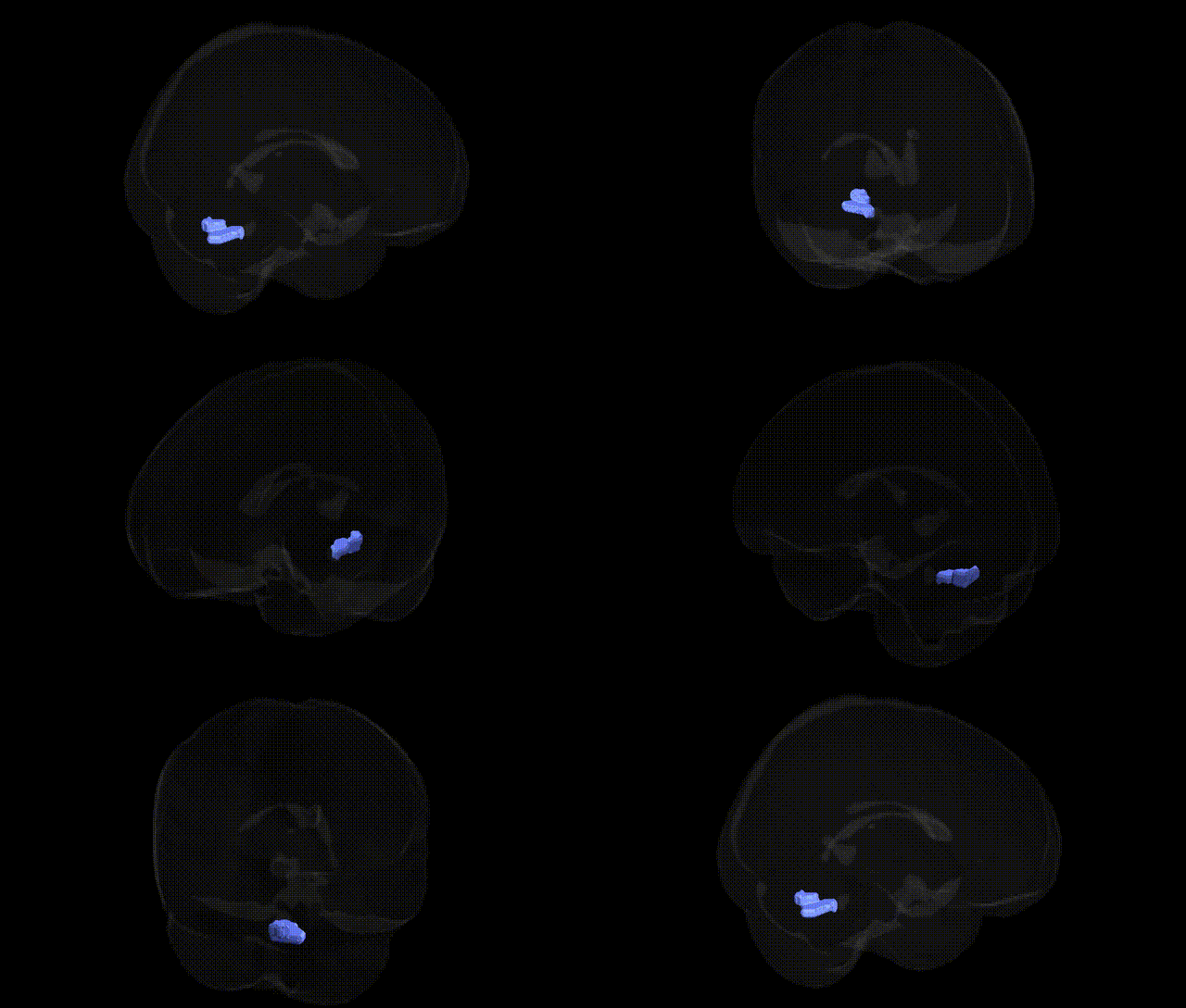
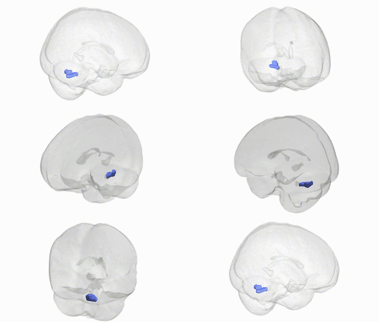
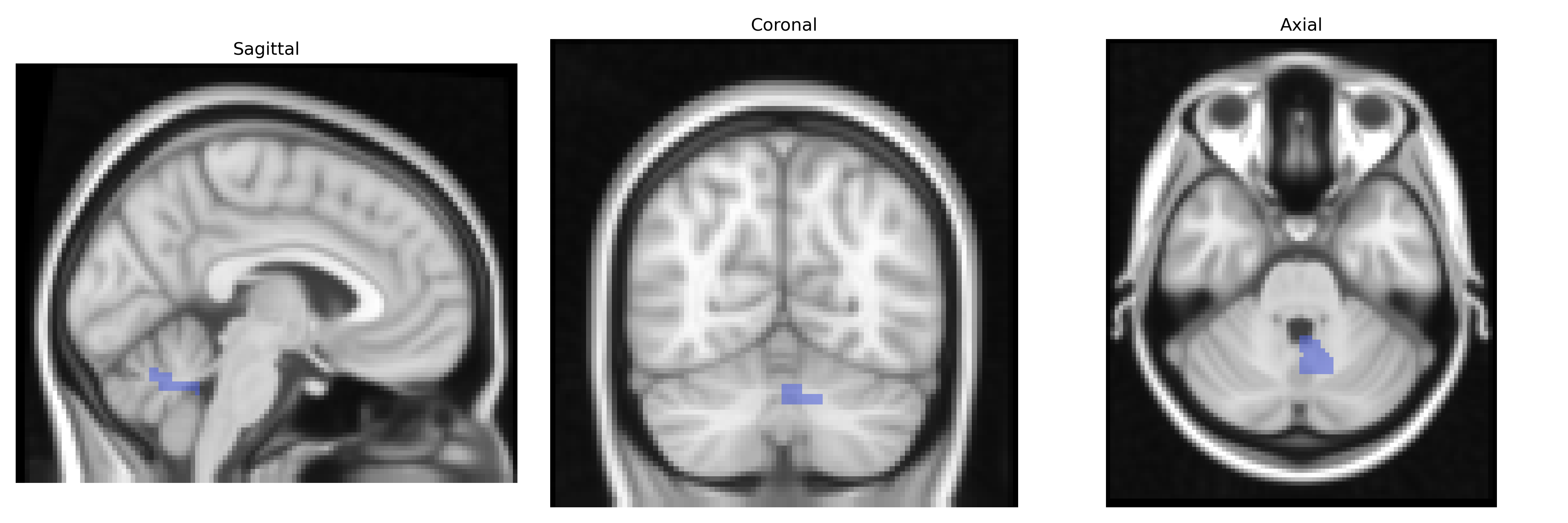
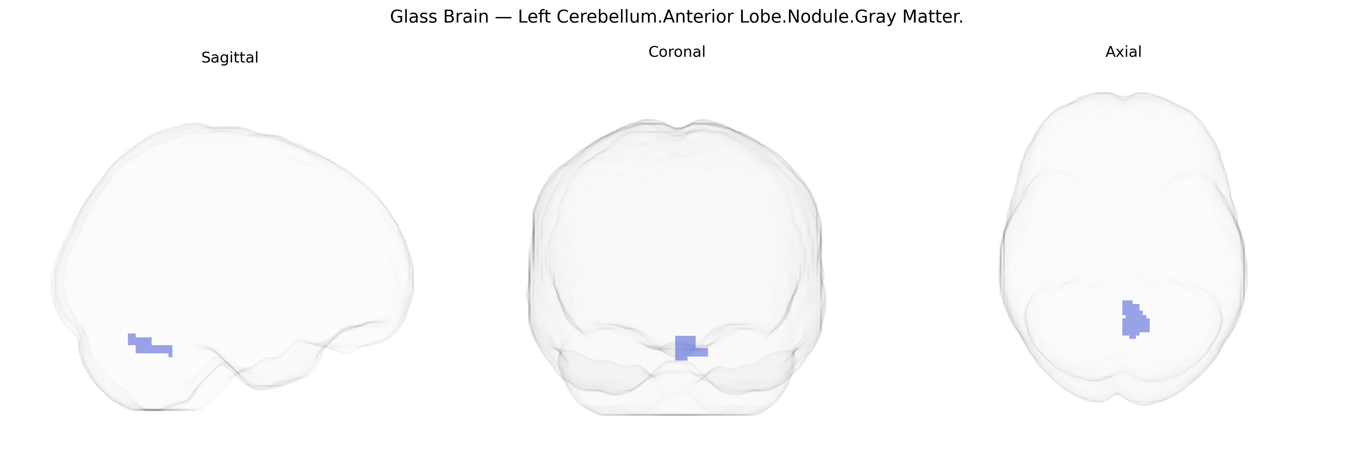
The bilateral Left Cerebellum.Anterior Lobe.Nodule.Gray Matter corresponds to gray matter within the cerebellar nodulus, a midline structure of the vestibulocerebellum located in the inferior portion of the vermis and functionally associated with the anterior lobe region in the Talairach 2 mm Atlas. The nodulus plays a key role in processing vestibular and oculomotor information, contributing to balance, posture, and coordination of eye movements through its connections with the vestibular nuclei and brainstem circuits. It participates in fine-tuning reflexes such as the vestibulo-ocular reflex and in integrating proprioceptive and vestibular inputs for stable gait and orientation in space. There is no direct Wikipedia article for this composite label; a closely related and encompassing structure is the Cerebellum.
The bilateral Left Cerebellum.Anterior Lobe.Nodule.Gray Matter region, corresponding roughly to vermis and adjacent anterior cerebellar structures in the Talairach 2mm atlas, has been implicated indirectly in genetic studies of cerebellar volume, motor control, and neurodevelopmental conditions rather than through region-specific GWAS hits. Large-scale imaging genetics consortia (e.g., ENIGMA, UK Biobank) have identified polygenic influences on total cerebellar and lobular volumes, with common variants in genes involved in neurodevelopment (such as AUTS2, CADM2, and various synaptic and axon guidance genes) contributing to cerebellar morphology, but these studies typically report lobular or global cerebellar measures rather than nodule-specific associations. The anterior cerebellum and vermis nodule are functionally linked to posture, gait, and oculomotor control, and structural or functional abnormalities in this region have been described in disorders with known genetic components, including autism spectrum disorder (where multiple risk genes converge on cerebellar development), attention-deficit/hyperactivity disorder, schizophrenia, and hereditary ataxias; however, these conditions are usually associated with broad cerebellar or vermal alterations rather than a discrete genetic signature for the nodule gray matter. GWAS of balance, motor coordination, and neuropsychiatric traits (such as depression, bipolar disorder, and cognitive performance) show cerebellar involvement in imaging follow-ups, but current evidence supports a highly polygenic, distributed pattern of genetic influence across the cerebellum, with no robust, replicated variant-to-region mapping specifically targeting the Left Cerebellum.Anterior Lobe.Nodule.Gray Matter label in the Talairach atlas.
Overview generated by GPT-4o (2026).
Region ID: 84
Hemisphere: bilateral
Atlas: Talairach labels 2mm
Full Quality Version: Download MP4
Full Quality Version: Download MP4
Wali Sidiqyar*, Gaurav Rudravaram*, Elyssa M. McMaster, Trent M. Schwartz, Adam M. Saunders, Kurt G. Schilling, Bennett A. Landman "Introducing SPINS: A Shared Public Visualization Library of Neuroanatomical Structures." Medical Imaging with Deep Learning- short paper
This resource is licensed under CC0 1.0 Universal (Public Domain).