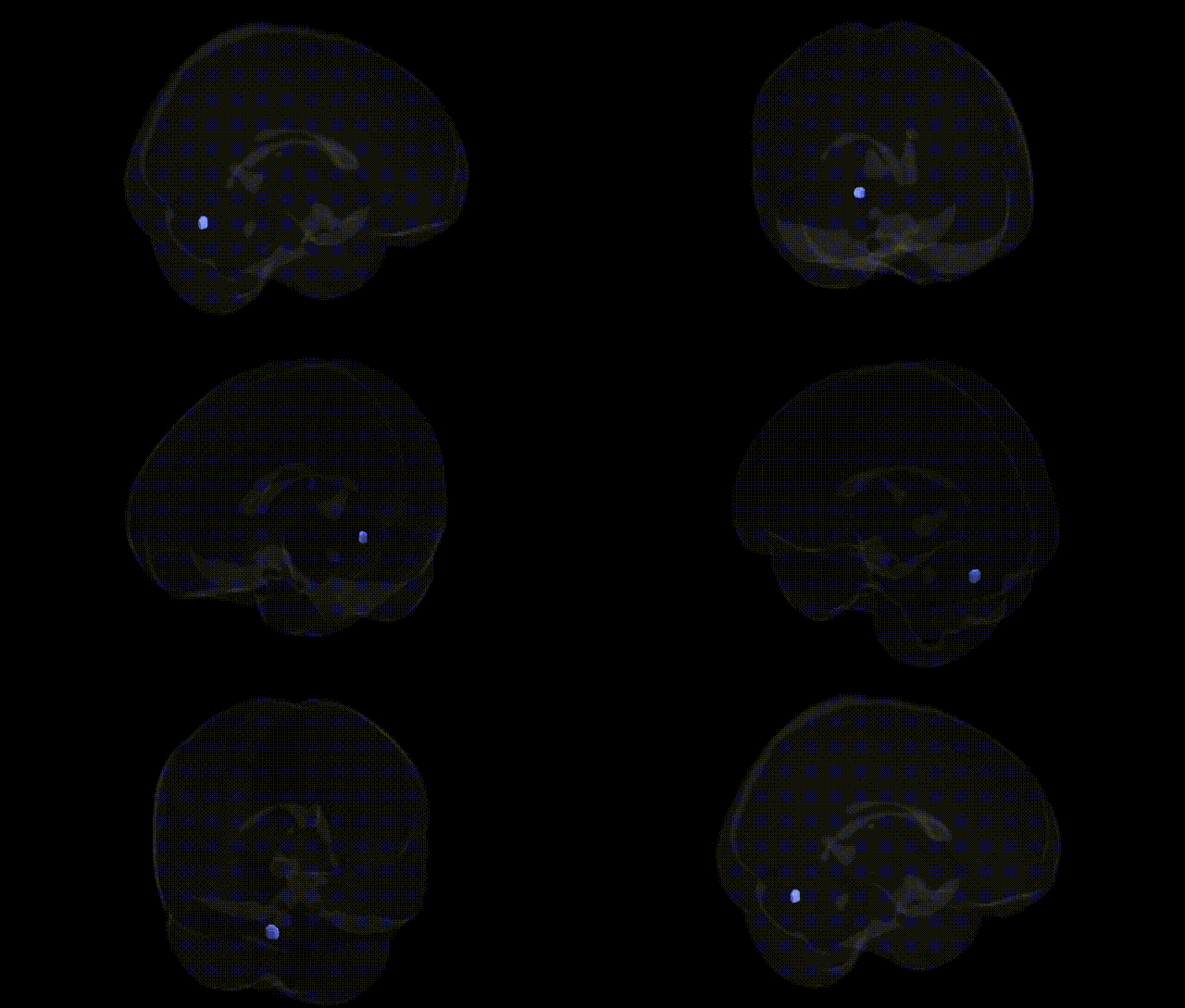
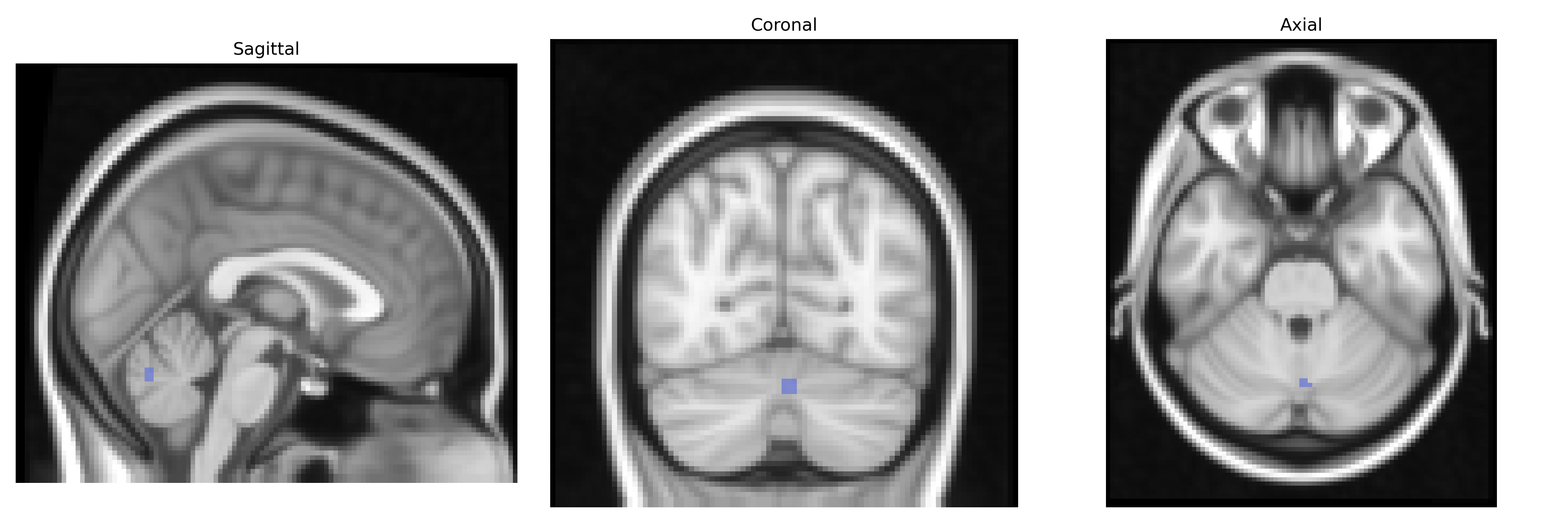
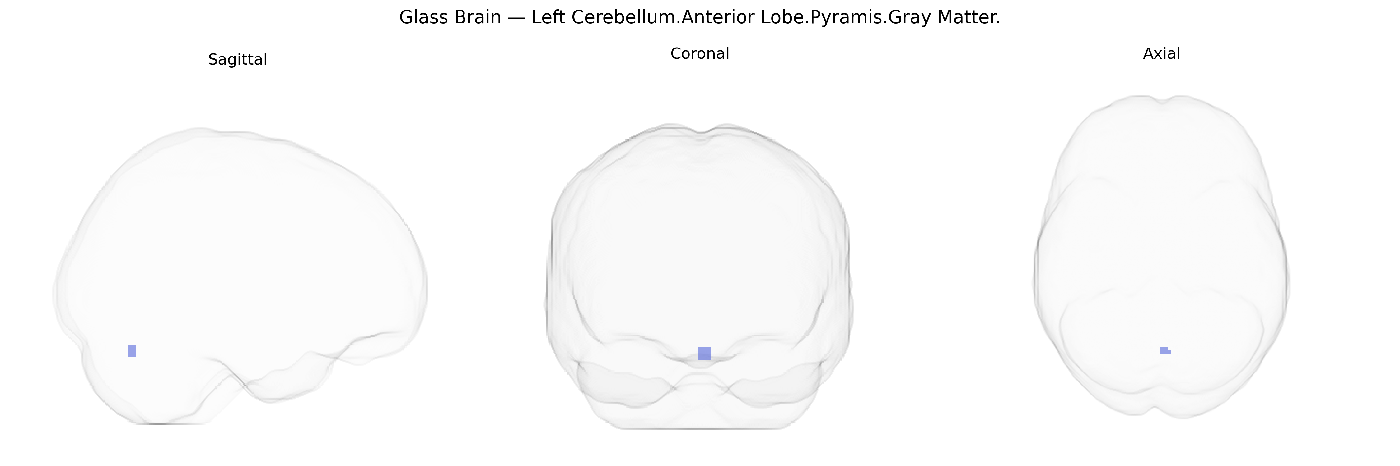
The bilateral Left Cerebellum.Anterior Lobe.Pyramis.Gray Matter refers to gray matter within the pyramis, a midline lobular structure of the anterior lobe of the cerebellum, located in the vermis and extending symmetrically into both cerebellar hemispheres on the left side in Talairach coordinate space. This region is part of the spinocerebellum, receiving extensive proprioceptive and somatosensory input via spinal and brainstem pathways, and contributes to the regulation of posture, muscle tone, and the coordination and timing of limb and trunk movements. Functionally, the pyramis participates in error correction and fine-tuning of ongoing motor activity, integrating sensory feedback with motor commands to maintain smooth, accurate movement execution. There is no direct link for the cerebellar pyramis; a related structure is the Cerebellum.
The bilateral Left Cerebellum.Anterior Lobe.Pyramis.Gray Matter region, as defined in the Talairach 2 mm atlas, lies within the anterior cerebellar vermis and adjacent hemispheric tissue, a zone repeatedly implicated in motor coordination, posture, and affective regulation; however, few genetic studies report associations specifically at this fine-grained label. Large-scale brain imaging GWAS (e.g., ENIGMA, UK Biobank) have identified polygenic influences on cerebellar gray matter volume, with variants near genes involved in neurodevelopment, synaptic function, and axonal guidance (such as variants near KIAA0586, RELN, and CACNA1A) associated with global or regional cerebellar structure, though localization typically distinguishes anterior vs posterior lobes or vermis rather than the pyramis label itself. In psychiatric and neurodevelopmental genetics, cerebellar vermis and anterior lobe abnormalities have been linked to autism spectrum disorder, attention-deficit/hyperactivity disorder, major depressive disorder, and schizophrenia in imaging–genetics work, where polygenic risk scores for these disorders correlate with altered cerebellar volume or connectivity, suggesting shared genetic influences on this territory. Mendelian and rare-variant conditions affecting cerebellar development (such as certain spinocerebellar ataxias, Joubert syndrome, and other cerebellar malformation syndromes) often involve genes critical for Purkinje cell function and cerebellar patterning, which can include structural changes encompassing the anterior lobe and pyramis, though studies usually describe macroscopic vermis hypoplasia or generalized cerebellar atrophy rather than this specific atlas-defined region. Overall, genetic associations for the Left Cerebellum.Anterior Lobe.Pyramis.Gray Matter label are inferred from broader cerebellar and vermis-focused imaging-genetic findings, with converging evidence that common and rare variants affecting cerebellar development and synaptic signaling contribute to individual differences in its structure and vulnerability to motor and psychiatric phenotypes.
Overview generated by GPT-4o (2026).
Region ID: 116
Hemisphere: bilateral
Atlas: Talairach labels 2mm
Full Quality Version: Download MP4
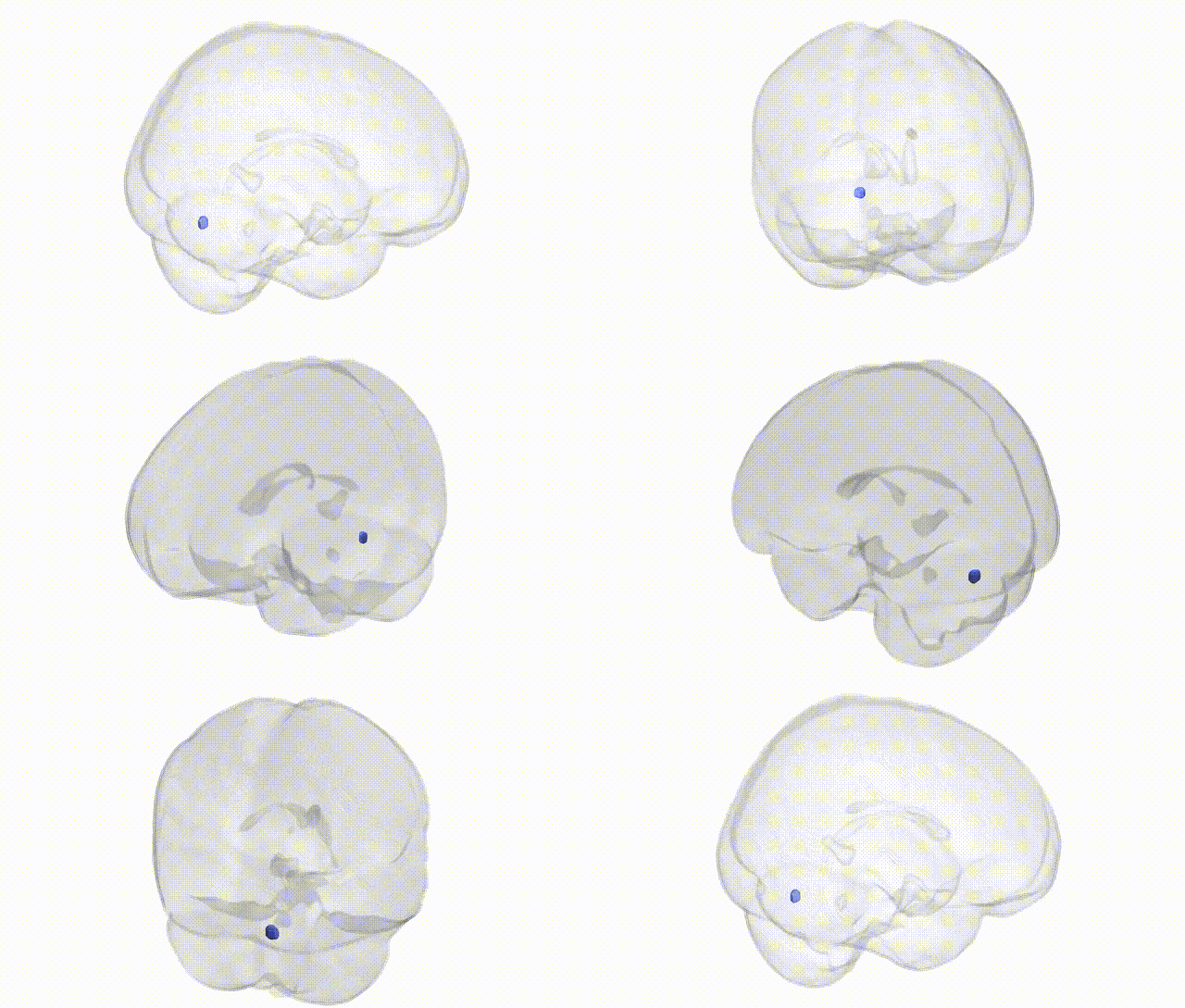
Full Quality Version: Download MP4
Wali Sidiqyar*, Gaurav Rudravaram*, Elyssa M. McMaster, Trent M. Schwartz, Adam M. Saunders, Kurt G. Schilling, Bennett A. Landman "Introducing SPINS: A Shared Public Visualization Library of Neuroanatomical Structures." Medical Imaging with Deep Learning- short paper
This resource is licensed under CC0 1.0 Universal (Public Domain).