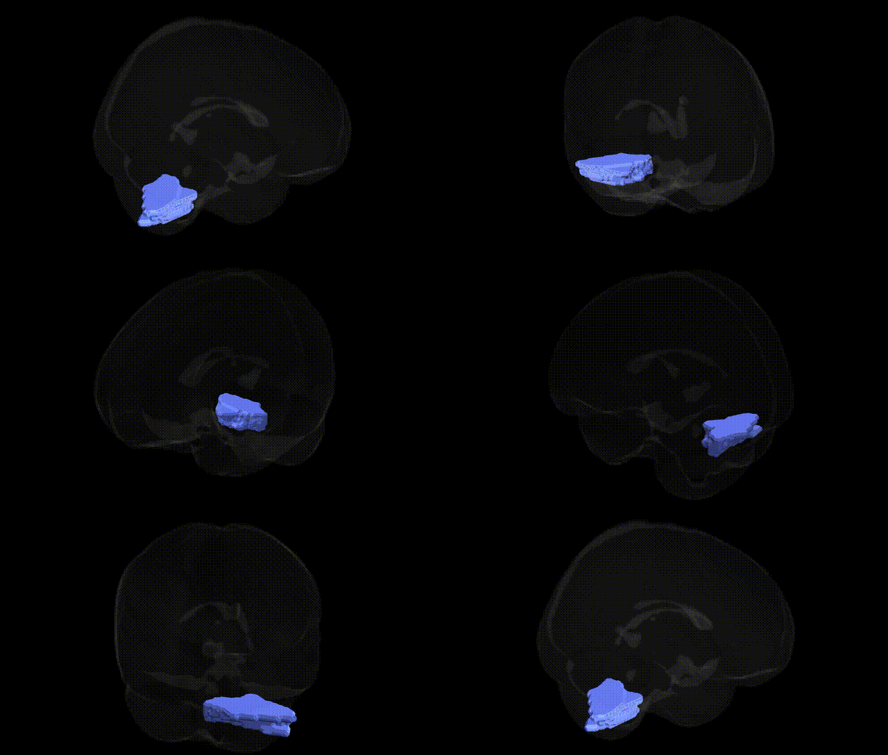
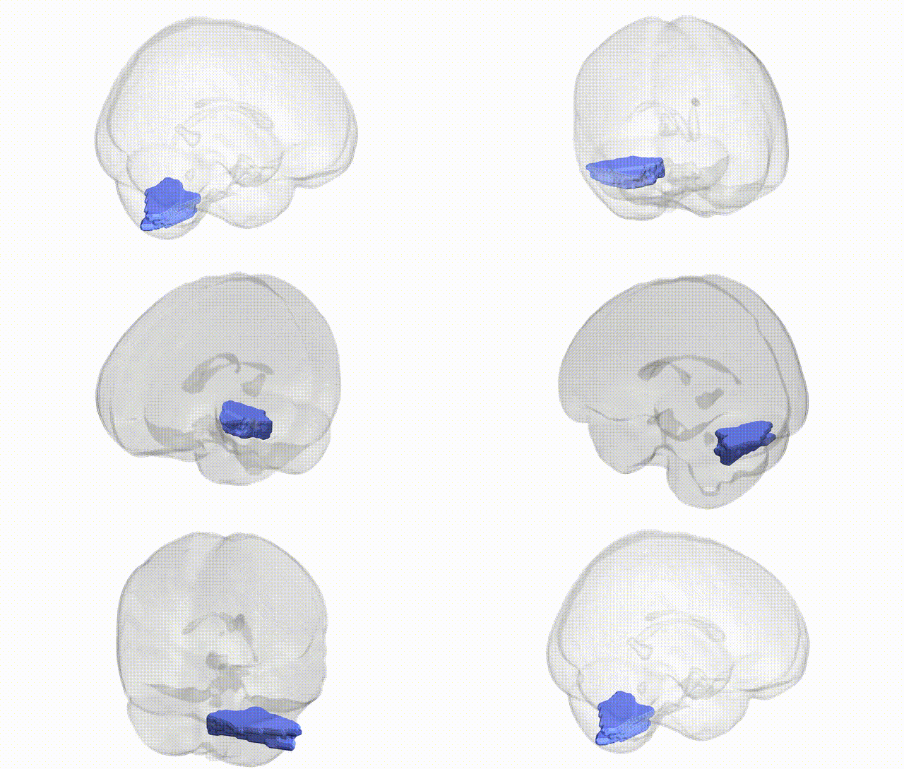
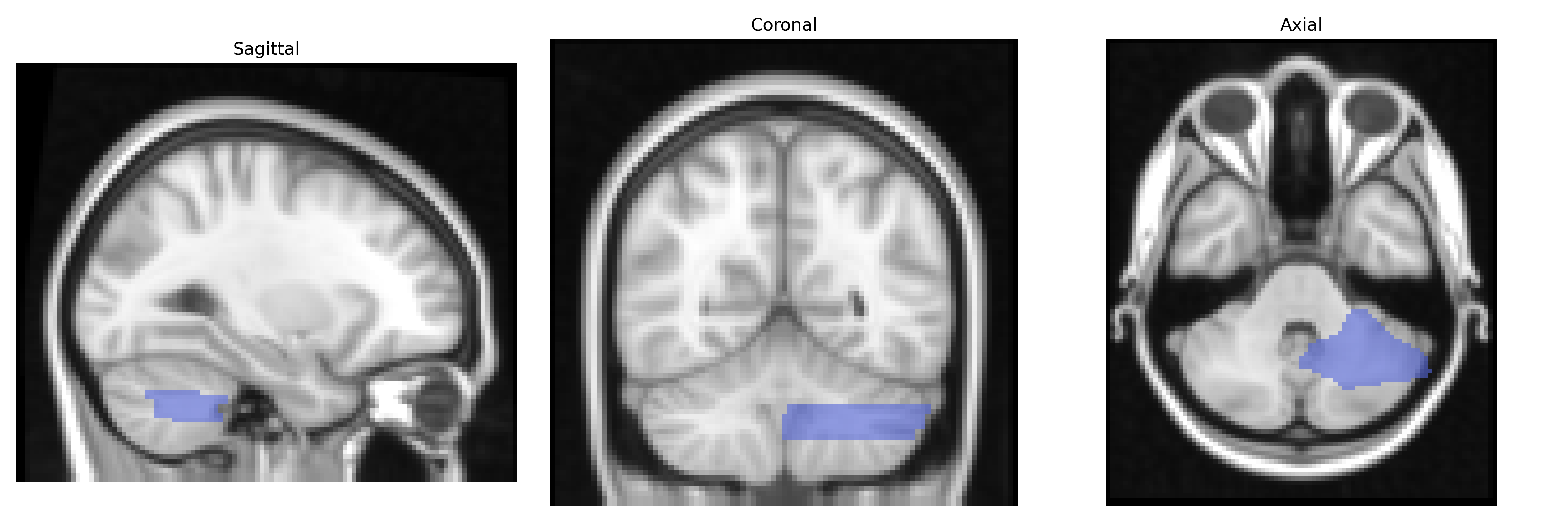
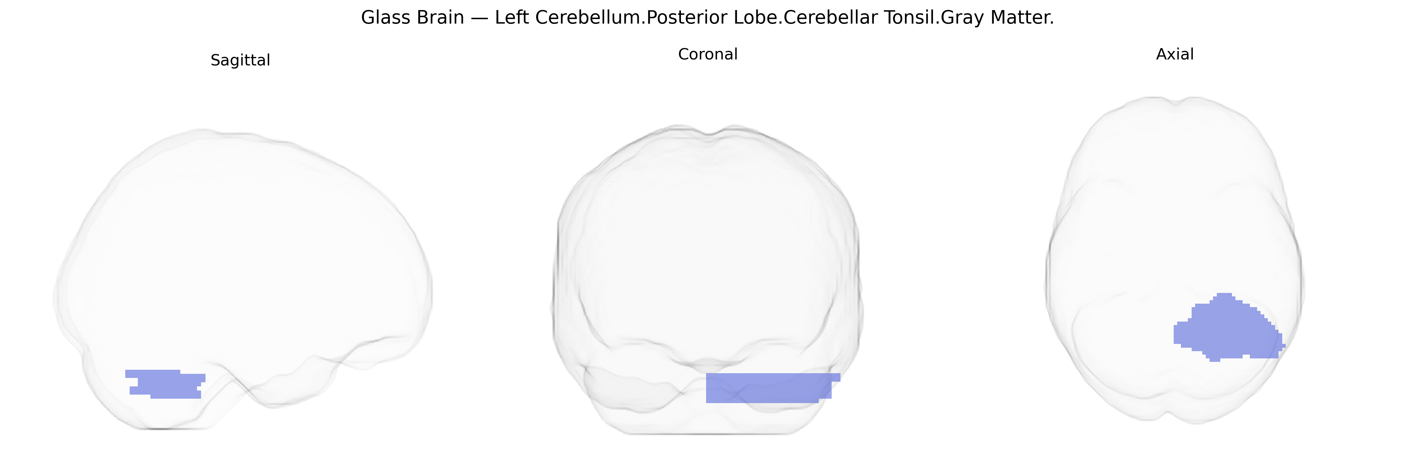
The bilateral Left Cerebellum Posterior Lobe Cerebellar Tonsil Gray Matter refers to paired nodular structures of cerebellar cortex located on the inferior aspect of the posterior lobe of the cerebellum, adjacent to the medulla and near the foramen magnum. These gray matter masses are part of the cerebellar hemispheres and contribute to coordination of movement, balance, and posture through integration of proprioceptive and vestibular inputs. Clinically, the cerebellar tonsils are notable because downward herniation through the foramen magnum can compress the brainstem and upper cervical spinal cord, as seen in conditions such as Chiari malformation and posterior fossa mass lesions. There is no direct Wikipedia article specifically for the cerebellar tonsil as a standalone entry; see the related structure Cerebellum.
The bilateral Left Cerebellum.Posterior Lobe.Cerebellar Tonsil.Gray Matter region in the Talairach 2mm Atlas falls within the posterior cerebellar territory broadly implicated by imaging genetics and GWAS of brain structure, though few studies have isolated the tonsillar subdivision specifically. Large-scale GWAS of cerebellar volume and cortical thickness have identified polygenic influences across many loci, including variants near genes involved in neurodevelopment, synaptic function, and cytoskeletal dynamics (e.g., KIAA0586, MAPT region, and multiple noncoding regulatory loci), with posterior cerebellar gray matter showing heritability and genetically correlated variation with cognitive performance, educational attainment, and general intelligence. Imaging genetics studies in psychiatric and neurodevelopmental disorders—such as schizophrenia, bipolar disorder, autism spectrum disorder, and ADHD—have linked risk variants in genes including CACNA1C, GRM3, and synaptic/neurite-related genes to altered cerebellar morphology or activation patterns in posterior lobules, which encompass or border the cerebellar tonsils. Cerebellar structural variation in this territory has also been associated with genetic risk scores for depression and anxiety, and with GWAS-identified loci for motor coordination and balance, reflecting the region’s role in sensorimotor and cognitive-affective integration. However, current GWAS typically report effects at the level of whole cerebellar lobules or total cerebellar volume, so genetic associations should be interpreted as affecting broader posterior cerebellar circuitry rather than uniquely targeting the cerebellar tonsil gray matter subdivision.
Overview generated by GPT-4o (2026).
Region ID: 3
Hemisphere: bilateral
Atlas: Talairach labels 2mm
Full Quality Version: Download MP4
Full Quality Version: Download MP4
Wali Sidiqyar*, Gaurav Rudravaram*, Elyssa M. McMaster, Trent M. Schwartz, Adam M. Saunders, Kurt G. Schilling, Bennett A. Landman "Introducing SPINS: A Shared Public Visualization Library of Neuroanatomical Structures." Medical Imaging with Deep Learning- short paper
This resource is licensed under CC0 1.0 Universal (Public Domain).