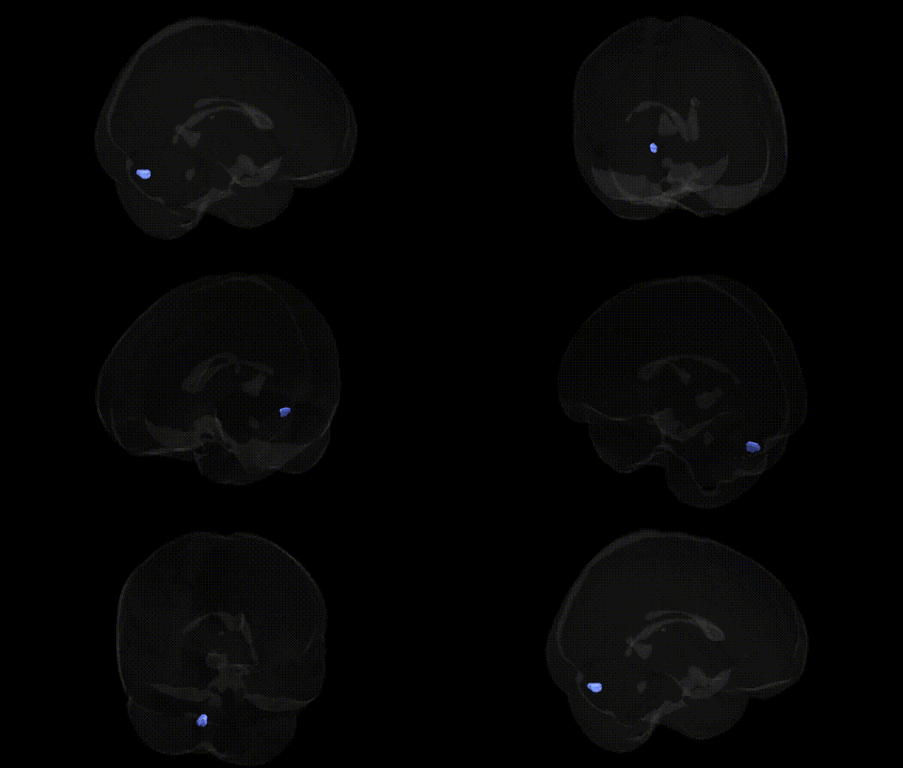
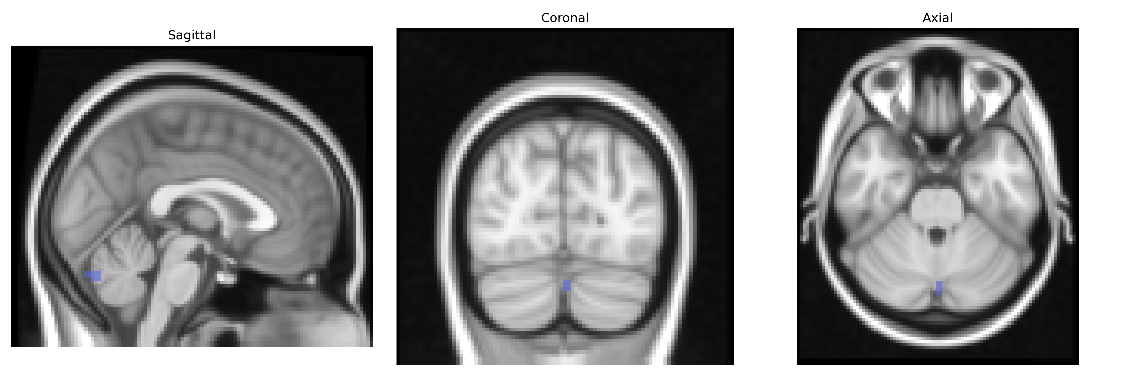
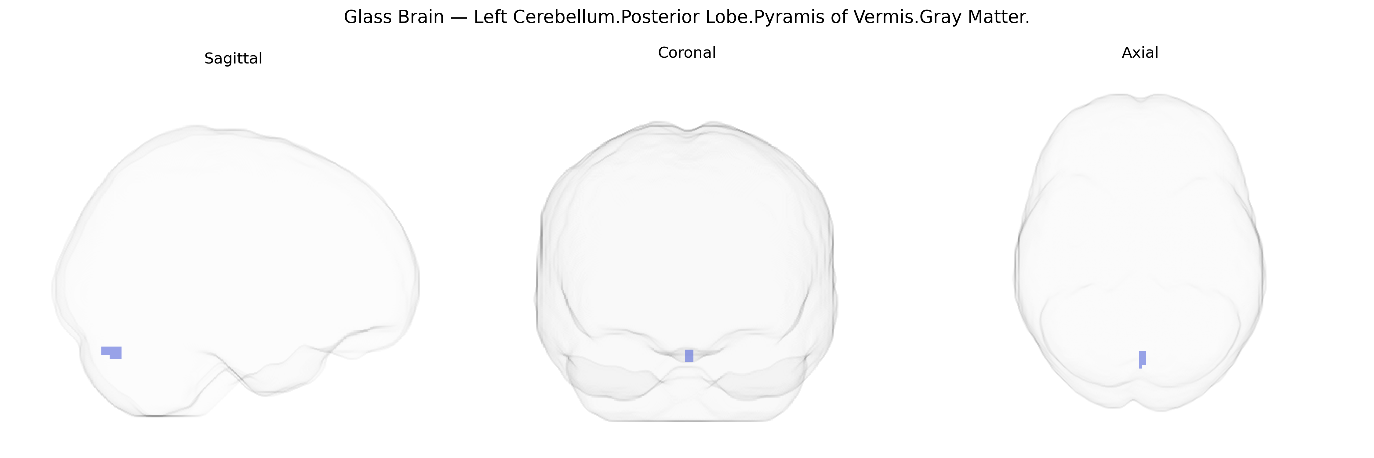
The bilateral Left Cerebellum Posterior Lobe Pyramis of Vermis Gray Matter corresponds to a midline cerebellar vermal structure located within the posterior lobe, composed primarily of cortical gray matter that overlies underlying white matter and deep cerebellar nuclei. The pyramis is part of the spinocerebellum, receiving somatosensory input mainly from the spinal cord and contributing to the regulation of posture, muscle tone, and coordination of axial and proximal limb movements. Functionally, it is involved in fine-tuning motor execution and maintaining balance through integration of proprioceptive information and efferent motor signals. As a vermal region, it may also play a role in affective and cognitive processes via cerebello-cortical loops, although its primary role is motor. There is no direct Wikipedia article for this specific substructure; a related and encompassing article is Cerebellar vermis.
The bilateral left cerebellar posterior lobe, including the pyramis of the vermis gray matter as defined in the Talairach 2 mm atlas, is implicated in genetic studies largely through broader cerebellar or posterior fossa measures rather than fine-grained regional GWAS. Polygenic influences on total cerebellar volume and lobular morphology have been reported in large-scale neuroimaging GWAS (e.g., ENIGMA, UK Biobank), with variants in or near genes involved in neurodevelopment (such as KIAA0319, DLG2, and common neurodevelopmental loci on chromosomes 3p and 6p) contributing to inter-individual variability in cerebellar structure, including posterior lobes. The pyramis/vermis region is frequently implicated in genetic studies of neurodevelopmental and psychiatric disorders where cerebellar vermis abnormalities are observed, such as autism spectrum disorder, attention-deficit/hyperactivity disorder, and schizophrenia, often via genes regulating synaptic formation and neuronal migration (e.g., RELN, CNTNAP2, and SHANK family genes), though these associations are typically reported at the level of disorder or total cerebellar/vermis measures rather than this specific subregion. Cerebellar posterior lobe and vermis gray matter variation has also been linked in imaging-genetic work to motor coordination, cognitive control, and affective traits, and appears in studies of hereditary cerebellar ataxias where pathogenic mutations (e.g., in ATXN1, ATXN2, ATXN3, CACNA1A) produce characteristic volumetric loss including vermis structures, suggesting that the pyramis of the vermis participates in genetically influenced circuits underpinning motor, cognitive, and affective functions, albeit without region-specific GWAS signals uniquely assigned to this Talairach-defined area.
Overview generated by GPT-4o (2026).
Region ID: 82
Hemisphere: bilateral
Atlas: Talairach labels 2mm
Full Quality Version: Download MP4
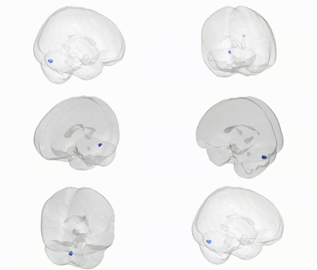
Full Quality Version: Download MP4
Wali Sidiqyar*, Gaurav Rudravaram*, Elyssa M. McMaster, Trent M. Schwartz, Adam M. Saunders, Kurt G. Schilling, Bennett A. Landman "Introducing SPINS: A Shared Public Visualization Library of Neuroanatomical Structures." Medical Imaging with Deep Learning- short paper
This resource is licensed under CC0 1.0 Universal (Public Domain).