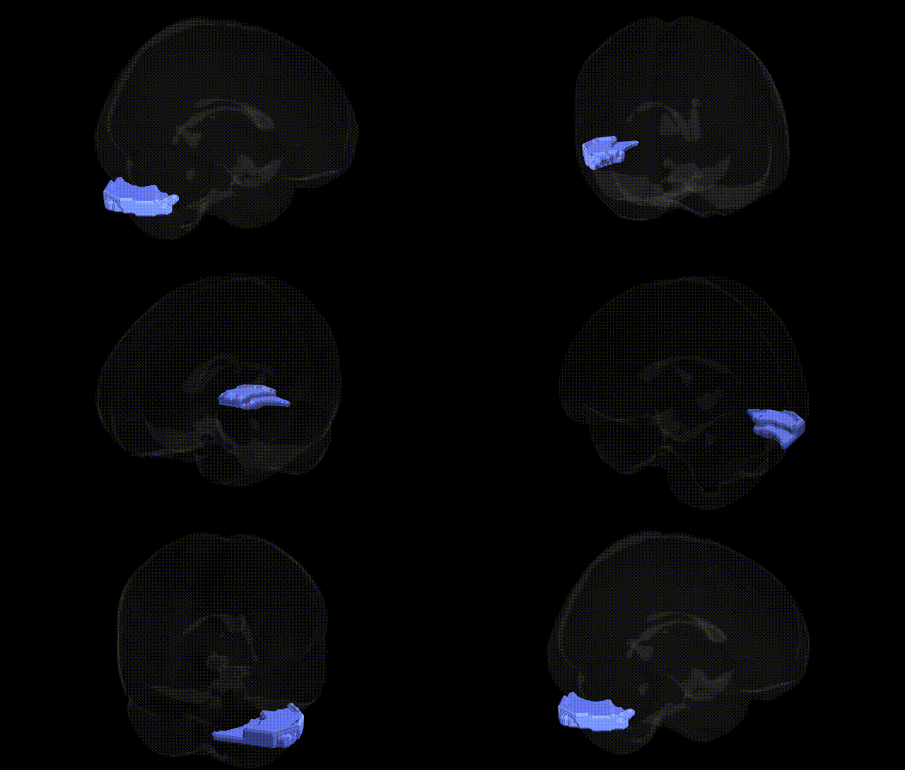
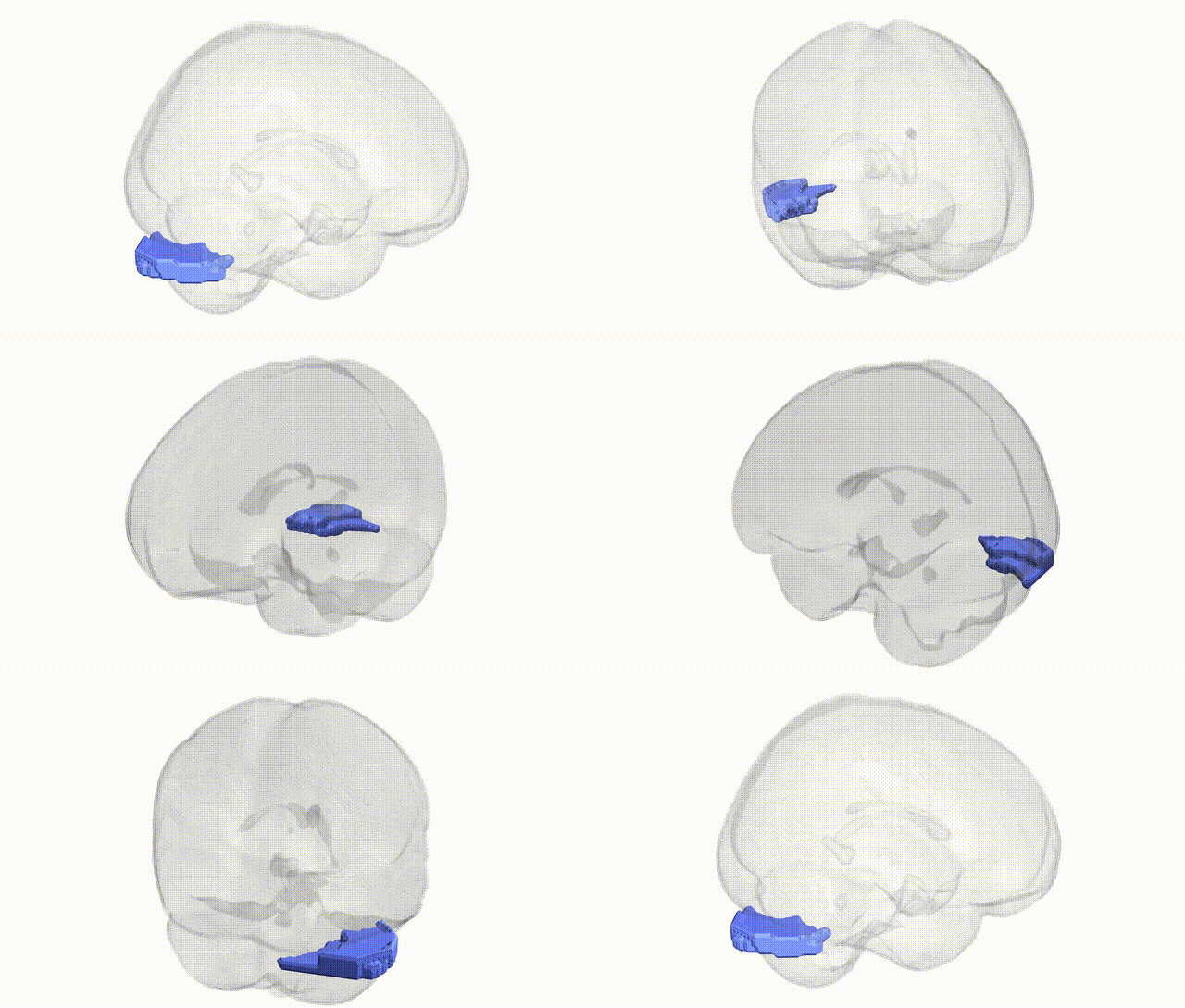
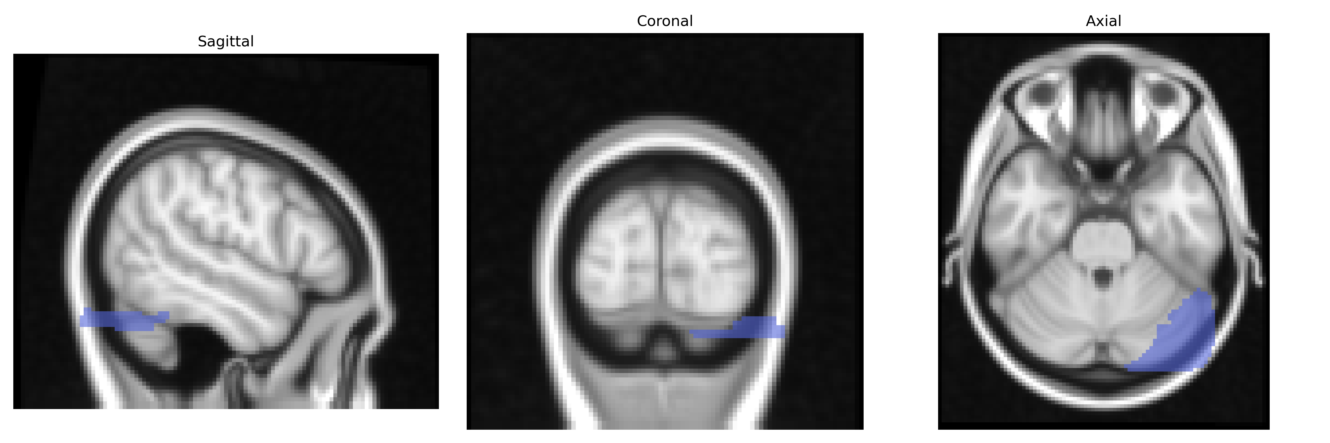
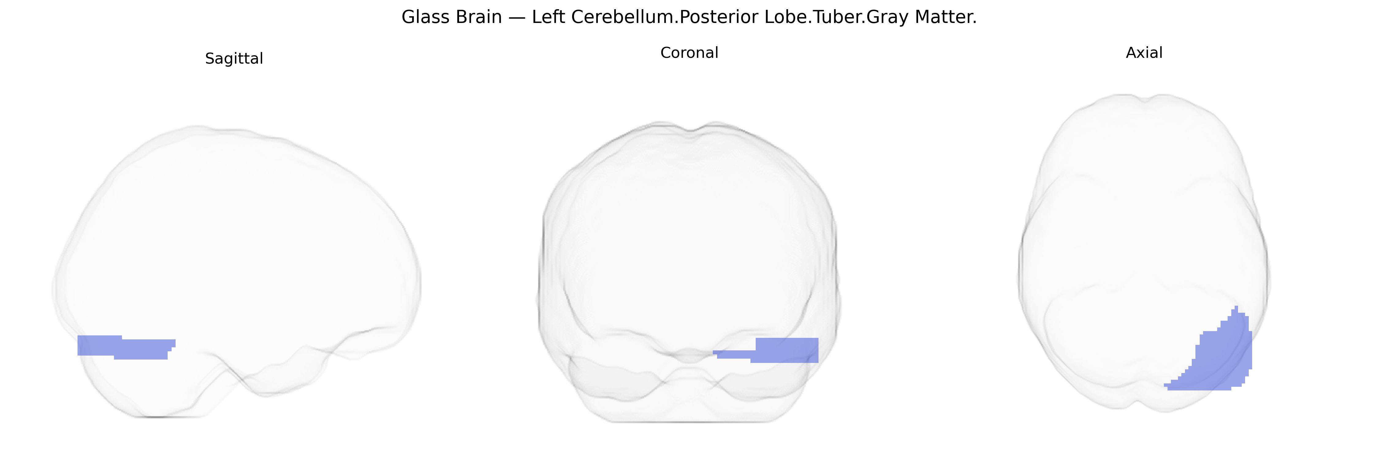
The bilateral Left Cerebellum Posterior Lobe Tuber Gray Matter refers to a cortical region on the inferior surface of the cerebellar hemisphere within the posterior lobe, corresponding to the “tuber” of the vermis and adjacent hemispheric cortex composed predominantly of cerebellar gray matter (Purkinje cells, granular layer, and molecular layer). This area participates in fine-tuning motor coordination, balance, and timing of movements, and, via extensive connections with the cerebral cortex and brainstem, contributes to motor learning and the integration of sensorimotor information. Functional imaging and clinical lesion studies suggest that posterior cerebellar regions, including the tuber-related cortex, may also play roles in higher-order cognitive and affective modulation. There is no direct link for this specific subregion; see the related structure Cerebellum.
The bilateral Left Cerebellum Posterior Lobe Tuber Gray Matter region, as defined in the Talairach 2 mm atlas, has not been the direct focus of region-specific GWAS, but convergent genetic and imaging-genetic evidence implicates the posterior cerebellum more broadly in a range of neurodevelopmental, cognitive, and psychiatric traits. Large neuroimaging GWAS (e.g., ENIGMA and UK Biobank studies) have identified multiple common variants associated with cerebellar volume and cortical thickness—particularly within genes involved in neurodevelopment, synaptic function, and axon guidance (such as CACNA1C, MAPT, and variants near FOXP1/FOXP2)—which in turn show genetic correlations with intelligence, educational attainment, and working memory, domains strongly supported by posterior cerebellar circuitry. The cerebellar posterior lobe, including the tuber region, has been implicated in autism spectrum disorder, schizophrenia, bipolar disorder, and major depression through imaging-genetic work demonstrating that polygenic risk scores for these disorders predict variation in cerebellar structure and connectivity; rare variant and CNV studies (e.g., 22q11.2 deletion, SHANK3, and other synaptic genes) also show cerebellar morphological changes overlapping this area. In motor and coordination disorders, including ataxias and dystonias, mutations in genes such as ATXN1–3, CACNA1A, and PRKCG frequently alter posterior cerebellar anatomy as quantified in voxel-based morphometry, although these studies typically report regional clusters spanning larger lobular territories rather than the tuber specifically. Overall, genetic associations point to the posterior cerebellum as a polygenic hub integrating cognitive, affective, and motor influences, with the tuber gray matter region likely participating in these broader genetically mediated structural and functional patterns, even though no GWAS has yet isolated this exact Talairach-defined parcel as a primary locus of association.
Overview generated by GPT-4o (2026).
Region ID: 80
Hemisphere: bilateral
Atlas: Talairach labels 2mm
Full Quality Version: Download MP4
Full Quality Version: Download MP4
Wali Sidiqyar*, Gaurav Rudravaram*, Elyssa M. McMaster, Trent M. Schwartz, Adam M. Saunders, Kurt G. Schilling, Bennett A. Landman "Introducing SPINS: A Shared Public Visualization Library of Neuroanatomical Structures." Medical Imaging with Deep Learning- short paper
This resource is licensed under CC0 1.0 Universal (Public Domain).