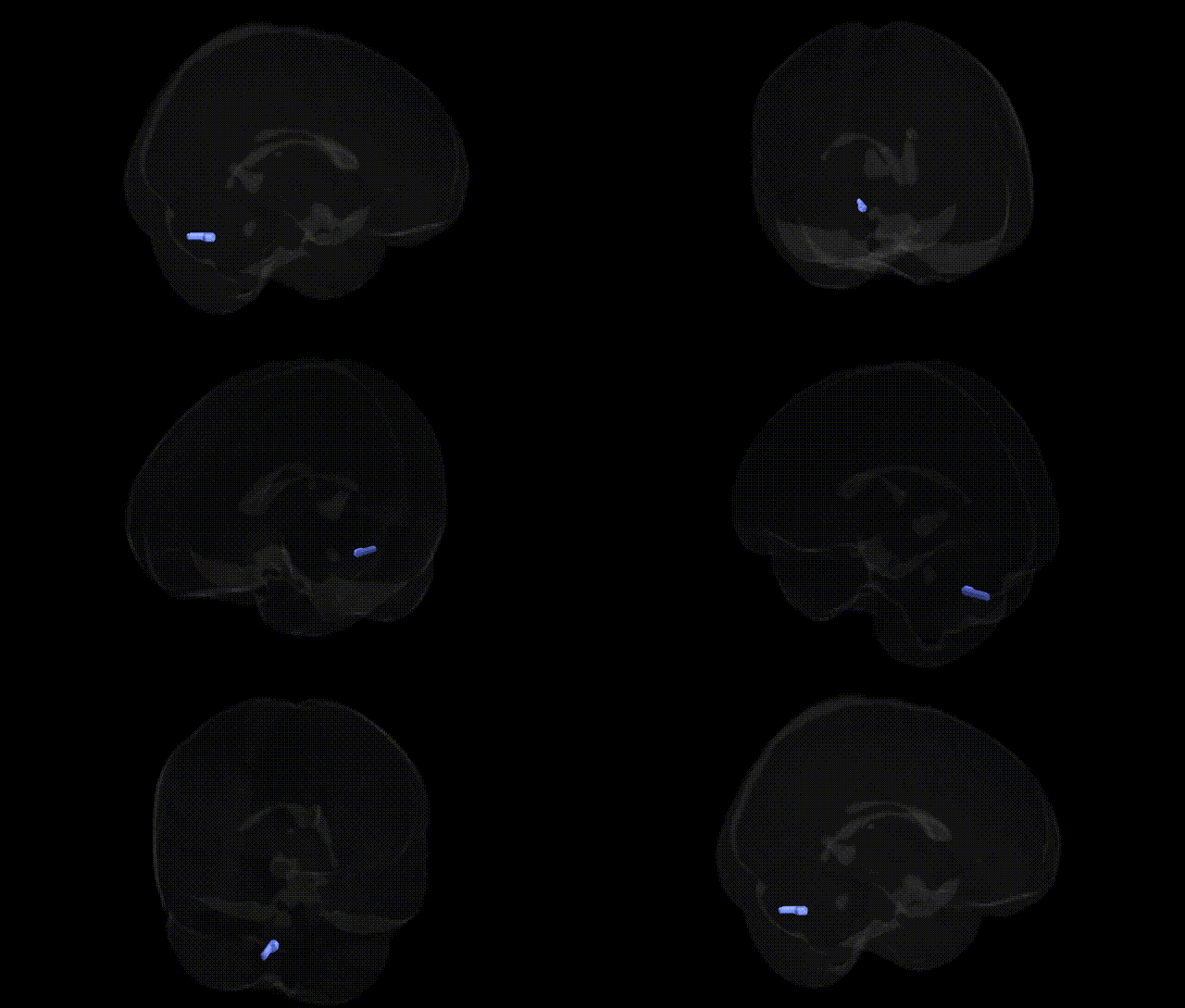
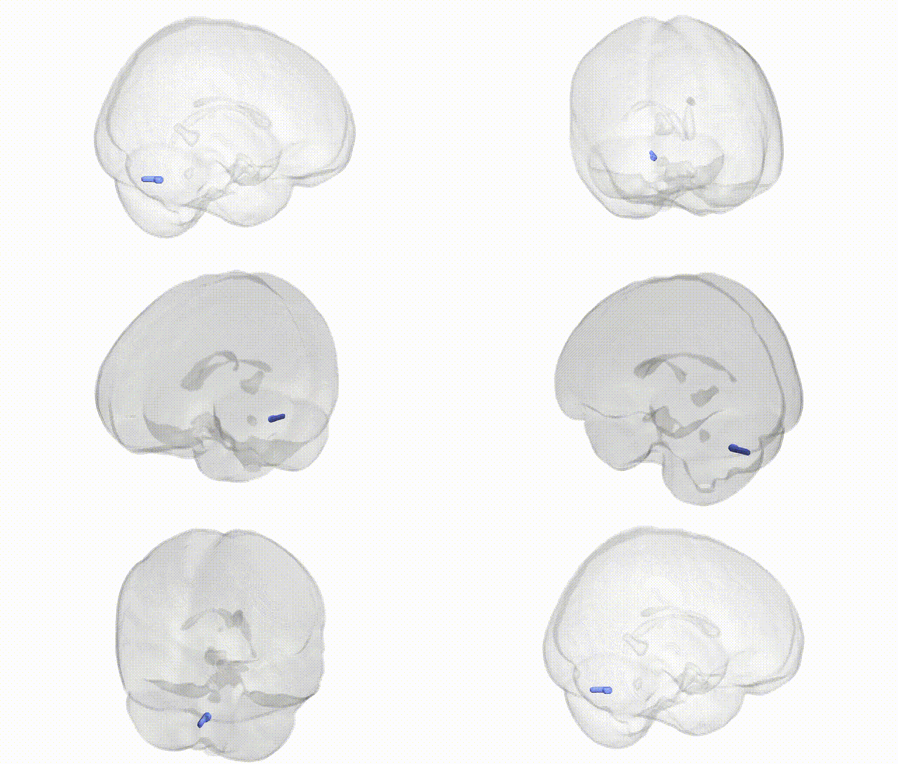
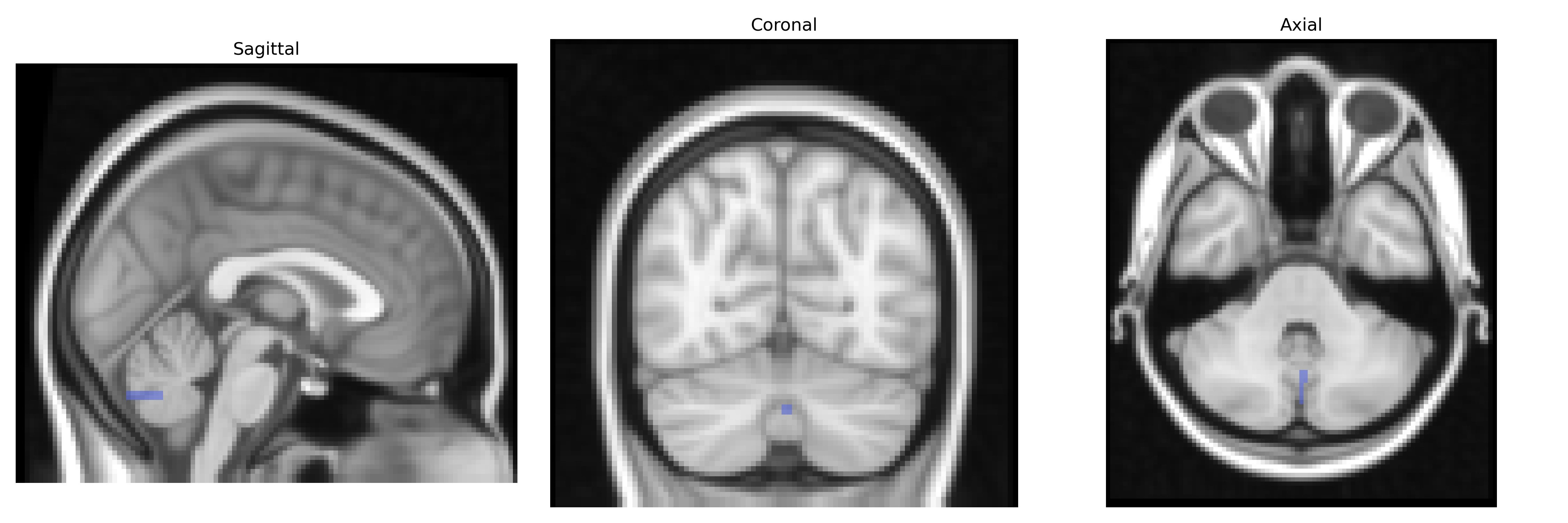
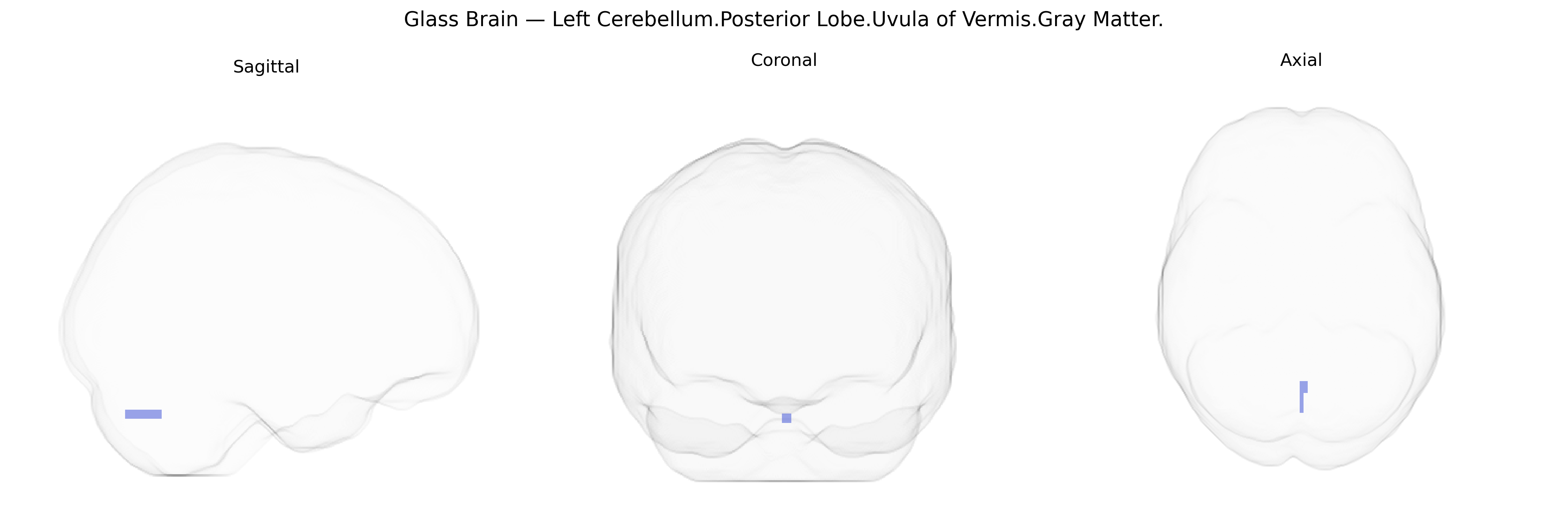
The bilateral Left Cerebellum Posterior Lobe, Uvula of Vermis, Gray Matter corresponds to a midline cerebellar structure located within the posterior lobe of the cerebellum, forming part of the inferior vermis. The uvula is involved in the coordination of posture, balance, and eye movements, integrating vestibular, proprioceptive, and visual inputs to fine-tune motor control, particularly of axial musculature. As gray matter, it consists predominantly of cerebellar cortex (including Purkinje cells and interneurons) that project to deep cerebellar nuclei, contributing to the modulation of motor output and adaptive motor learning. There is no direct link for the uvula of vermis; a related structure is the cerebellar vermis: Cerebellar vermis
The bilateral Left Cerebellum.Posterior Lobe.Uvula of Vermis.Gray Matter region, as defined in the Talairach 2 mm Atlas, corresponds to the posterior vermis, a midline cerebellar structure implicated in affective regulation, motor timing, and cognitive-emotional integration; genetic association evidence is largely indirect, as GWAS typically report broader cerebellar or vermal measures rather than this subregion specifically. Large neuroimaging GWAS (e.g., ENIGMA, UK Biobank) have identified multiple loci (including variants near genes such as KCNK2, DLG2, and MAPT) associated with total cerebellar and vermal gray matter volumes, some of which overlap with risk loci for psychiatric conditions such as schizophrenia, bipolar disorder, major depression, and autism spectrum disorder, where structural and functional abnormalities of the posterior vermis have been documented. In addition, polygenic risk scores for neurodevelopmental and mood disorders show associations with altered cerebellar morphology and connectivity, suggesting that shared genetic architectures influencing synaptic development, neuroplasticity, and neuroinflammatory pathways may contribute to vermal gray matter variation. Cerebellar vermis abnormalities have also been reported in genetic ataxias (e.g., spinocerebellar ataxias with CAG repeat expansions in genes such as ATXN1, ATXN2, and CACNA1A), where midline cerebellar degeneration can include the uvula and adjacent lobules, and in copy number variant syndromes (e.g., 22q11.2 deletion) that show consistent cerebellar volumetric changes. Overall, while no GWAS has isolated the uvula of vermis gray matter as a distinct phenotype, converging genetic and imaging findings implicate common neurodevelopmental and synaptic genes in shaping structure and vulnerability of the posterior vermis, linking this region to motor coordination, affective and cognitive symptoms, and cerebellar ataxia phenotypes.
Overview generated by GPT-4o (2026).
Region ID: 67
Hemisphere: bilateral
Atlas: Talairach labels 2mm
Full Quality Version: Download MP4
Full Quality Version: Download MP4
Wali Sidiqyar*, Gaurav Rudravaram*, Elyssa M. McMaster, Trent M. Schwartz, Adam M. Saunders, Kurt G. Schilling, Bennett A. Landman "Introducing SPINS: A Shared Public Visualization Library of Neuroanatomical Structures." Medical Imaging with Deep Learning- short paper
This resource is licensed under CC0 1.0 Universal (Public Domain).