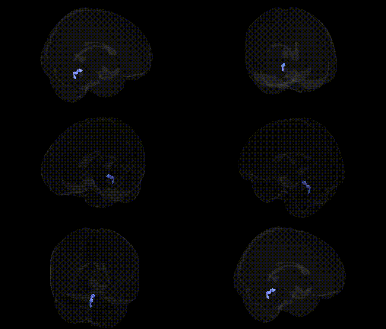
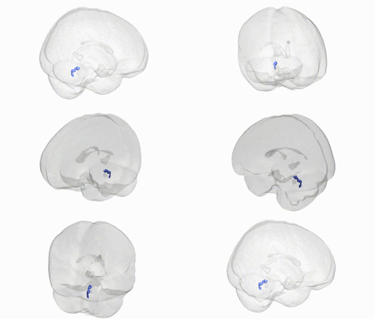
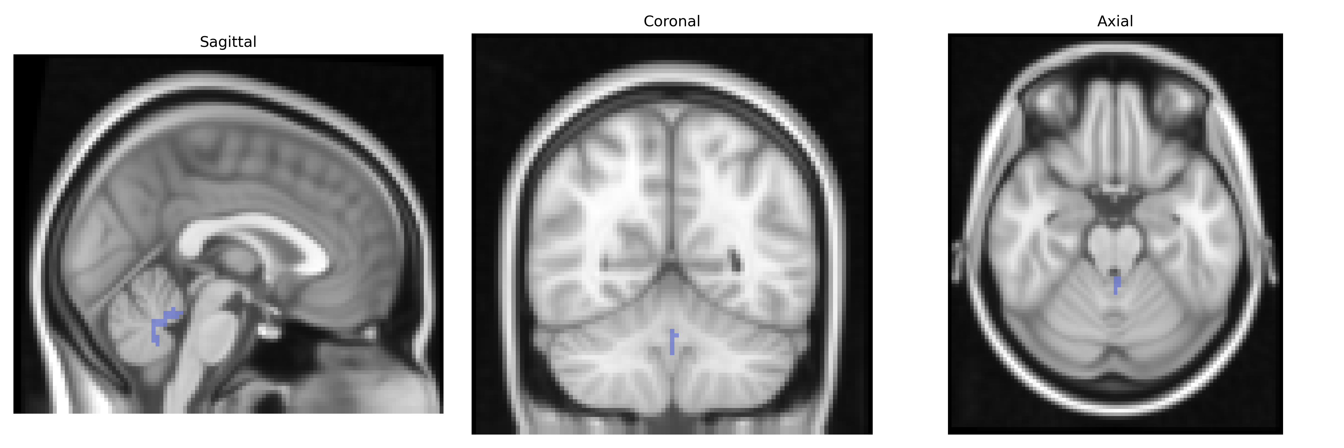
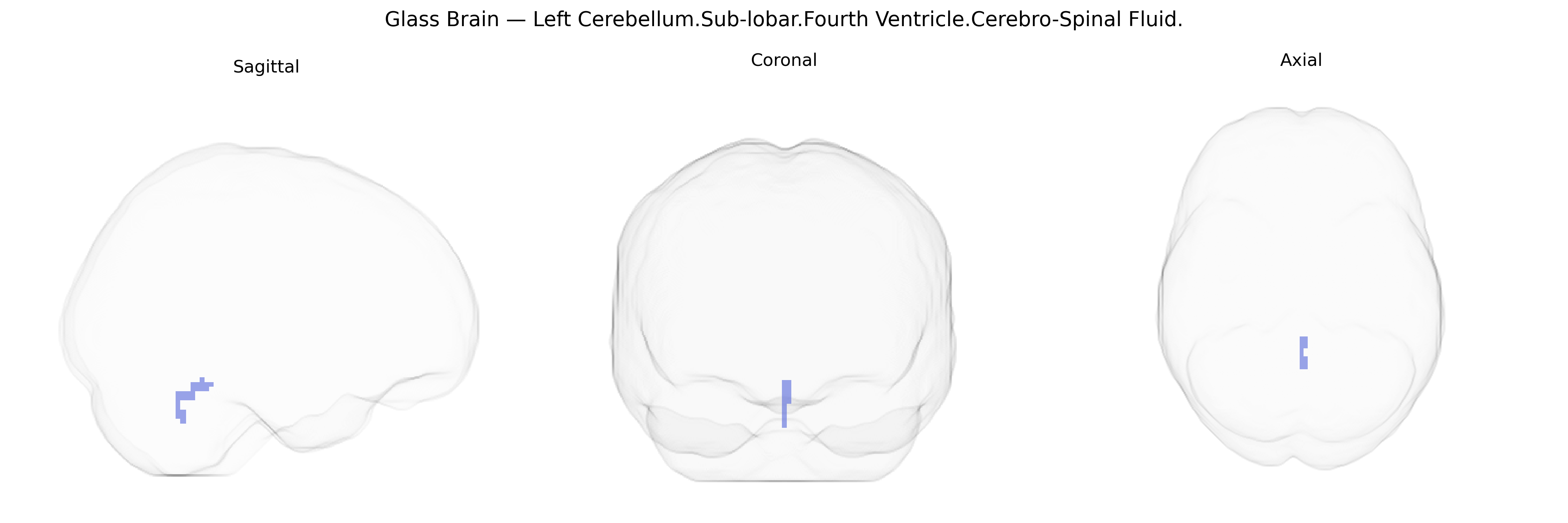
The bilateral Left Cerebellum.Sub-lobar.Fourth Ventricle.Cerebro-Spinal Fluid region in the Talairach 2 mm Atlas corresponds to cerebrospinal fluid (CSF) occupying the central cavity adjacent to the cerebellum known as the fourth ventricle. The fourth ventricle is a diamond-shaped space located in the posterior fossa, dorsal to the pons and medulla and ventral to the cerebellum, forming part of the ventricular system that circulates CSF produced by the choroid plexus. CSF within this compartment contributes to mechanical cushioning of neural tissue, maintenance of intracranial pressure, and transport of metabolites and signaling molecules. In neuroimaging atlases, this region is typically classified as non-parenchymal, CSF-filled space rather than functional gray matter, and serves as an anatomical landmark for hindbrain structures and midline symmetry. There is no direct link for “Left Cerebellum.Sub-lobar.Fourth Ventricle.Cerebro-Spinal Fluid,” but it is part of the Fourth ventricle.
Genetic association studies rarely target the bilateral Left Cerebellum.Sub-lobar.Fourth Ventricle.Cerebro-Spinal Fluid region itself, as it is primarily a cerebrospinal fluid (CSF) compartment rather than neural parenchyma; instead, GWAS and other imaging-genetics work typically examine global or regional CSF volume, ventricular size, or total intracranial volume as proxy measures of brain structure. Large-scale neuroimaging GWAS (e.g., ENIGMA, UK Biobank) have identified loci in or near genes such as HMGA2, ASTN2, TESC, and others associated with intracranial volume, brain size, and ventricular or CSF space expansion, which indirectly encompasses the fourth ventricle and surrounding cerebellar-CSF interface. These genetic variants are often also implicated in neurodevelopmental and neuropsychiatric conditions—such as schizophrenia, bipolar disorder, autism spectrum disorder, and cognitive traits—where atypical brain and ventricular volumes are observed, suggesting shared biological pathways in brain growth, neurodevelopment, and neurodegeneration. However, no robust, region-specific GWAS has isolated genetic effects uniquely tied to the Talairach-defined “Left Cerebellum.Sub-lobar.Fourth Ventricle.CSF” label, and current evidence largely reflects general associations with brain and CSF volume rather than a discrete functional or disease-related genetic signature for this exact anatomical region.
Overview generated by GPT-4o (2026).
Region ID: 70
Hemisphere: bilateral
Atlas: Talairach labels 2mm
Full Quality Version: Download MP4
Full Quality Version: Download MP4
Wali Sidiqyar*, Gaurav Rudravaram*, Elyssa M. McMaster, Trent M. Schwartz, Adam M. Saunders, Kurt G. Schilling, Bennett A. Landman "Introducing SPINS: A Shared Public Visualization Library of Neuroanatomical Structures." Medical Imaging with Deep Learning- short paper
This resource is licensed under CC0 1.0 Universal (Public Domain).