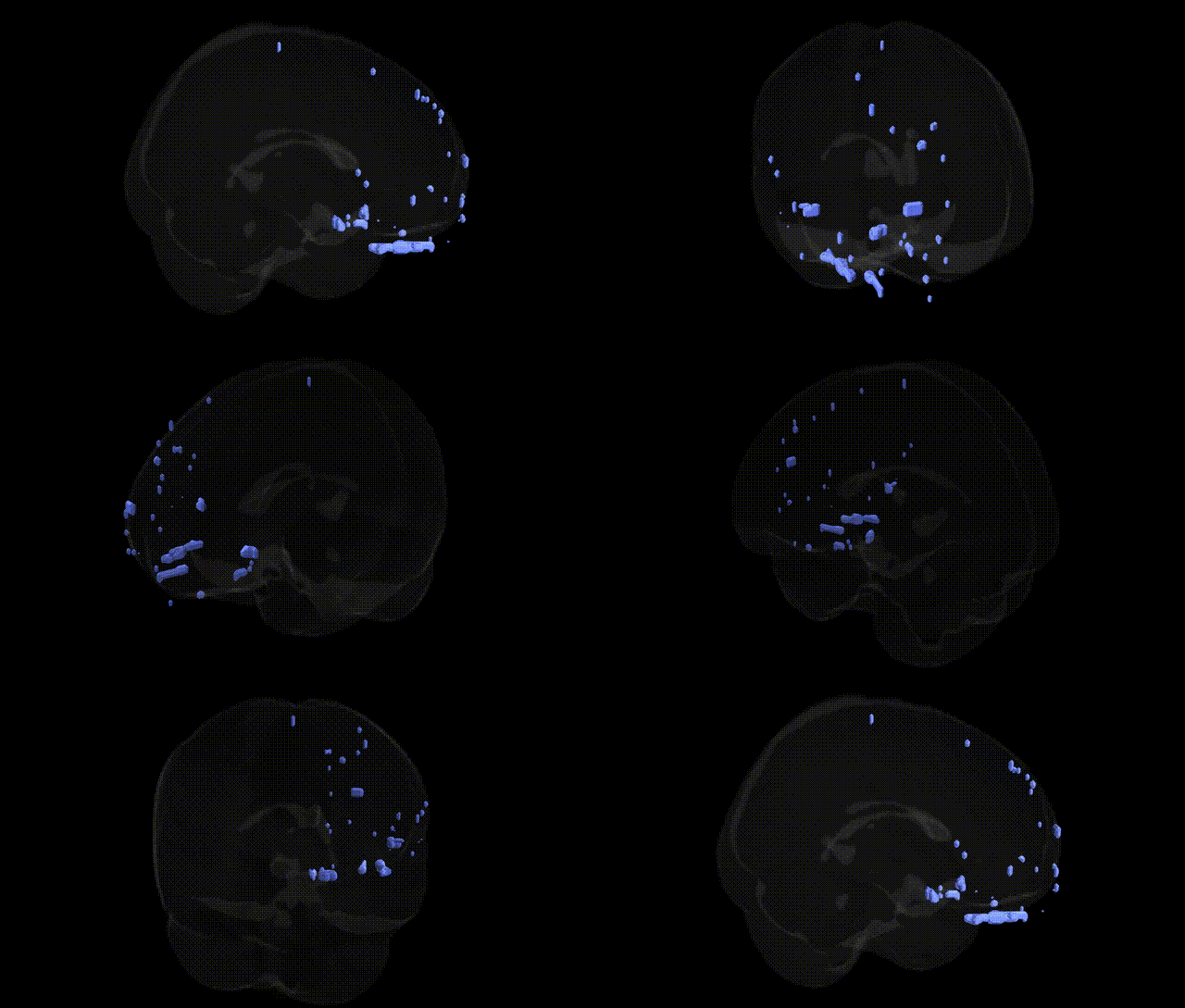
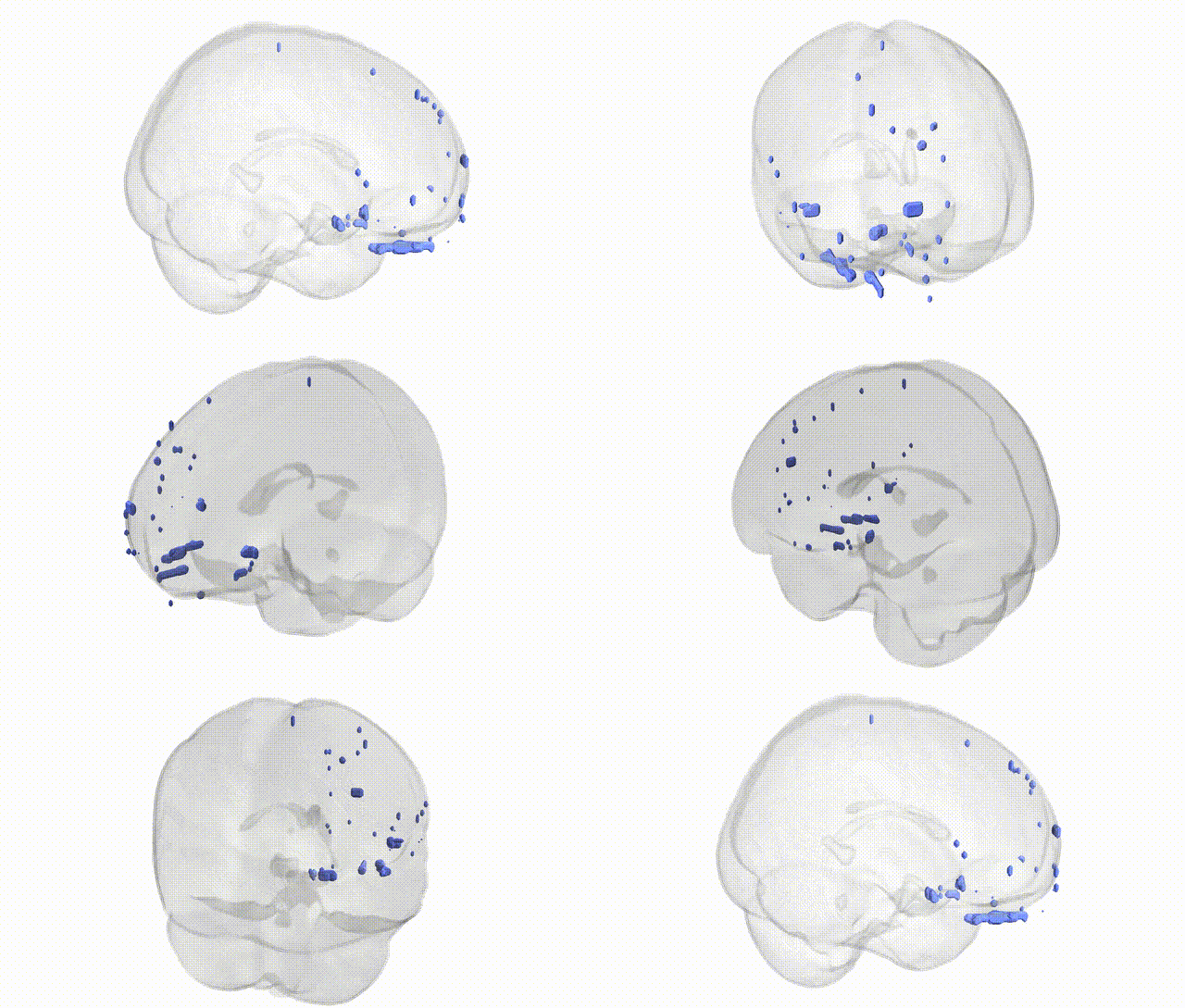
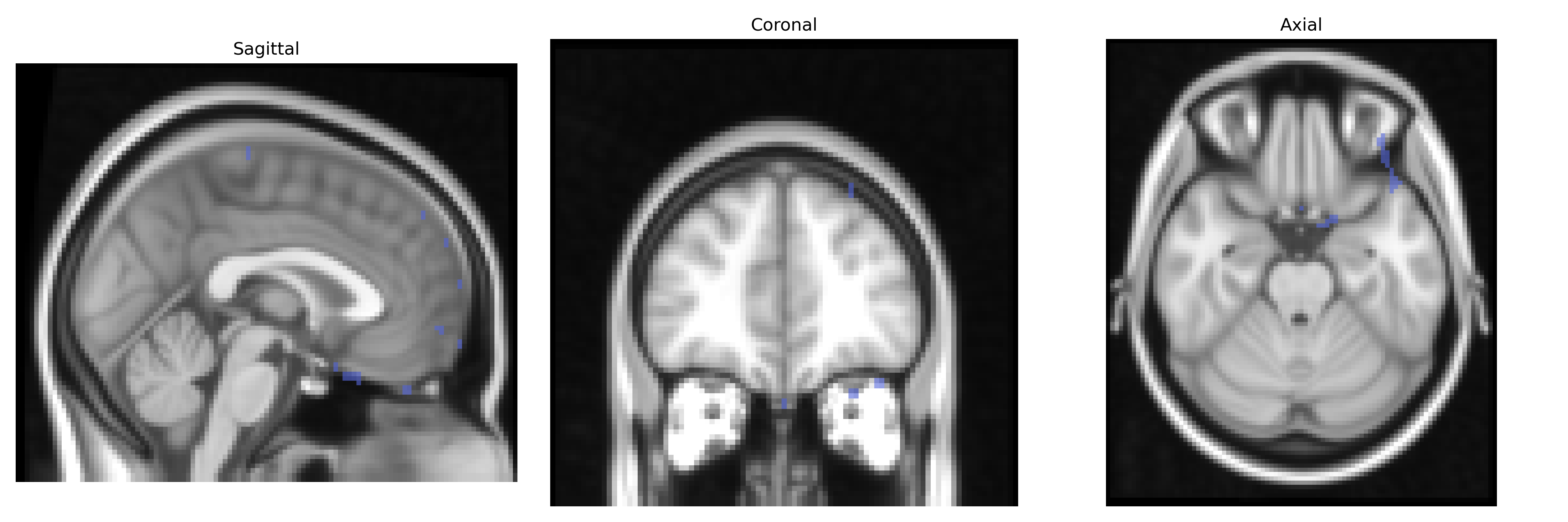
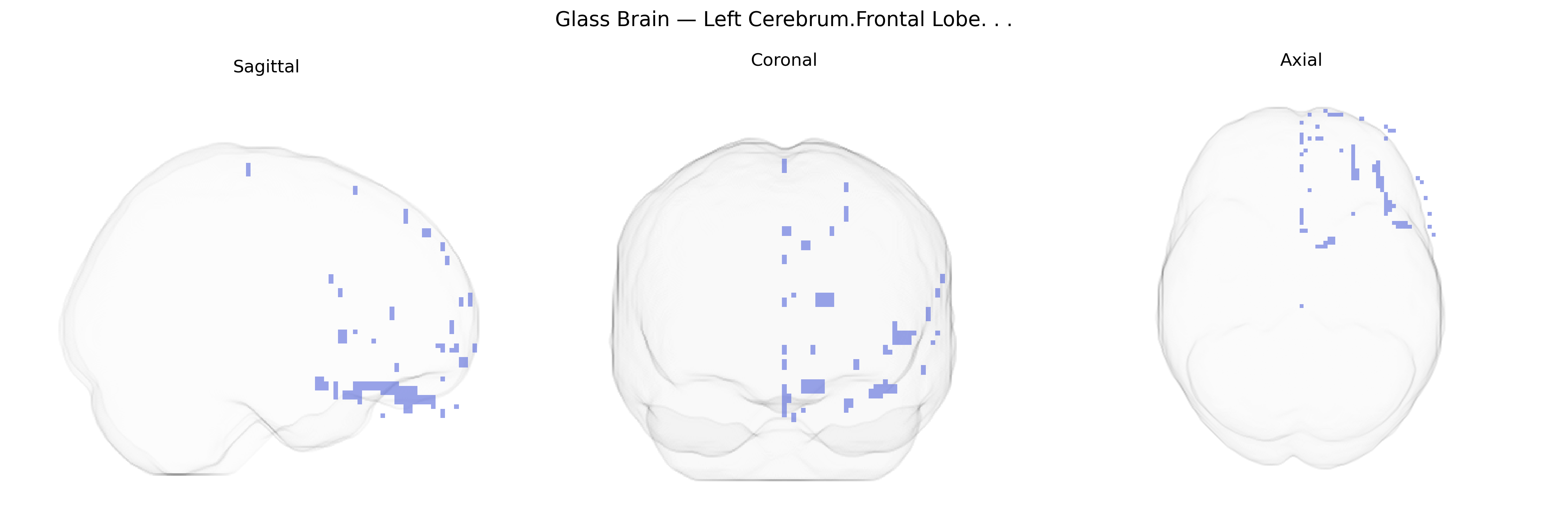
The bilateral Left Cerebrum.Frontal Lobe, as defined in the Talairach 2 mm atlas, encompasses the anterior portion of the left cerebral hemisphere, extending from the central sulcus to the frontal pole and comprising multiple cytoarchitectonic regions including premotor, supplementary motor, and prefrontal cortices. This lobe participates in higher-order executive functions such as planning, decision-making, working memory, behavioral inhibition, and the regulation of social and emotional behavior, while also contributing substantially to voluntary motor control via primary and premotor motor areas. In the dominant (typically left) hemisphere, frontal regions are critically involved in language production (e.g., Broca’s area in the inferior frontal gyrus), speech articulation, and syntactic processing, as well as in complex cognitive operations requiring attention, problem solving, and cognitive flexibility. Through extensive reciprocal connectivity with parietal, temporal, limbic, and subcortical structures, the left frontal lobe integrates sensory, mnemonic, and affective information to guide goal-directed behavior and adaptive responses.
Frontal lobe
The bilateral left frontal lobe (Talairach 2 mm atlas label: Left Cerebrum.Frontal Lobe) has been repeatedly implicated in genetic studies of cognition, psychiatric risk, and brain structure, with many findings driven by large-scale GWAS of cortical morphology and functional networks rather than single-region studies. Common variants near genes involved in neurodevelopment and synaptic function—such as CNTNAP2, DISC1, CACNA1C, GRIN2B, and those in the 16p11.2 and 22q11.2 CNV regions—have been associated with alterations in frontal cortical thickness, surface area, and activation patterns, which in turn relate to disorders including schizophrenia, bipolar disorder, major depressive disorder, ADHD, and autism spectrum disorder. GWAS of brain imaging phenotypes (e.g., ENIGMA, UK Biobank) have identified loci in or near genes such as FOXP2, KIAA0586, CENPW, RSPO3, and TCF4 that influence global and regional frontal lobe volumes and cortical measures, and polygenic scores for educational attainment and general cognitive ability show robust associations with structural and functional variation in left frontal regions supporting language, executive function, and working memory. Additionally, risk alleles in genes regulating neurotransmission (e.g., DRD2, COMT, SLC6A4) have been linked to differences in prefrontal activation and connectivity during tasks engaging left frontal circuits, while Alzheimer’s disease and frontotemporal dementia risk loci (e.g., APOE, MAPT, GRN) are associated with atrophy patterns that prominently involve frontal regions, including left frontal lobe subareas like dorsolateral and ventrolateral prefrontal cortex. Overall, genetic influences on the left frontal lobe reflect a polygenic architecture bridging neurodevelopmental, psychiatric, cognitive, and neurodegenerative traits rather than region-specific single-gene effects.
Overview generated by GPT-4o (2026).
Region ID: 106
Hemisphere: bilateral
Atlas: Talairach labels 2mm
Full Quality Version: Download MP4
Full Quality Version: Download MP4
Wali Sidiqyar*, Gaurav Rudravaram*, Elyssa M. McMaster, Trent M. Schwartz, Adam M. Saunders, Kurt G. Schilling, Bennett A. Landman "Introducing SPINS: A Shared Public Visualization Library of Neuroanatomical Structures." Medical Imaging with Deep Learning- short paper
This resource is licensed under CC0 1.0 Universal (Public Domain).