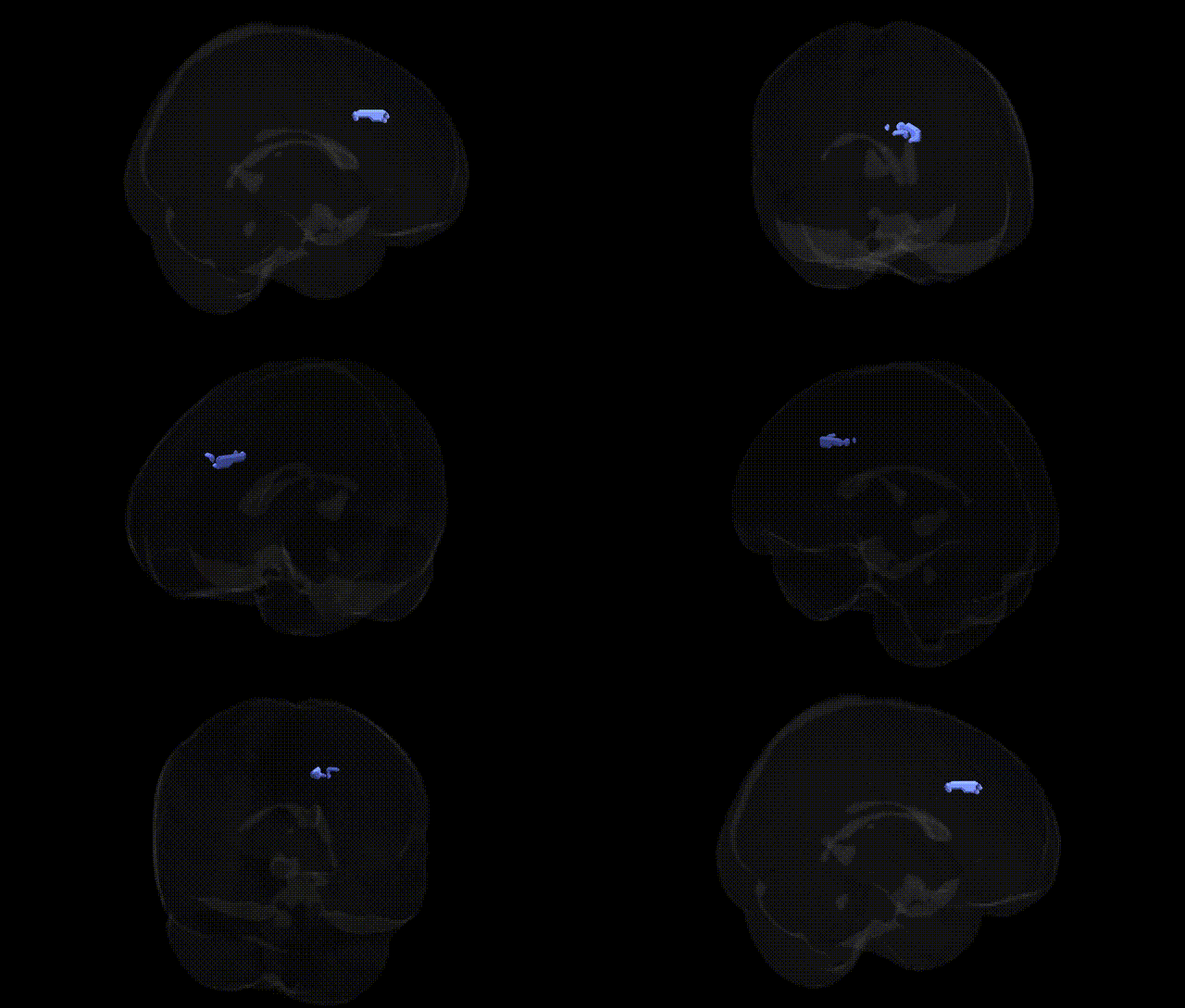
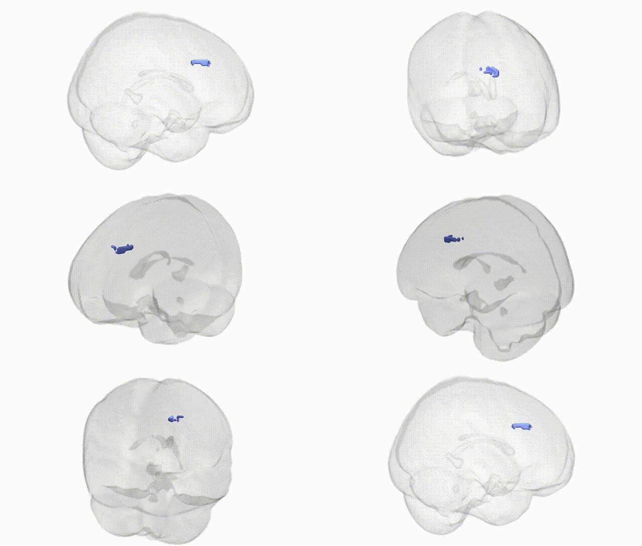
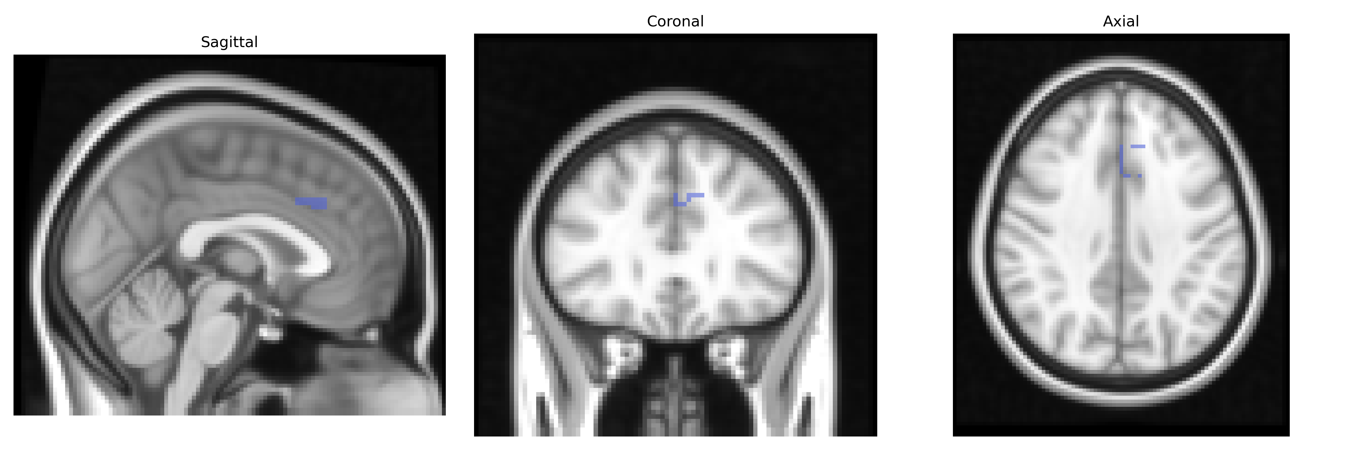
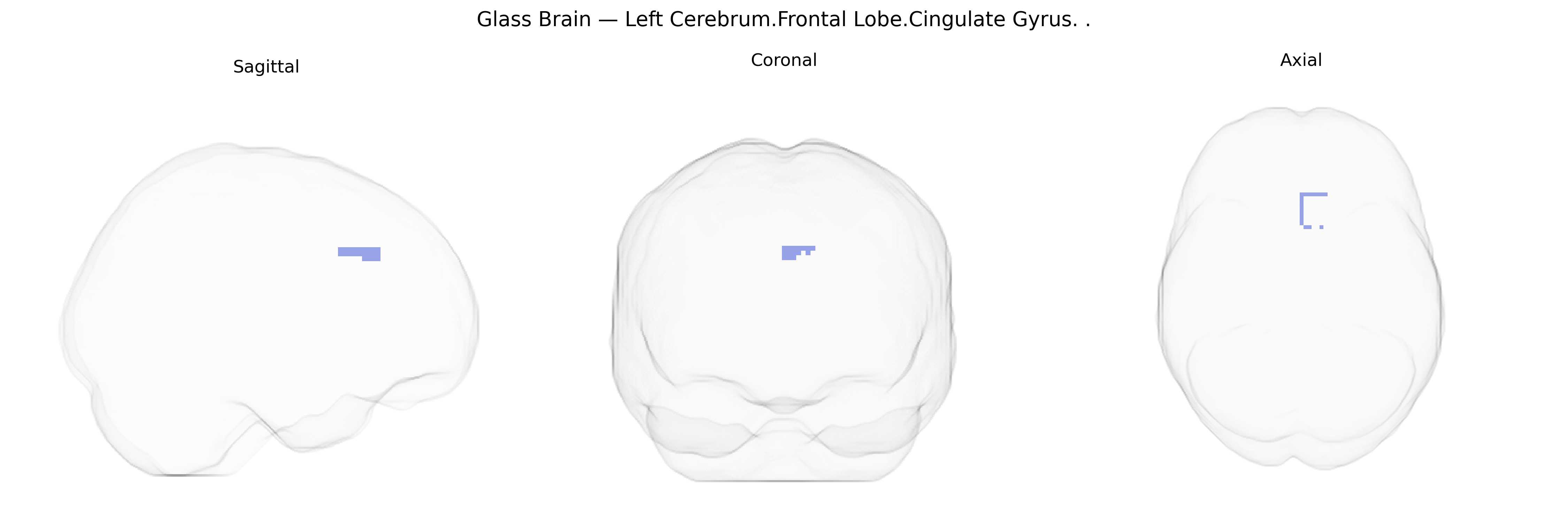
The bilateral Left Cerebrum.Frontal Lobe.Cingulate Gyrus corresponds to cortical tissue in the anterior portion of the cingulate gyrus situated within the medial frontal lobe, arching above the corpus callosum. This region participates in a wide array of functions including regulation of emotion, motivation, autonomic control, and aspects of cognitive control such as conflict monitoring and decision-making. It forms part of the limbic system and is heavily interconnected with prefrontal, parietal, and limbic structures, integrating affective and cognitive information to influence behavior. Functionally, it is implicated in pain processing, error detection, reward-based learning, and modulation of attention, with dysfunction associated with mood disorders, anxiety, obsessive–compulsive disorder, and chronic pain syndromes. Direct article: Cingulate gyrus.
The bilateral left cerebrum frontal lobe cingulate gyrus (roughly encompassing portions of anterior/mid-cingulate cortex in the Talairach 2 mm atlas) has been implicated in numerous genetic and GWAS findings, largely through imaging genetics and disorder-related studies rather than direct atlas-specific analyses. Variants in genes involved in synaptic function, neurodevelopment, and glutamatergic and dopaminergic signaling (e.g., COMT, DRD2, GRM3, DISC1, BDNF, and 5-HTTLPR/SLC6A4) have been associated with cingulate structure or activation, often in tasks engaging cognitive control, error monitoring, and affect regulation. Large neuroimaging GWAS consortia (such as ENIGMA and UK Biobank–based studies) show that common variants across the genome contribute polygenically to cortical thickness and surface area in medial frontal and cingulate regions, with notable signals near genes regulating neuronal migration, axon guidance, and myelination; however, these findings usually reference broader anterior cingulate or medial frontal parcels rather than specifically the Talairach bilateral left frontal cingulate label. The region figures prominently in genetic studies of psychiatric and neurodevelopmental disorders—schizophrenia, major depressive disorder, bipolar disorder, attention-deficit/hyperactivity disorder, autism spectrum disorder, obsessive–compulsive disorder, and post-traumatic stress disorder—where risk variants and polygenic risk scores often predict altered cingulate volume, connectivity, or task-related activation. Additionally, cingulate measures show heritability and genetic correlations with traits such as neuroticism, cognitive performance, pain sensitivity, and substance use, indicating that genetic architecture influencing this region overlaps with that of personality, cognition, and addiction-related phenotypes, though these relationships remain substantially polygenic and distributed across the cortex.
Overview generated by GPT-4o (2026).
Region ID: 1005
Hemisphere: bilateral
Atlas: Talairach labels 2mm
Full Quality Version: Download MP4
Full Quality Version: Download MP4
Wali Sidiqyar*, Gaurav Rudravaram*, Elyssa M. McMaster, Trent M. Schwartz, Adam M. Saunders, Kurt G. Schilling, Bennett A. Landman "Introducing SPINS: A Shared Public Visualization Library of Neuroanatomical Structures." Medical Imaging with Deep Learning- short paper
This resource is licensed under CC0 1.0 Universal (Public Domain).