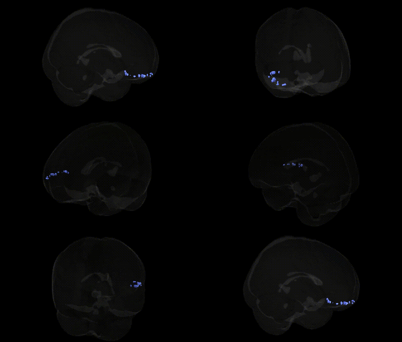
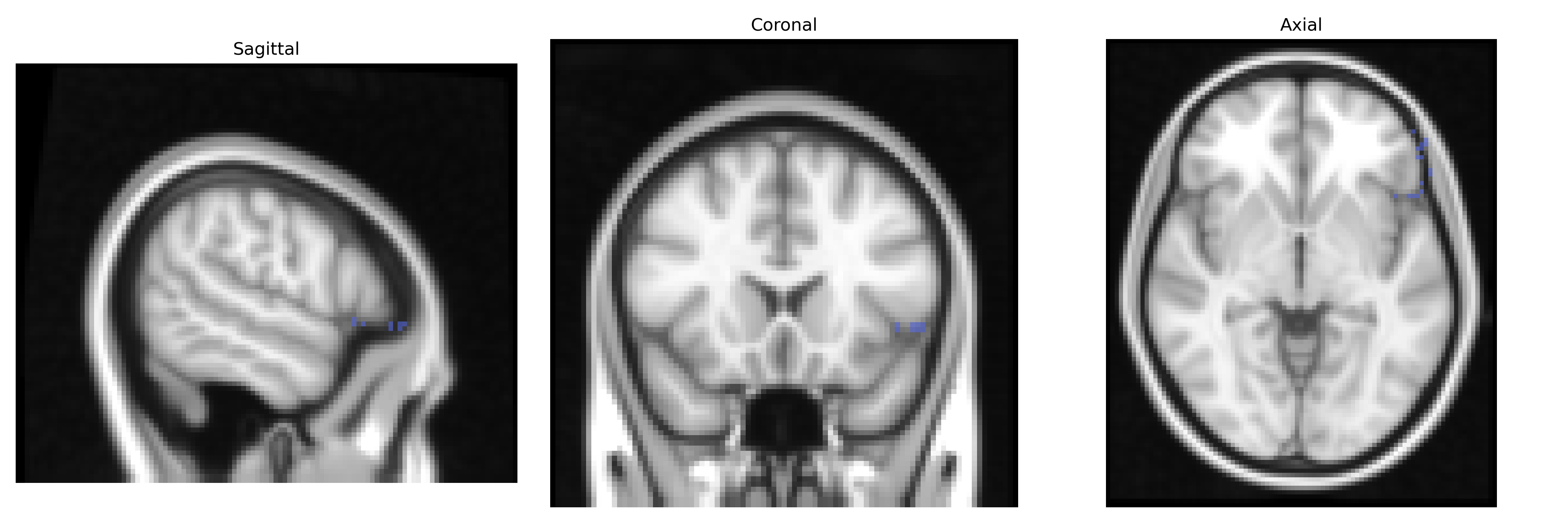
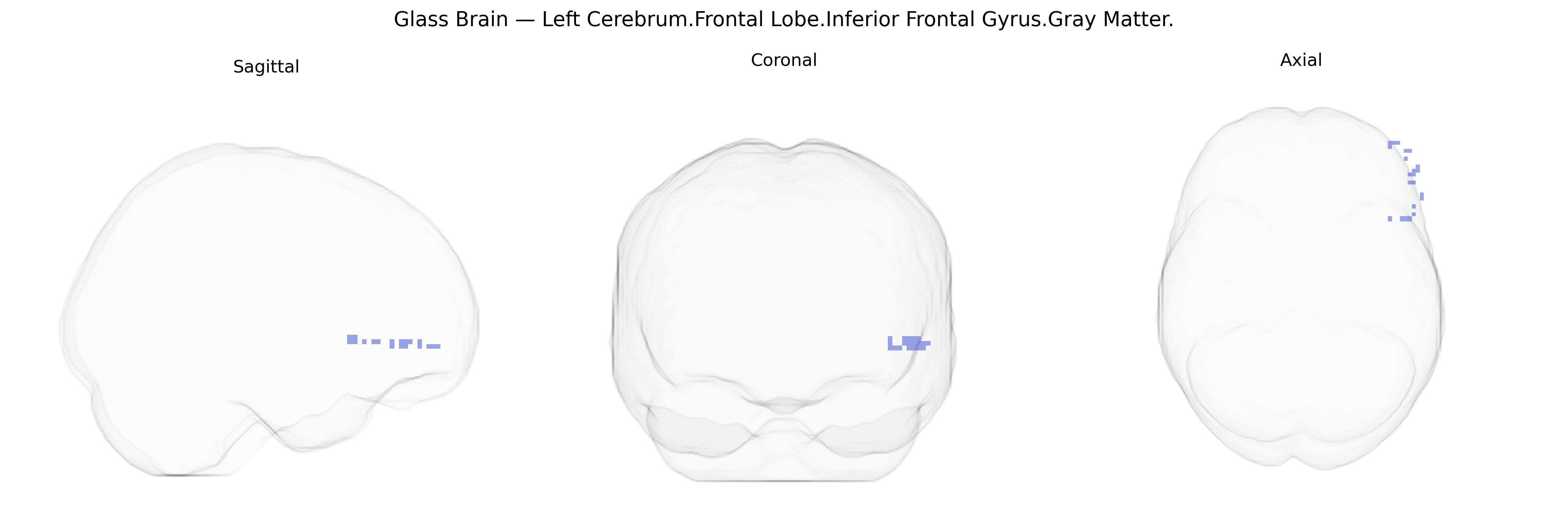
The bilateral Left Cerebrum.Frontal Lobe.Inferior Frontal Gyrus.Gray Matter corresponds primarily to cortical tissue within the left inferior frontal gyrus, a key region of the frontal lobe implicated in language, speech production, and higher-order cognitive control. This region includes parts of Brodmann areas 44 and 45 (classically associated with Broca’s area), which contribute to syntactic processing, verbal fluency, and the planning and execution of complex motor programs related to articulation. Functionally, the gray matter here is involved in integrating semantic and phonological information, selecting among competing representations, and modulating behavior via executive functions such as response inhibition and working memory. As part of the dominant (usually left) hemisphere language network, it interacts heavily with temporal and parietal cortices and subcortical structures to support coherent speech and language comprehension. There is no single Wikipedia article precisely matching the full Talairach label; a closely related and encompassing structure is the Inferior frontal gyrus.
The bilateral inferior frontal gyrus (IFG) gray matter in the left frontal lobe, corresponding to Brodmann areas 44/45 in the Talairach 2 mm atlas, has been repeatedly implicated in genetic studies of language, executive function, and psychiatric risk. Structural and functional GWAS of frontal cortical thickness, surface area, and activation have linked common variants in genes such as FOXP2 and CNTNAP2 to IFG-related language processing and speech production, while polygenic scores for educational attainment, cognitive ability, and reading skill show associations with IFG volume and activation during verbal tasks. Large-scale imaging genetics consortia (e.g., ENIGMA) have reported that common variants in neurodevelopmental and synaptic genes (including those related to glutamatergic signaling and neuronal migration) influence gray matter measures across frontal regions, with the left IFG often emerging in meta-analytic maps of genetic effects on language and working memory networks. Psychiatric GWAS and follow-up imaging genetics studies have linked schizophrenia, major depression, bipolar disorder, and ADHD polygenic risk scores to altered IFG structure and function, suggesting that genetic vulnerability for these conditions partly acts through fronto-limbic circuitry. Additionally, alcohol and substance use GWAS have associated risk variants with reduced IFG volume and impaired inhibitory control, while autism and specific language impairment studies implicate variants in FOXP1/FOXP2 pathways and other neurodevelopmental genes in altered IFG morphology and connectivity.
Overview generated by GPT-4o (2026).
Region ID: 599
Hemisphere: bilateral
Atlas: Talairach labels 2mm
Full Quality Version: Download MP4
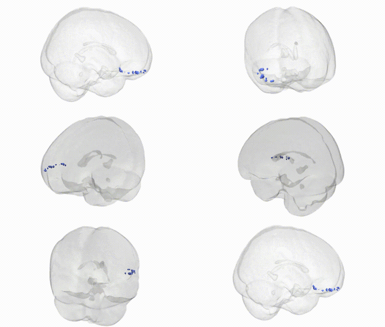
Full Quality Version: Download MP4
Wali Sidiqyar*, Gaurav Rudravaram*, Elyssa M. McMaster, Trent M. Schwartz, Adam M. Saunders, Kurt G. Schilling, Bennett A. Landman "Introducing SPINS: A Shared Public Visualization Library of Neuroanatomical Structures." Medical Imaging with Deep Learning- short paper
This resource is licensed under CC0 1.0 Universal (Public Domain).