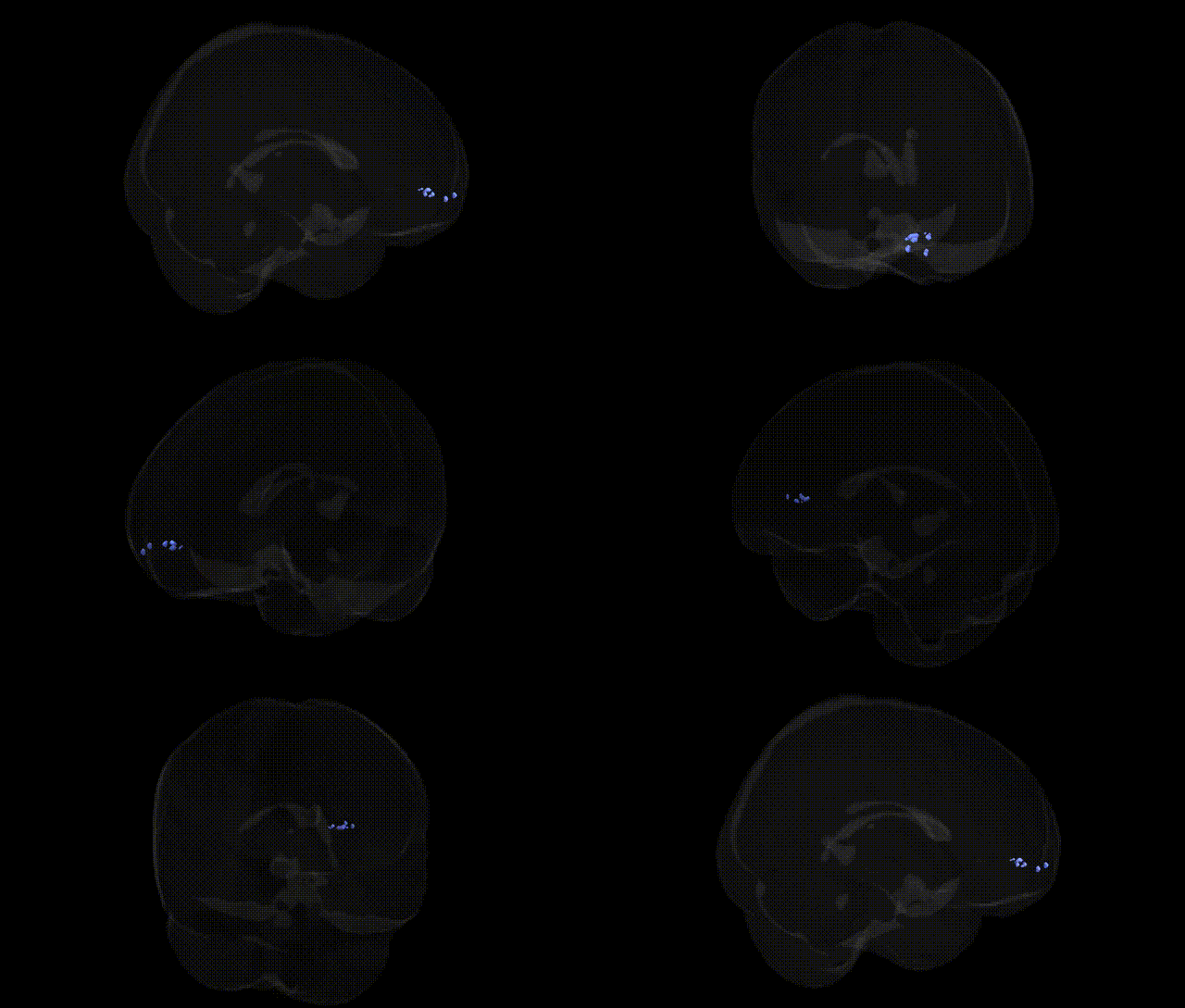
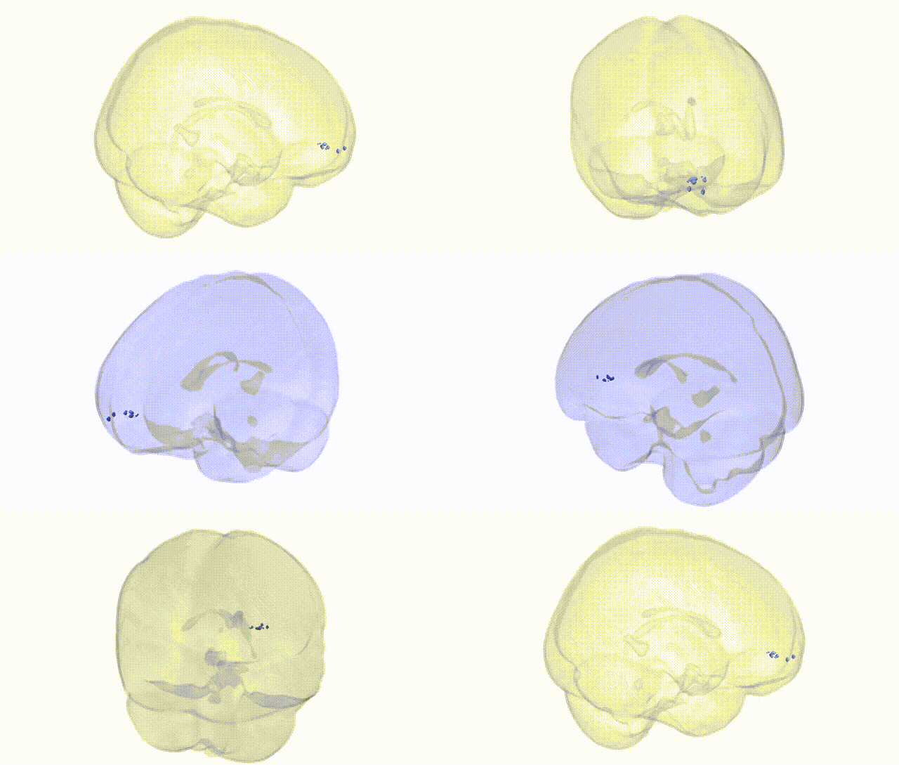
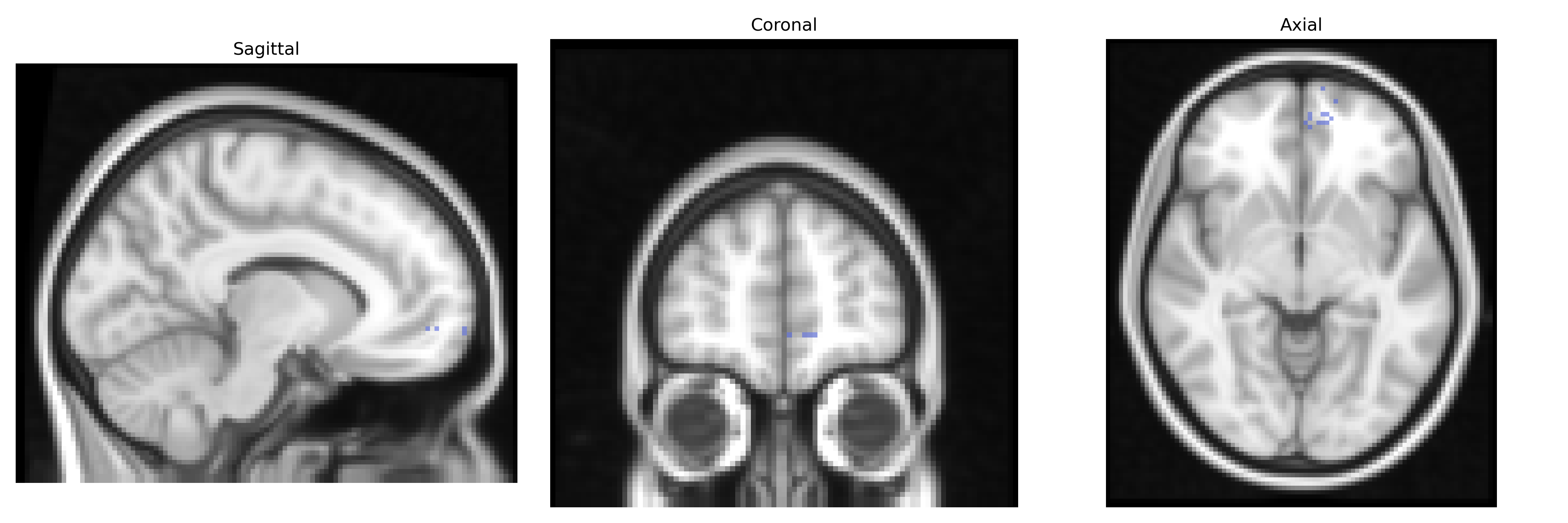
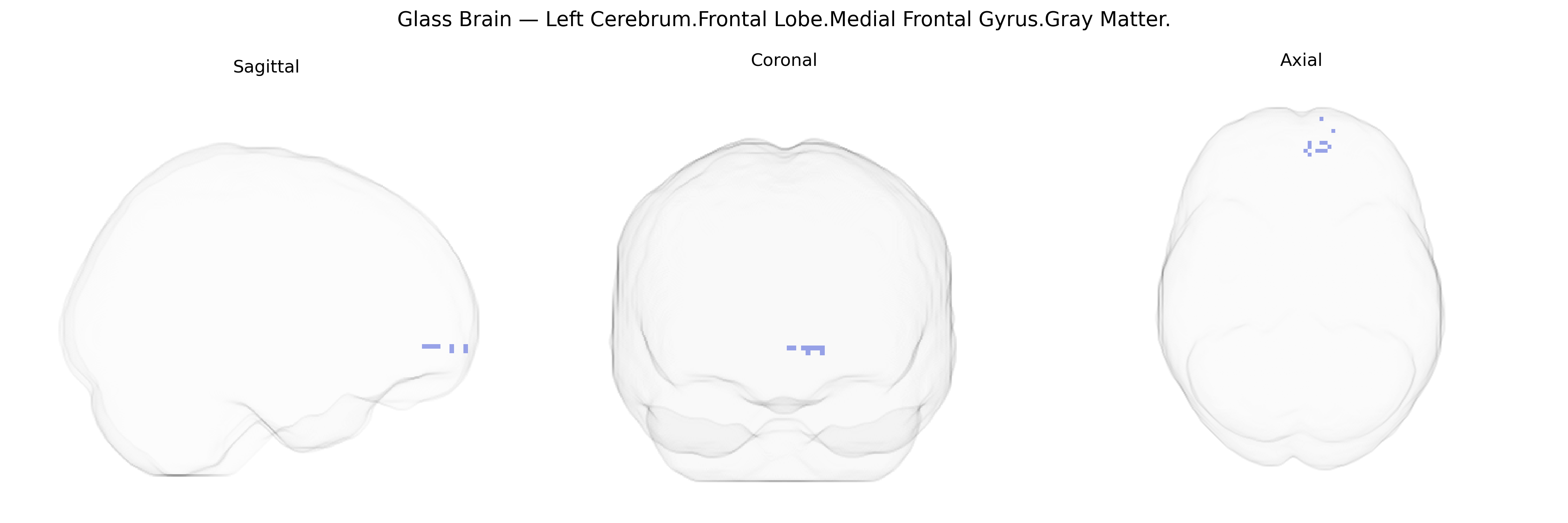
The bilateral Left Cerebrum.Frontal Lobe.Medial Frontal Gyrus.Gray Matter, as defined in the Talairach 2 mm atlas, corresponds largely to the medial portion of the superior frontal gyrus on the left hemisphere, extending along the medial wall of the frontal lobe. This region contains association cortex involved in higher-order executive functions, including voluntary action planning, decision-making, aspects of working memory, and regulation of attention, and it contributes to motivational and affective processing through connections with limbic and paralimbic structures. Functionally and cytoarchitectonically, this area overlaps with medial prefrontal and supplementary motor territories, integrating cognitive control with motor preparation and affective evaluation. There is no direct link for “medial frontal gyrus”; a closely related structure is the Medial prefrontal cortex.
Genetic associations with gray matter volume in the bilateral Left Cerebrum.Frontal Lobe.Medial Frontal Gyrus, as defined in the Talairach 2 mm atlas, largely overlap with loci implicated in broader medial prefrontal and anterior cingulate circuitry. GWAS of cortical thickness and surface area have identified variants near genes such as CENPW, RSPO3, and TSPAN9 that influence frontal lobe morphology, with some studies reporting specific effects in medial or superior frontal regions. This area is consistently implicated in GWAS and imaging–genetics analyses of psychiatric disorders, including schizophrenia (e.g., common risk loci in the MHC region on chromosome 6 and CACNA1C), major depressive disorder (variants near SIRT1 and NEGR1), bipolar disorder (CACNA1C, ANK3), and attention-deficit/hyperactivity disorder, where polygenic risk scores correlate with reduced medial frontal gray matter volume and altered connectivity. Medial frontal regions encompassing the medial frontal gyrus show structural and functional alterations associated with genetic risk for autism spectrum disorder (e.g., loci near CHD8 and CNTNAP2 in broader frontal networks), obsessive–compulsive disorder, and anxiety, often via genes involved in synaptic plasticity and glutamatergic signaling such as GRIN2A and GRM3. Imaging–genetics studies also link common variants in dopaminergic and serotonergic genes (COMT Val158Met, DRD2, 5-HTTLPR) to medial frontal activation patterns during cognitive control and decision-making, and polygenic scores for cognitive ability and educational attainment correlate with medial frontal cortical measures, suggesting that this region is a key anatomical substrate through which widely distributed neuropsychiatric and cognitive GWAS loci exert their effects.
Overview generated by GPT-4o (2026).
Region ID: 611
Hemisphere: bilateral
Atlas: Talairach labels 2mm
Full Quality Version: Download MP4
Full Quality Version: Download MP4
Wali Sidiqyar*, Gaurav Rudravaram*, Elyssa M. McMaster, Trent M. Schwartz, Adam M. Saunders, Kurt G. Schilling, Bennett A. Landman "Introducing SPINS: A Shared Public Visualization Library of Neuroanatomical Structures." Medical Imaging with Deep Learning- short paper
This resource is licensed under CC0 1.0 Universal (Public Domain).