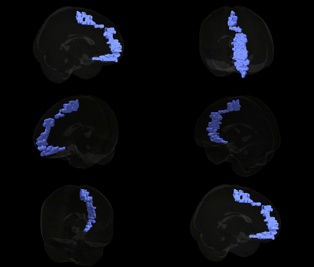
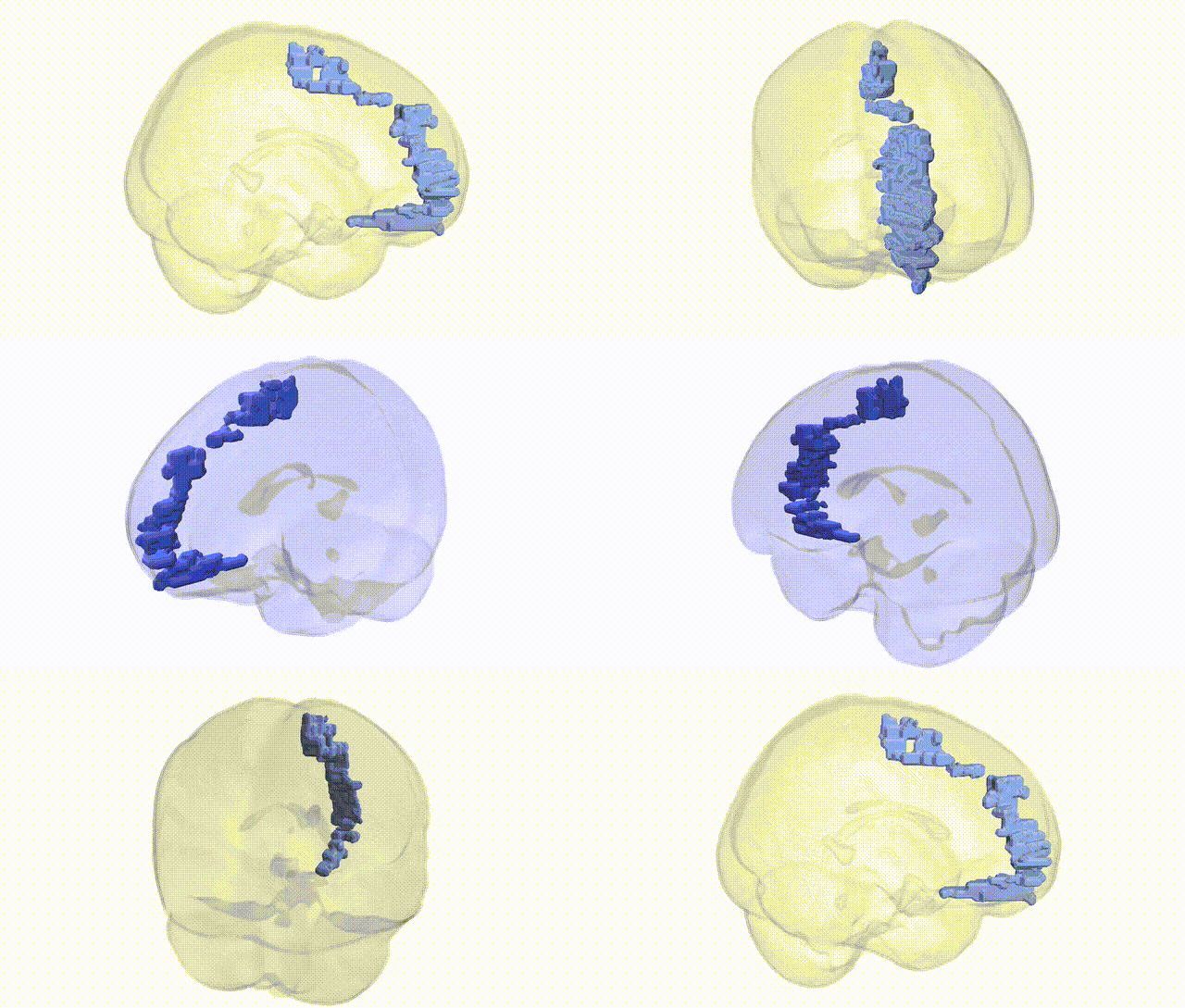
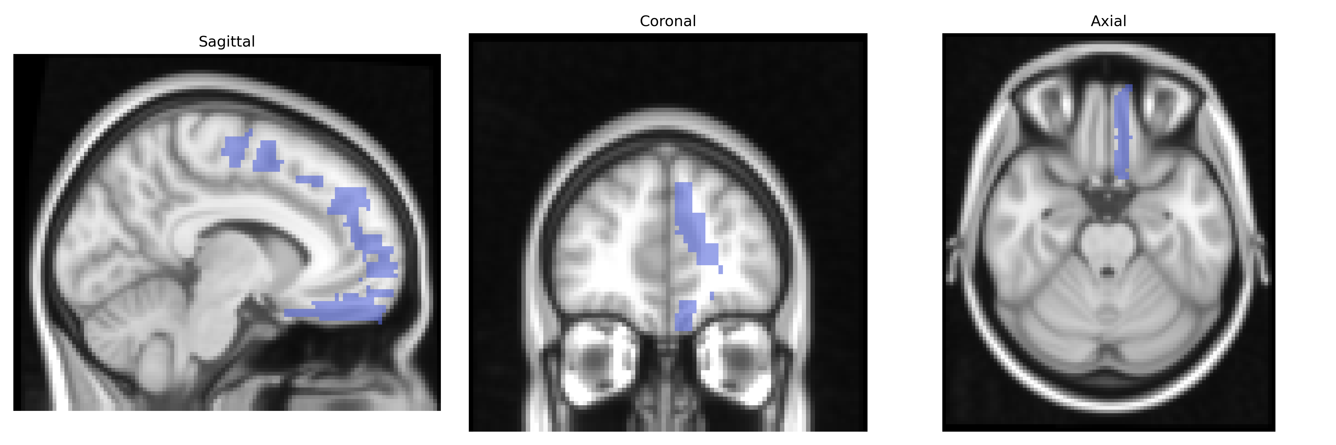
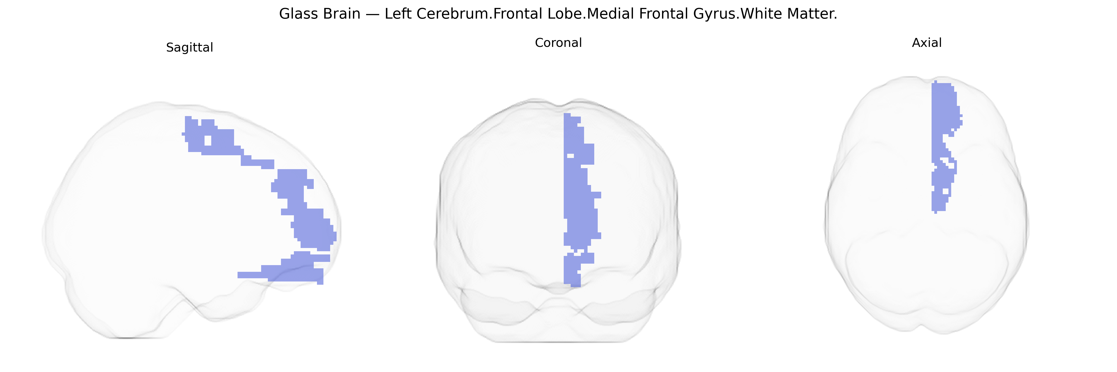
The bilateral Left Cerebrum.Frontal Lobe.Medial Frontal Gyrus.White Matter corresponds to subcortical fiber tracts underlying the medial frontal gyrus within the frontal lobe, primarily encompassing association and projection pathways that interconnect medial prefrontal cortical areas with other frontal, limbic, and subcortical structures. These white matter bundles contribute to higher-order executive functions, motivation, decision-making, and aspects of social cognition by facilitating communication between the medial prefrontal cortex, anterior cingulate cortex, basal ganglia, and thalamus. As part of the frontal lobe’s medial surface, this region participates in networks involved in action selection, performance monitoring, and regulation of goal-directed behavior. There is no direct Wikipedia article for the “medial frontal gyrus white matter”; a closely related cortical region is the Medial prefrontal cortex.
The bilateral medial frontal gyrus white matter (Left Cerebrum.Frontal Lobe.Medial Frontal Gyrus.White Matter in the Talairach 2 mm atlas) lies beneath medial prefrontal and anterior cingulate cortex, regions repeatedly implicated in imaging genetics and GWAS of brain structure and cognition. Large-scale neuroimaging GWAS (e.g., ENIGMA, UK Biobank) have identified common variants near genes involved in axonal development, myelination, and synaptic signaling (such as NRG1, CNTNAP2, DISC1, and white-matter–related loci like those near VCAN and PLEKHG1) that influence frontal white matter volume, fractional anisotropy, and connectivity, including tracts traversing medial frontal territories (e.g., forceps minor, cingulum bundle). Polygenic scores for schizophrenia, bipolar disorder, major depressive disorder, and ADHD show associations with reduced integrity or altered microstructure in medial frontal white matter, supporting a shared genetic basis for fronto-limbic dysconnectivity in these conditions. Variants in serotonin and dopamine system genes (e.g., SLC6A4, DRD2–ANKK1, COMT) have been linked to individual differences in medial prefrontal/ACC white matter and related traits such as impulsivity, cognitive control, and harm avoidance. GWAS of cognitive performance and educational attainment highlight frontal white matter–related loci that overlap with genes influencing cortical surface area and thickness in medial frontal regions, suggesting that genetic influences on intelligence and executive function are partly mediated by the structural integrity of this white matter. Additionally, risk alleles for neurodevelopmental disorders (including autism spectrum conditions and 22q11.2 deletion–related loci) have been associated with atypical development of medial frontal connectivity, while APOE and other neurodegeneration-related variants affect frontal white matter in aging and Alzheimer’s disease, indicating that genetic effects on this region span neurodevelopmental, psychiatric, cognitive, and neurodegenerative domains.
Overview generated by GPT-4o (2026).
Region ID: 230
Hemisphere: bilateral
Atlas: Talairach labels 2mm
Full Quality Version: Download MP4
Full Quality Version: Download MP4
Wali Sidiqyar*, Gaurav Rudravaram*, Elyssa M. McMaster, Trent M. Schwartz, Adam M. Saunders, Kurt G. Schilling, Bennett A. Landman "Introducing SPINS: A Shared Public Visualization Library of Neuroanatomical Structures." Medical Imaging with Deep Learning- short paper
This resource is licensed under CC0 1.0 Universal (Public Domain).