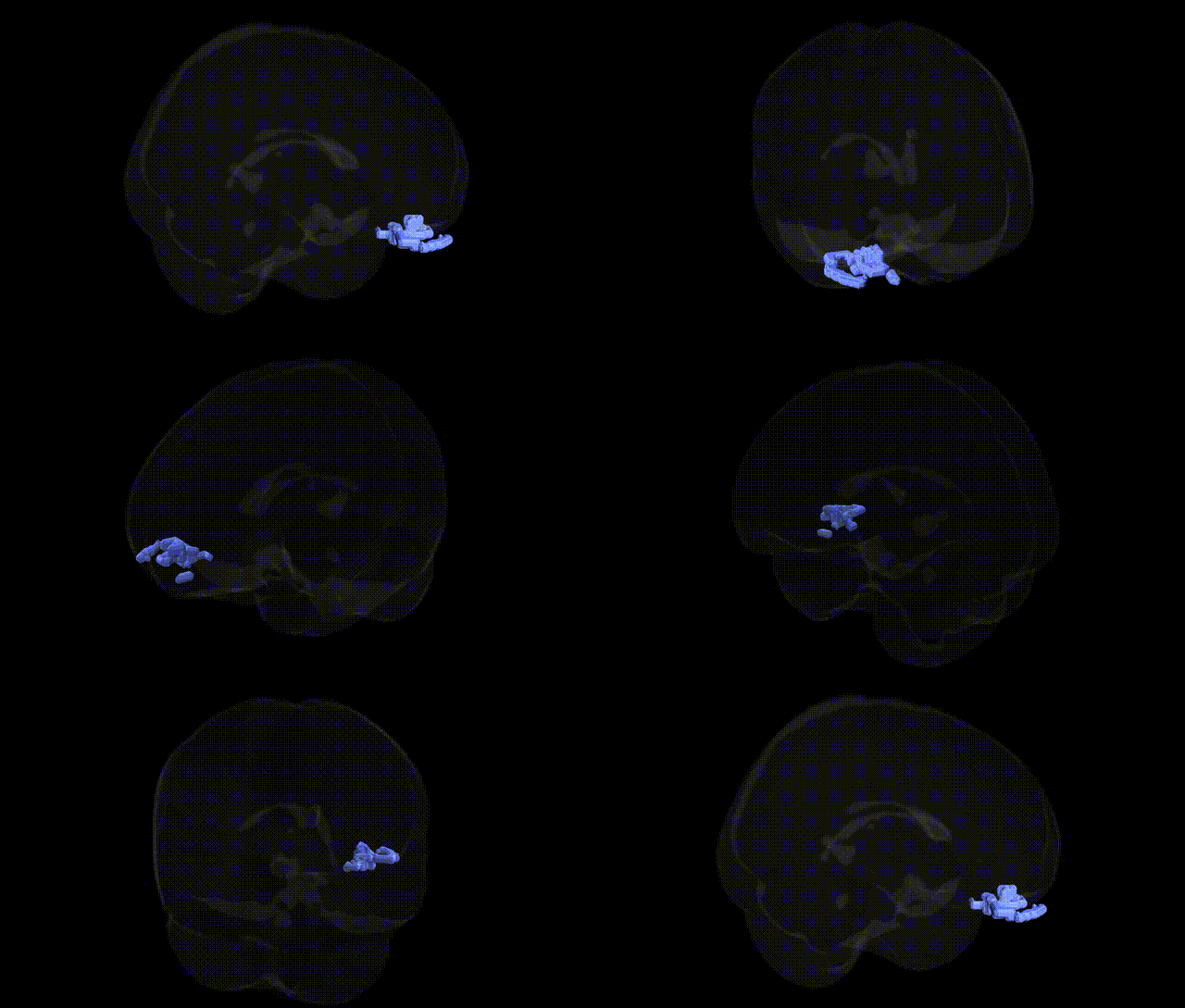
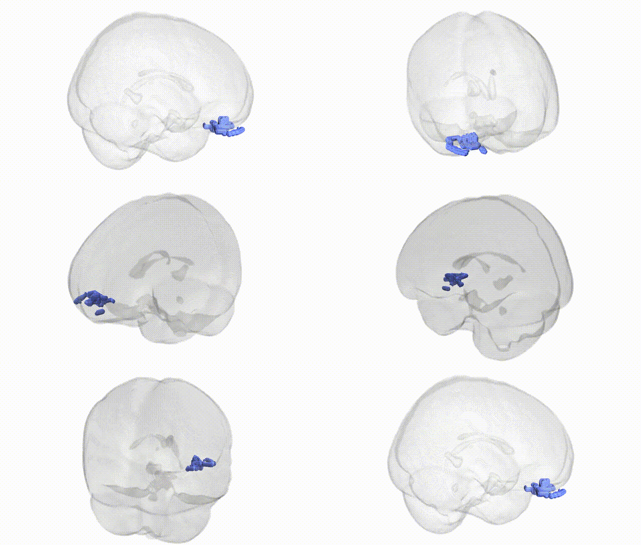
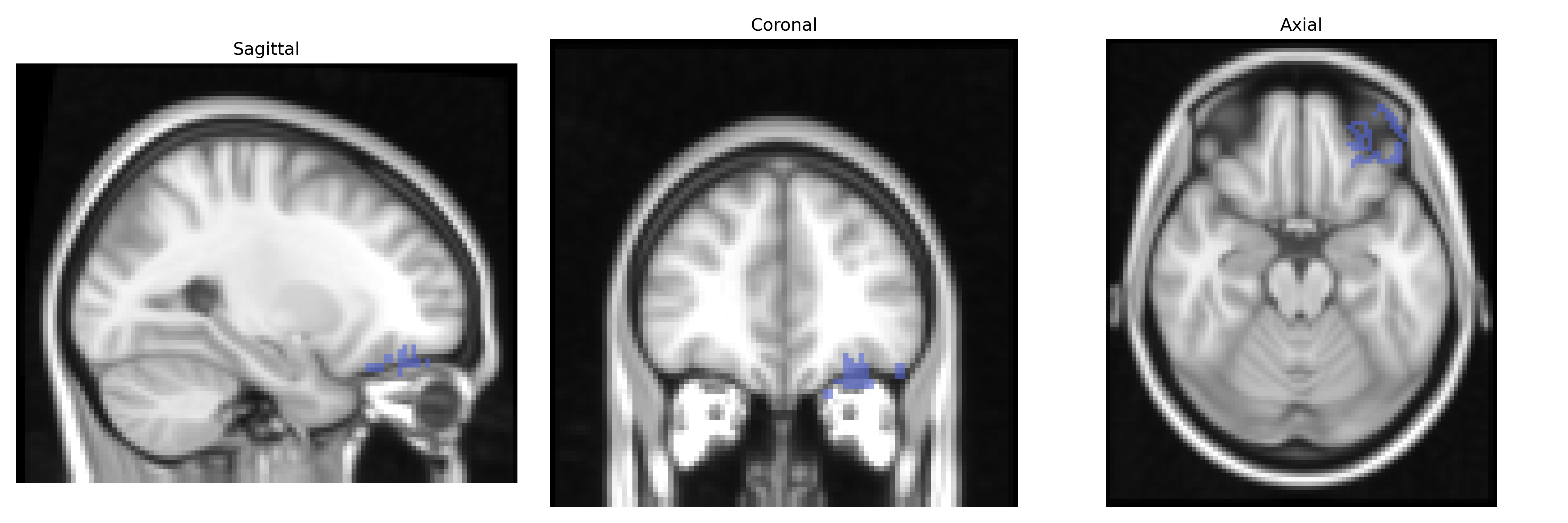
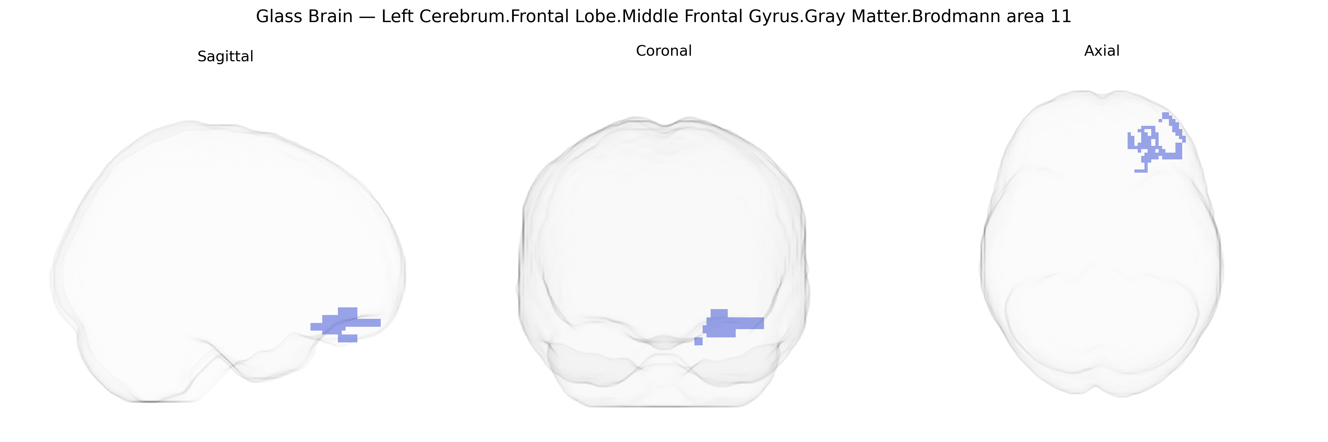
Bilateral Left Cerebrum Frontal Lobe Middle Frontal Gyrus Gray Matter Brodmann area 11 corresponds to a subdivision of the orbitofrontal cortex situated on the ventral aspect of the frontal lobe, involved in higher-order integrative processes linking sensory, affective, and reward-related information to decision-making and behavioral regulation. Neuronal populations in this region participate in valuation of outcomes, expectation of rewards and punishments, emotional modulation of choices, and adaptive updating of stimulus–reinforcement associations. Brodmann area 11 exhibits dense connections with limbic structures (including the amygdala and hippocampus), ventromedial prefrontal regions, and multimodal sensory association cortices, enabling integration of internal states and external cues to guide goal-directed behavior, social cognition, and inhibitory control. Functional alterations and structural abnormalities in this area have been implicated in mood and anxiety disorders, obsessive–compulsive traits, and disturbances of decision-making and impulse regulation. There is no direct link; a closely related structure is the Orbitofrontal cortex.
The bilateral left middle frontal gyrus within Brodmann area 11 (orbitofrontal/ventromedial prefrontal territory in the Talairach 2 mm atlas) has been repeatedly implicated in genetic studies of higher-order cognition, mood, and psychiatric risk, largely via imaging genetics and GWAS of brain structure and function. Large-scale neuroimaging GWAS (e.g., ENIGMA, UK Biobank) have shown that cortical thickness and surface area in orbitofrontal and adjacent middle frontal regions are polygenic and influenced by common variants in genes related to synaptic function, neurodevelopment, and neuronal adhesion, including loci near genes such as HMGA2, IGF1, and multiple glutamatergic and GABAergic pathway genes, though specific signals often span broader prefrontal parcels rather than BA11 precisely. Variants in COMT (Val158Met) and dopamine-related genes have been associated with middle frontal activation and gray matter variation in tasks requiring executive control and decision‑making, overlapping BA11/ventromedial prefrontal circuitry. Genetic studies of major depressive disorder, bipolar disorder, and schizophrenia consistently report case–control and polygenic risk score associations with altered volume or thickness in orbitofrontal/middle frontal regions, with implicated loci in immune (e.g., MHC region), synaptic (CACNA1C, GRIN2A), and neurodevelopmental (DISC1, NRG1) pathways contributing to prefrontal morphometry and connectivity. GWAS of personality traits (e.g., neuroticism), risk‑taking, and impulsivity show convergent associations with structural and functional measures in BA11/ventromedial prefrontal cortex, linking polygenic architectures for affect regulation and valuation to this region. Additionally, imaging genetic studies in obsessive–compulsive disorder, substance use disorders, and attention‑deficit/hyperactivity disorder connect disorder‑related alleles (including in glutamate signaling and dopaminergic genes) to orbitofrontal/middle frontal gray matter changes and altered fronto‑striatal circuitry. Overall, heritable variation in BA11/middle frontal structure and function is strongly implicated in polygenic risk for mood, psychotic, and impulse‑control disorders, as well as in traits such as cognitive control, valuation, and emotional regulation, though most signals reflect distributed prefrontal networks rather than region‑exclusive genetic effects.
Overview generated by GPT-4o (2026).
Region ID: 197
Hemisphere: bilateral
Atlas: Talairach labels 2mm
Full Quality Version: Download MP4
Full Quality Version: Download MP4
Wali Sidiqyar*, Gaurav Rudravaram*, Elyssa M. McMaster, Trent M. Schwartz, Adam M. Saunders, Kurt G. Schilling, Bennett A. Landman "Introducing SPINS: A Shared Public Visualization Library of Neuroanatomical Structures." Medical Imaging with Deep Learning- short paper
This resource is licensed under CC0 1.0 Universal (Public Domain).