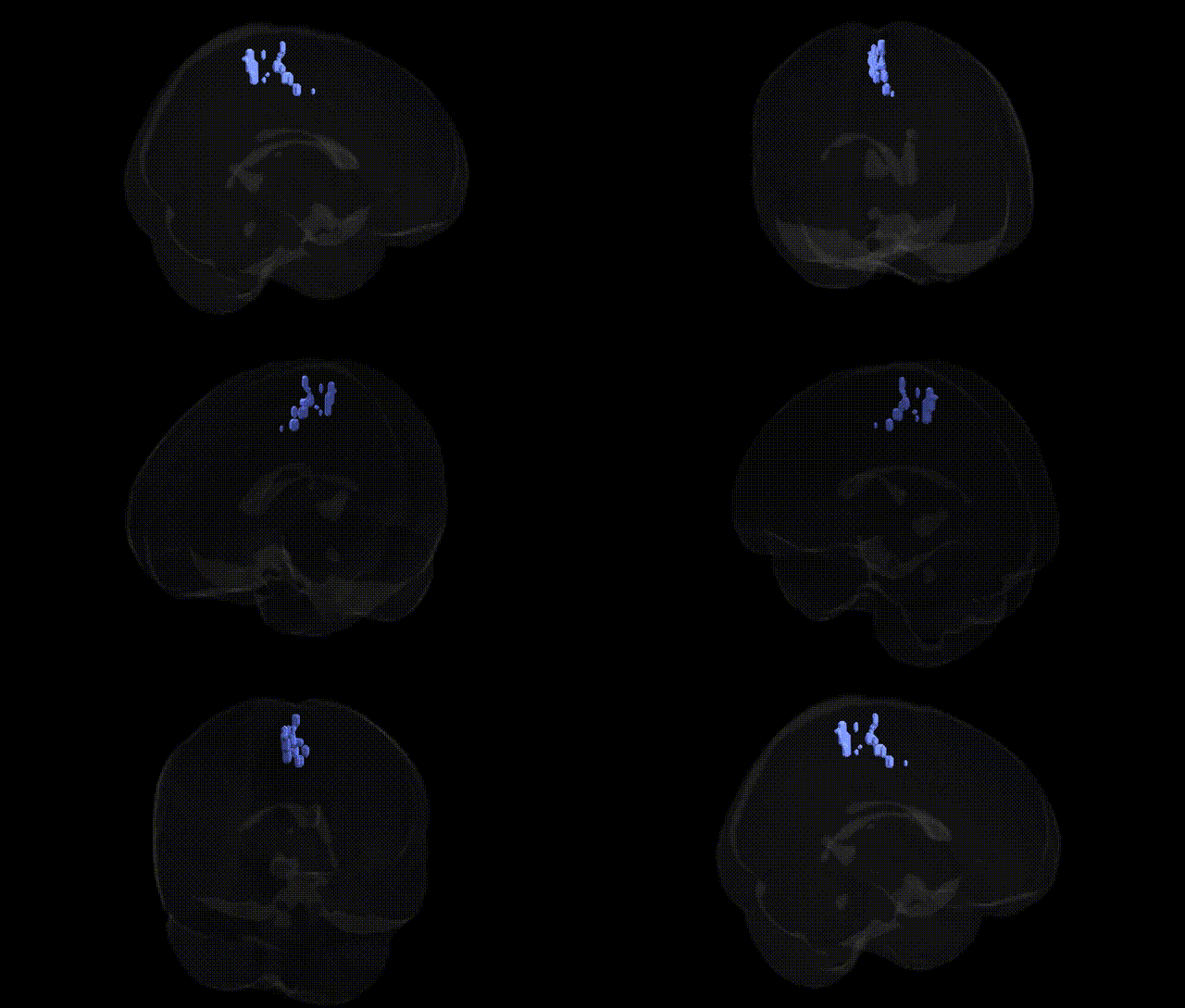
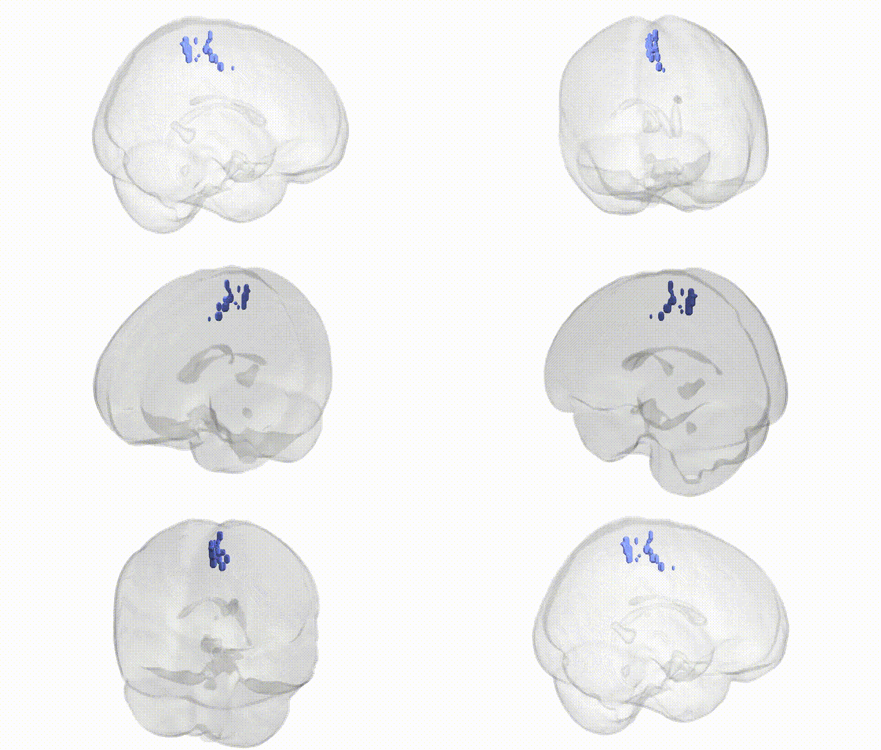
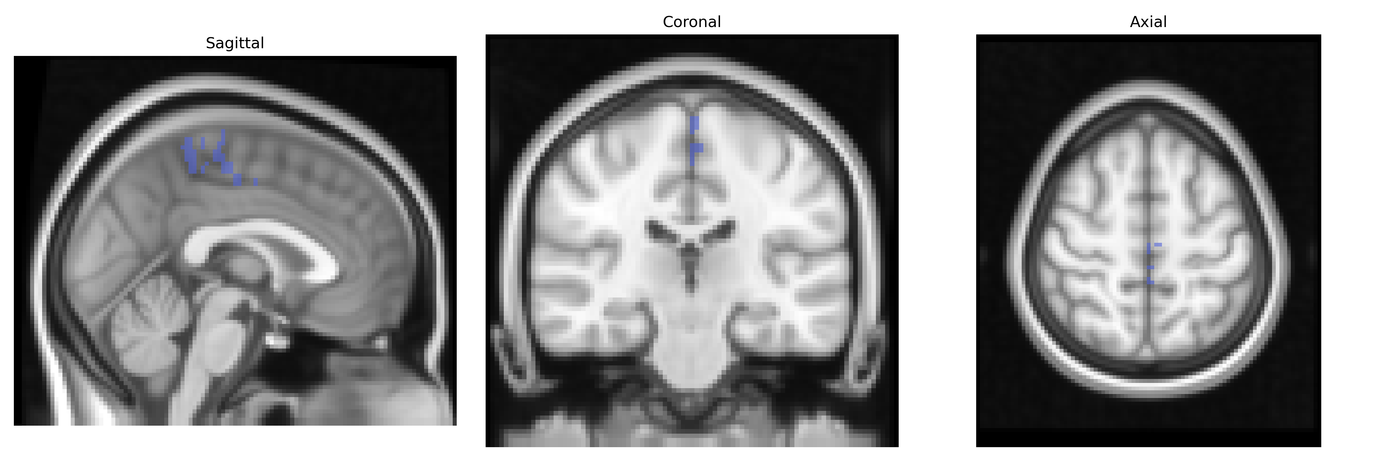
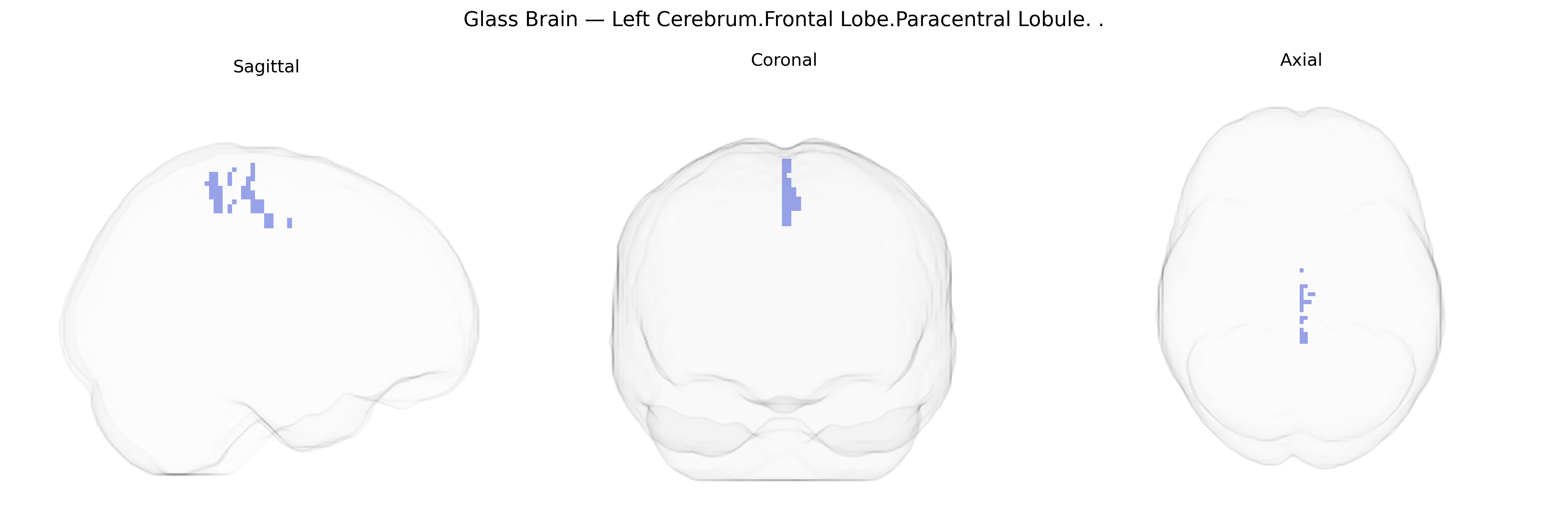
The bilateral Left Cerebrum.Frontal Lobe.Paracentral Lobule corresponds to the paracentral lobule region of the frontal-medial cortex, situated on the medial surface of the hemisphere around the upper part of the central sulcus, bridging portions of the precentral and postcentral gyri. Functionally, it contains parts of the primary motor and primary somatosensory cortices representing mainly the contralateral lower limb and pelvic floor, and is involved in voluntary motor control, sensory processing, and aspects of sphincter regulation. Its frontal component is particularly associated with motor planning and execution for leg movements, while the adjacent parietal portion contributes to integration of somatosensory input from the lower extremities. Developmentally and cytoarchitectonically, it is part of the sensorimotor strip and is highly interconnected with supplementary motor and premotor areas as well as subcortical motor circuits.
Paracentral lobule
The bilateral left frontal paracentral lobule, encompassing medial primary motor and somatosensory cortices for lower limb functions, has been implicated in several genetic and GWAS-based associations via imaging genetics and disorder-focused studies, despite limited work targeting this specific Talairach-defined region. Variants in genes influencing cortical structure and motor/somatosensory areas—such as those affecting neuronal development (e.g., CNTNAP2, BDNF, and genes in neurodevelopmental and synaptic pathways) and global measures of cortical thickness and surface area from large consortia (ENIGMA, UK Biobank)—have been associated with structural variation in adjacent medial frontal motor regions that include or border the paracentral lobule. GWAS of regional cortical measures have identified loci near genes involved in axon guidance, cytoskeletal dynamics, and synaptic signaling that correlate with motor strip and medial frontal morphology, indirectly implicating this area. Clinically, genetic studies in multiple sclerosis and other demyelinating or motor disorders, as well as hereditary spastic paraplegias, often show lesions or atrophy affecting the paracentral lobule’s corticospinal representation, with risk variants in immune-related genes (e.g., HLA region) and neurodegenerative genes associated with damage patterns involving this region. In epilepsy (particularly focal motor seizures) and neurodevelopmental conditions such as cerebral palsy, genetic risk factors in ion channel and cortical development genes can manifest as structural or functional abnormalities in motor and sensory cortical regions that include the paracentral lobule. Overall, genetic associations for this precise bilateral left paracentral lobule label are typically inferred from broader studies of medial frontal, primary motor/somatosensory cortex and lower-limb motor networks, rather than from region-specific GWAS focused explicitly on the Talairach 2 mm paracentral lobule parcel.
Overview generated by GPT-4o (2026).
Region ID: 1046
Hemisphere: bilateral
Atlas: Talairach labels 2mm
Full Quality Version: Download MP4
Full Quality Version: Download MP4
Wali Sidiqyar*, Gaurav Rudravaram*, Elyssa M. McMaster, Trent M. Schwartz, Adam M. Saunders, Kurt G. Schilling, Bennett A. Landman "Introducing SPINS: A Shared Public Visualization Library of Neuroanatomical Structures." Medical Imaging with Deep Learning- short paper
This resource is licensed under CC0 1.0 Universal (Public Domain).