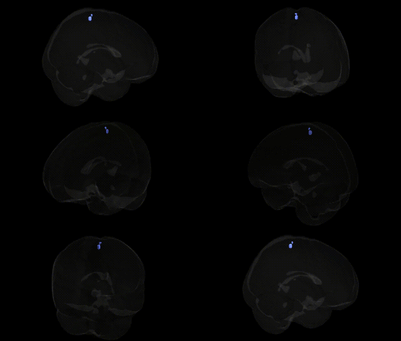
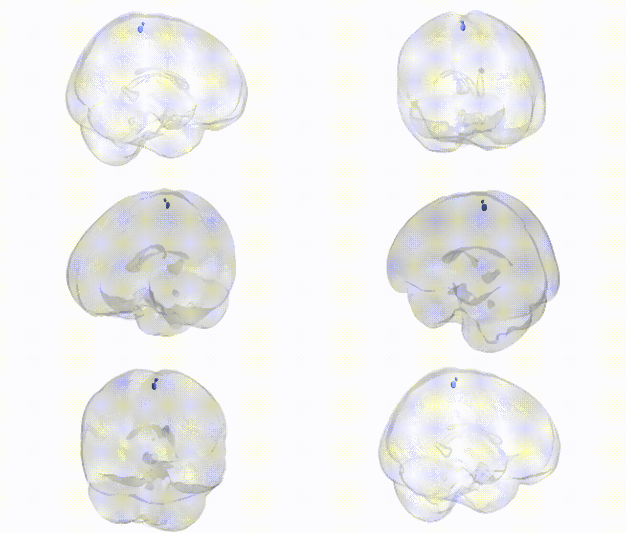
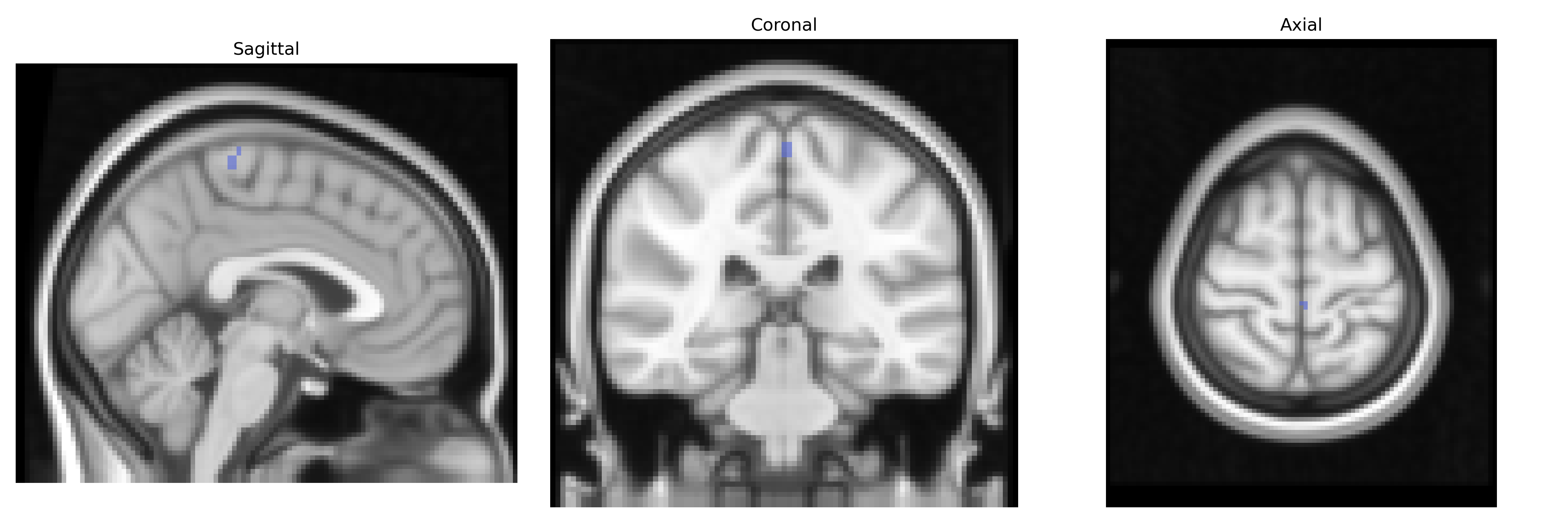
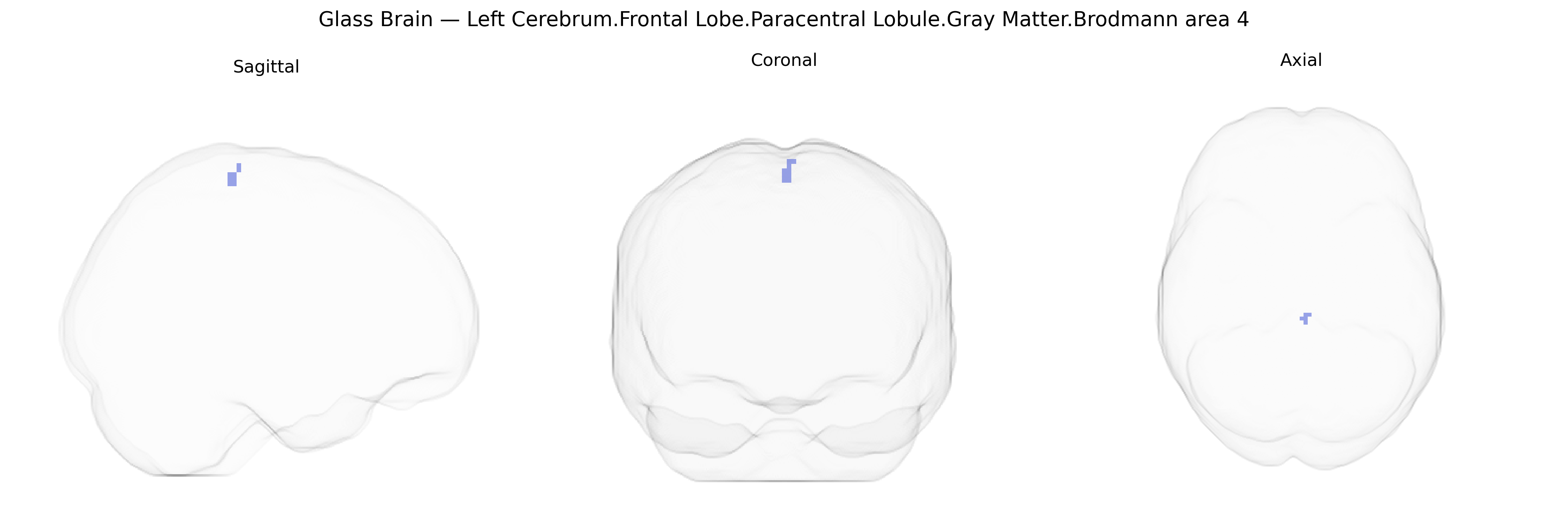
The bilateral Left Cerebrum.Frontal Lobe.Paracentral Lobule.Gray Matter.Brodmann area 4 corresponds to primary motor cortex located medially in the frontal lobe, within the paracentral lobule, representing predominantly the motor control of the lower extremities and parts of the trunk. This agranular cortical region is characterized cytoarchitectonically by thick pyramidal cell layers (especially layer V) containing large Betz cells that project via the corticospinal tract to spinal motor neurons, enabling execution of voluntary movements. Functionally, it integrates descending motor commands, contributes to somatotopic organization of the motor homunculus, and participates in fine control of posture and gait. Lesions in this area typically result in contralateral motor deficits, especially weakness or paralysis of the leg and foot. There is no direct link for this combined Talairach label; see the related area Brodmann area 4.
The bilateral paracentral lobule (BA4 gray matter in the frontal lobe) lies in primary motor cortex controlling lower limb and pelvic musculature and shows genetic associations largely through its role in motor function, somatosensation, and network-level traits. Twin and heritability studies using structural MRI indicate substantial genetic influence on cortical thickness and surface area in this region, often within global motor and sensorimotor cortex measures. Large GWAS of brain structure (e.g., ENIGMA consortium and UK Biobank–based analyses) have identified multiple loci affecting cortical morphology in primary motor areas, including variants in or near genes involved in neurodevelopment, synaptic function, and axon guidance (such as patterns implicating MAPT, MSR1, and other neurodevelopmental genes, though not always specific to BA4 alone). Functionally, GWAS of motor-related traits (e.g., gait speed, balance, and strength) and polygenic risk scoring link sensorimotor cortex activation, including paracentral lobule, with genetic risk for neurodegenerative disorders such as amyotrophic lateral sclerosis, Parkinson’s disease, and multiple sclerosis, all of which can involve lower-limb motor deficits corresponding to paracentral territories. In epilepsy genetics, focal motor seizures involving leg and foot representation have been associated with pathogenic variants and copy number changes affecting cortical development, with imaging and lesion mapping often implicating paracentral regions. Incontinence and bladder control studies have connected paracentral lobule activity to genetically influenced pelvic floor and autonomic control, although specific regional GWAS signals are limited. More broadly, polygenic architectures for traits such as general cognitive ability, educational attainment, and cortical excitability show distributed effects across the frontal cortex, and imaging-genetics work using activation and connectivity phenotypes has repeatedly implicated genetic modulation of sensorimotor network hubs that include the paracentral lobule, even if few findings single out this BA4 parcel from the Talairach 2 mm atlas as an isolated locus of association.
Overview generated by GPT-4o (2026).
Region ID: 1089
Hemisphere: bilateral
Atlas: Talairach labels 2mm
Full Quality Version: Download MP4
Full Quality Version: Download MP4
Wali Sidiqyar*, Gaurav Rudravaram*, Elyssa M. McMaster, Trent M. Schwartz, Adam M. Saunders, Kurt G. Schilling, Bennett A. Landman "Introducing SPINS: A Shared Public Visualization Library of Neuroanatomical Structures." Medical Imaging with Deep Learning- short paper
This resource is licensed under CC0 1.0 Universal (Public Domain).