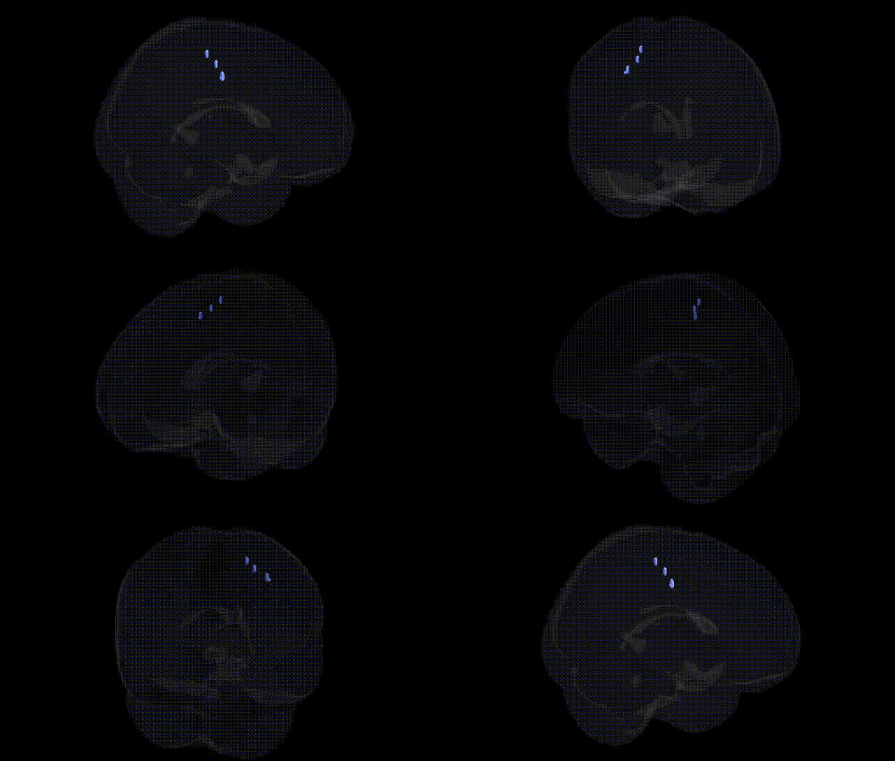
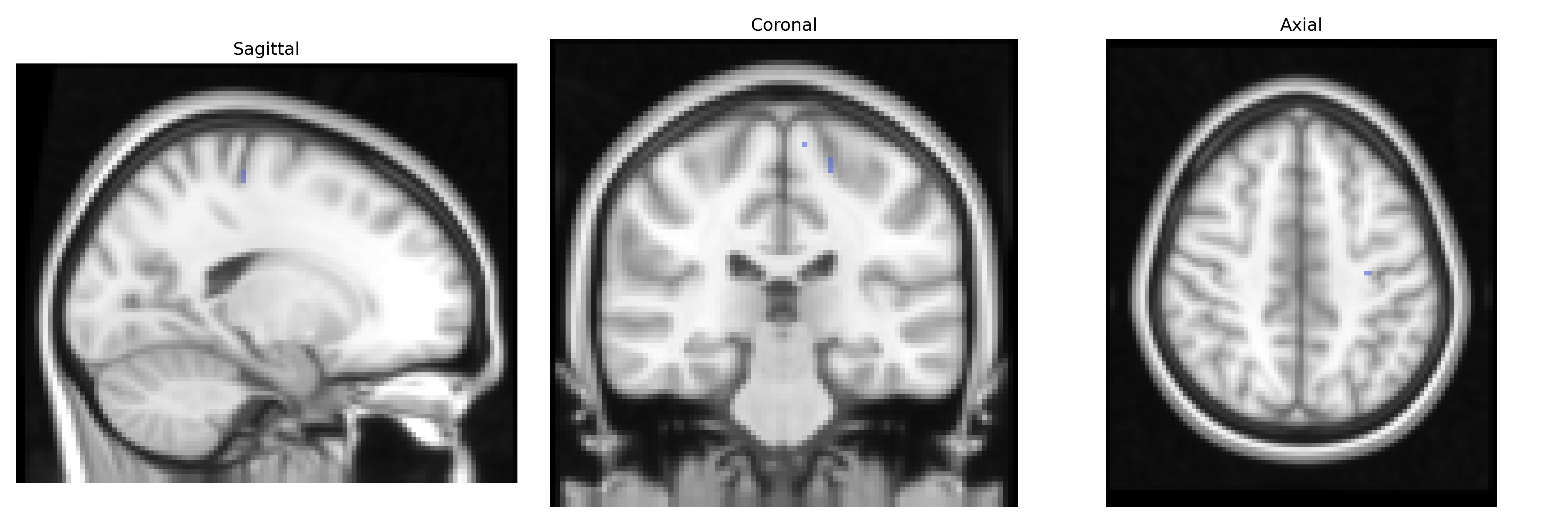
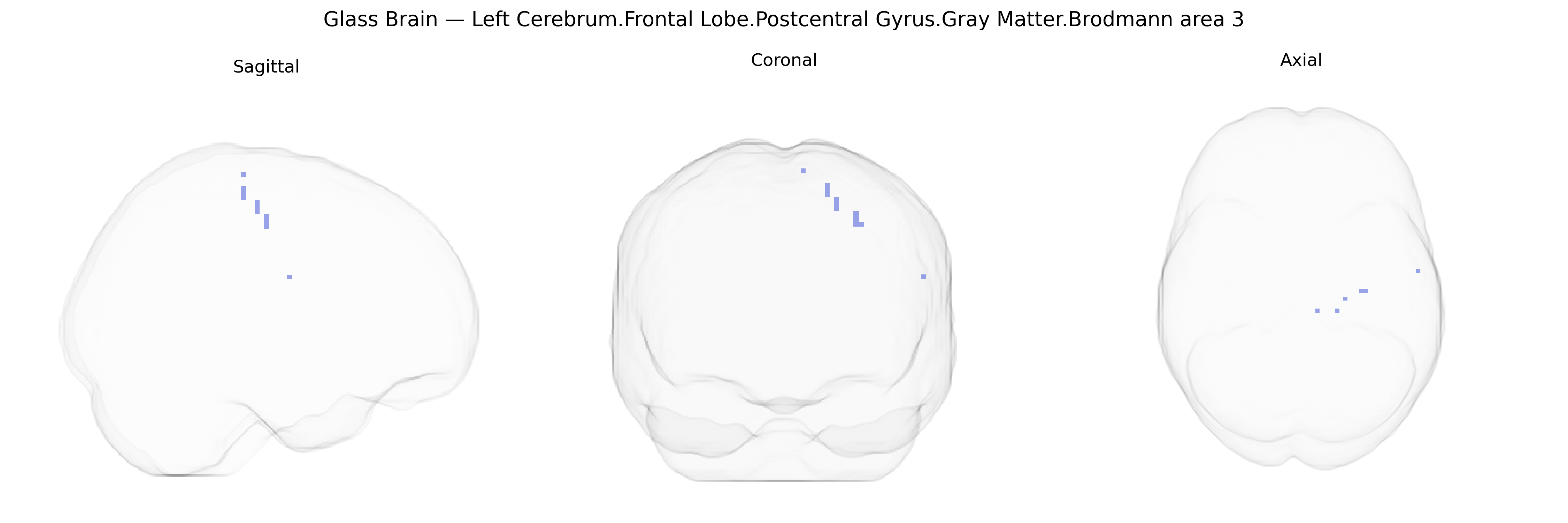
The bilateral Left Cerebrum.Frontal Lobe.Postcentral Gyrus.Gray Matter.Brodmann area 3 region, as defined in the Talairach 2 mm atlas, corresponds primarily to the anterior portion of the primary somatosensory cortex, traditionally located in the postcentral gyrus of the parietal lobe but here labeled within a frontal-lobe framework of the atlas nomenclature. Brodmann area 3 is cytoarchitectonically distinct and heavily granular, receiving dense thalamocortical input from the ventral posterior nucleus of the thalamus, and is critically involved in processing fine tactile, proprioceptive, and nociceptive information from the contralateral body. Neurons in area 3 exhibit small receptive fields and high spatial resolution, forming the initial cortical stage of somatosensory processing that is further elaborated in adjacent somatosensory areas (e.g., BA1 and BA2). This bilateral region contributes to the encoding of stimulus location, intensity, and texture, supporting higher-order functions such as object manipulation and sensorimotor integration. There is no direct Wikipedia article specifically for “Brodmann area 3,” but it is a subregion of the primary somatosensory cortex: Primary somatosensory cortex.
The bilateral Left Cerebrum.Frontal Lobe.Postcentral Gyrus.Gray Matter.Brodmann area 3 (primary somatosensory cortex) has been implicated in genetic studies largely through imaging genetics and GWAS of brain structure and neuropsychiatric traits rather than locus-specific association. Large-scale GWAS of cortical thickness and surface area (e.g., ENIGMA consortium studies) have identified polygenic influences on somatosensory regions, with significant heritability and variants near genes involved in neurodevelopment (such as microtubule, synaptic, and axon guidance pathways), though associations are typically reported at the level of broader parietal–sensorimotor cortices rather than BA3 alone. Somatosensory cortex, including BA3, shows altered structure and function in disorders with strong genetic components—such as autism spectrum disorder, schizophrenia, ADHD, and chronic pain syndromes—with risk loci (e.g., in synaptic genes like CACNA1C, GRIN2A, and others) linked in imaging genetics to abnormal sensorimotor activation or cortical morphology. GWAS on traits like pain sensitivity, tactile processing, and motor coordination have shown associations with genes influencing neuronal excitability and cortical development, which, through functional MRI and structural MRI endophenotypes, map to primary somatosensory regions including BA3. However, no single gene or variant is uniquely or specifically established as a “BA3 gene”; instead, highly polygenic risk architectures for neurodevelopmental, psychiatric, and pain-related disorders converge on altered structure and activation of this somatosensory area.
Overview generated by GPT-4o (2026).
Region ID: 897
Hemisphere: bilateral
Atlas: Talairach labels 2mm
Full Quality Version: Download MP4
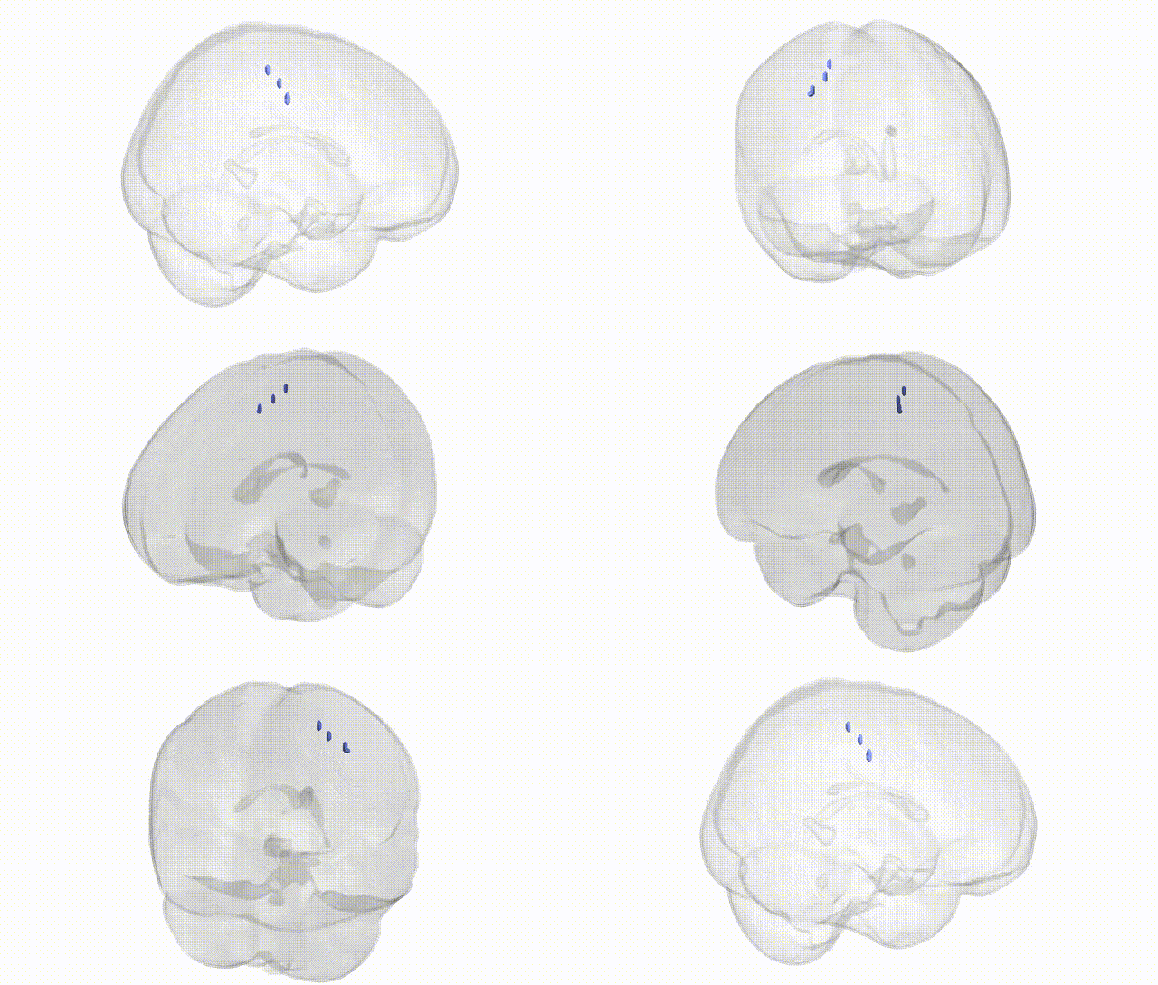
Full Quality Version: Download MP4
Wali Sidiqyar*, Gaurav Rudravaram*, Elyssa M. McMaster, Trent M. Schwartz, Adam M. Saunders, Kurt G. Schilling, Bennett A. Landman "Introducing SPINS: A Shared Public Visualization Library of Neuroanatomical Structures." Medical Imaging with Deep Learning- short paper
This resource is licensed under CC0 1.0 Universal (Public Domain).