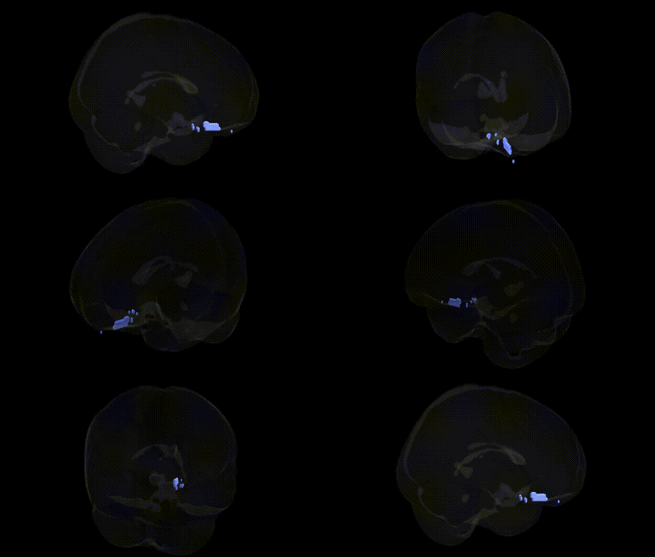
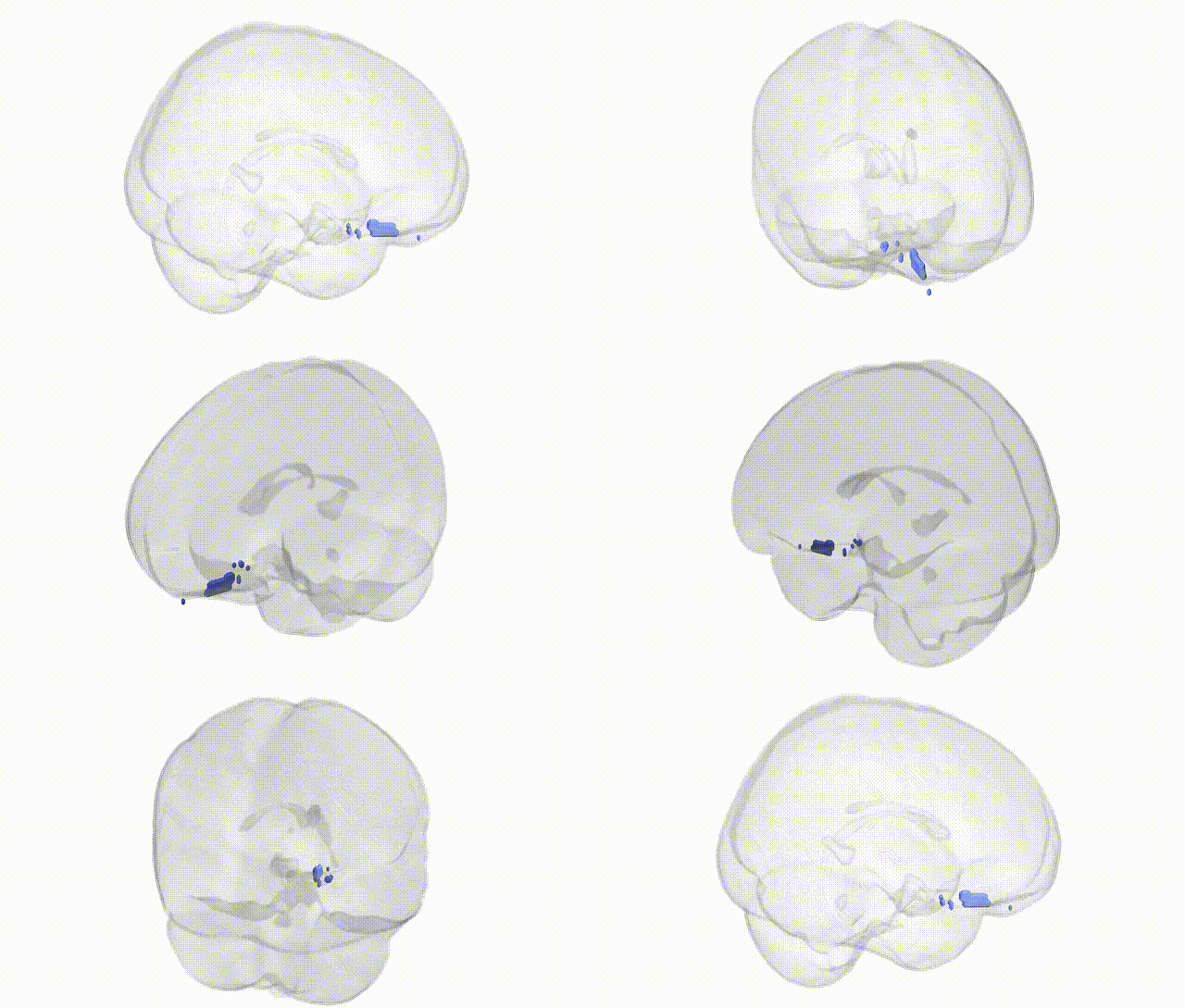
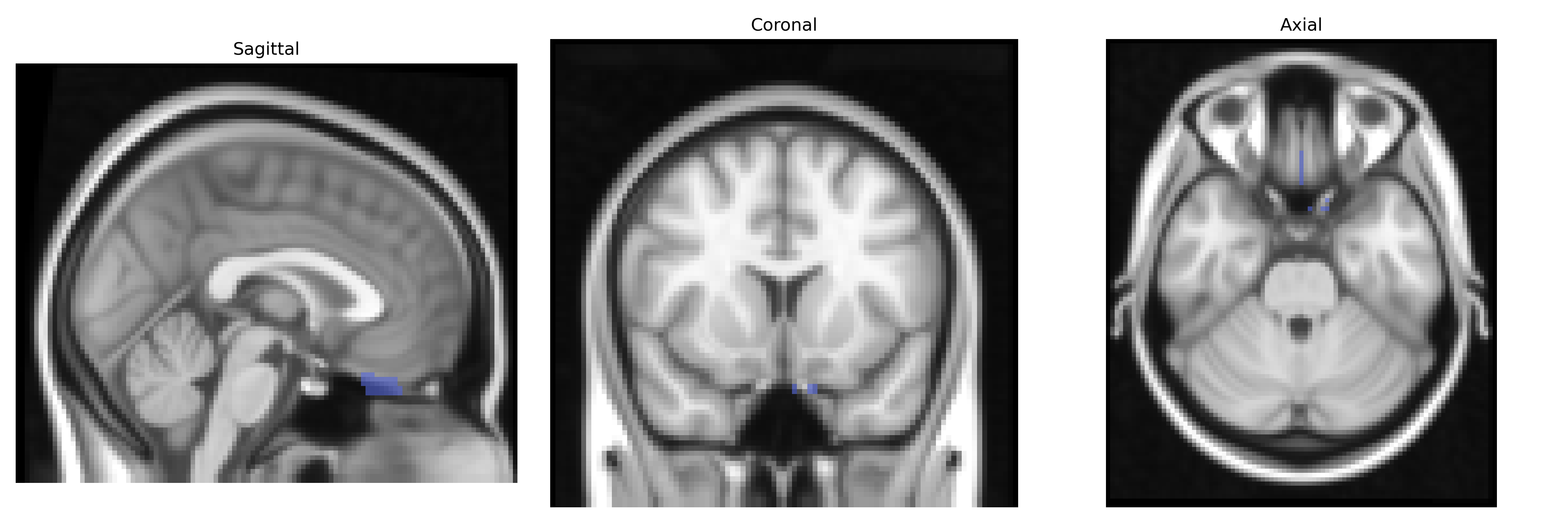
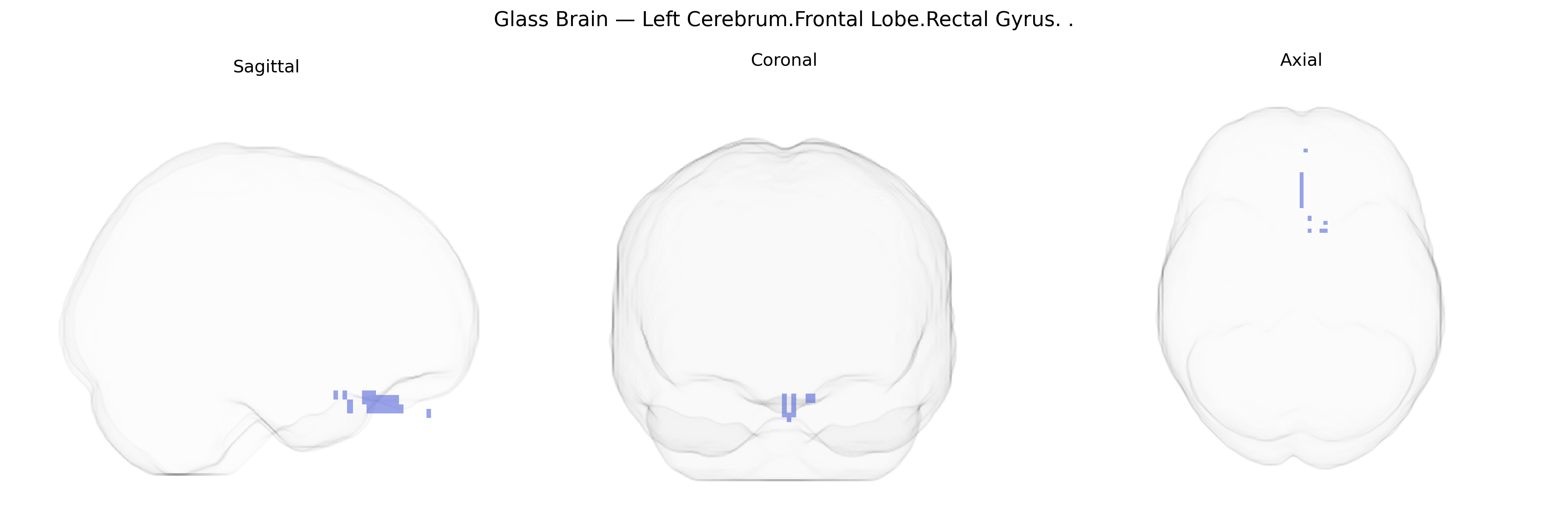
The bilateral Left Cerebrum.Frontal Lobe.Rectal Gyrus corresponds to the gyrus rectus (also called the straight gyrus), a medial frontal lobe structure situated on the ventral surface of the frontal lobe, adjacent to the olfactory sulcus and lying above the orbital plate of the frontal bone. It forms part of the orbitofrontal cortex and is implicated in higher-order cognitive and affective functions, including aspects of reward evaluation, emotional processing, and social behavior, although its specific functional role remains incompletely characterized. The gyrus rectus has reciprocal connections with limbic and paralimbic regions, such as the cingulate cortex and medial temporal structures, supporting integration of visceral, emotional, and executive information.
Gyrus rectus
The bilateral left rectal gyrus (often referred to as the gyrus rectus in the medial orbitofrontal/ventromedial prefrontal cortex) has no direct, extensively cataloged genetic association literature specifically under the Talairach 2 mm “Left Cerebrum.Frontal Lobe.Rectal Gyrus” label, but genetic and GWAS-based imaging and clinical studies implicate the broader region in several traits and disorders. Imaging genetics work using structural and functional MRI has shown that variants in genes involved in synaptic function and cortical development (e.g., BDNF, COMT, and serotonin-related genes) can modulate medial orbitofrontal/ventromedial prefrontal volumes or activation, which frequently include or overlap the gyrus rectus. Large-scale GWAS of brain morphology (such as ENIGMA consortia studies) have mapped common variants associated with frontal cortical thickness and surface area, and some of these frontal measures involve medial orbitofrontal territory that encompasses the rectal gyrus, although individual gyri are rarely isolated as specific GWAS targets. Clinically, genetic susceptibility loci for mood and anxiety disorders, obsessive–compulsive disorder, and substance use disorders—often in dopaminergic, glutamatergic, and neurodevelopmental pathways—have been linked, via imaging genetics and risk-allele carriers, to altered function or connectivity of ventromedial/orbitofrontal regions including the gyrus rectus, particularly in valuation, reward processing, and emotional regulation networks. However, no robust, region-specific GWAS or single-gene association is currently established that uniquely attributes genetic risk to the bilateral left rectal gyrus in isolation; instead, its genetic associations are inferred from broader orbitofrontal and medial prefrontal circuitry involvement in polygenic neuropsychiatric and cognitive traits.
Overview generated by GPT-4o (2026).
Region ID: 143
Hemisphere: bilateral
Atlas: Talairach labels 2mm
Full Quality Version: Download MP4
Full Quality Version: Download MP4
Wali Sidiqyar*, Gaurav Rudravaram*, Elyssa M. McMaster, Trent M. Schwartz, Adam M. Saunders, Kurt G. Schilling, Bennett A. Landman "Introducing SPINS: A Shared Public Visualization Library of Neuroanatomical Structures." Medical Imaging with Deep Learning- short paper
This resource is licensed under CC0 1.0 Universal (Public Domain).