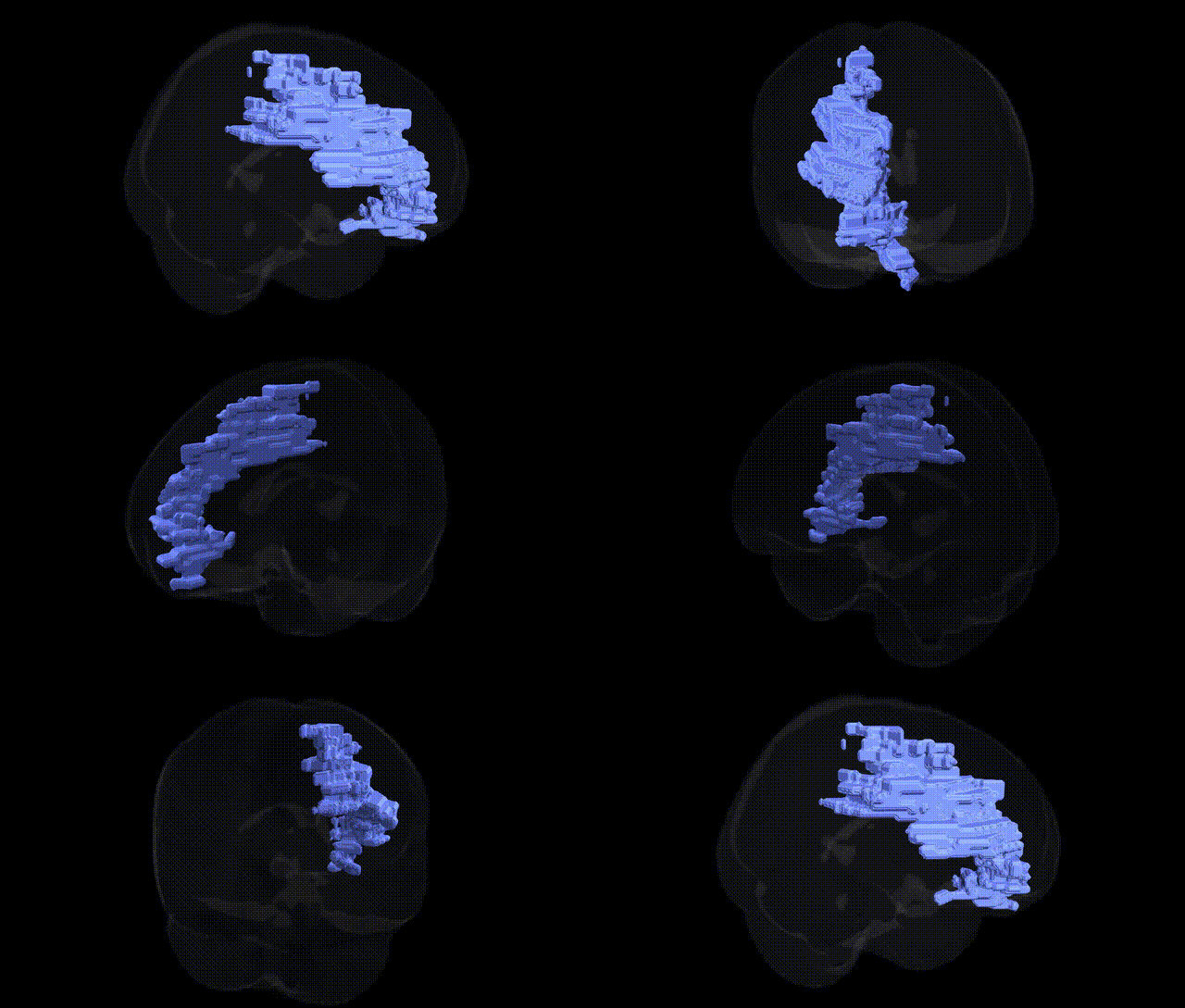
The bilateral Left Cerebrum.Frontal Lobe.Sub-Gyral.White Matter corresponds to deep frontal lobe white matter located beneath the cortical gyri of the left frontal hemisphere. This region consists primarily of myelinated axonal tracts that interconnect frontal cortical areas with one another (short and long association fibers), as well as with more posterior cortical regions and subcortical structures, including the basal ganglia and thalamus. Functionally, these sub-gyral white matter pathways support higher-order executive functions, working memory, language production, motor planning, and aspects of social cognition by enabling efficient communication within and beyond the frontal lobe. Disruption of this white matter—through ischemia, trauma, demyelination, or neurodegenerative processes—can lead to deficits in these domains, reflecting the integrative role of frontal connectivity in complex behavior. There is no direct Wikipedia article for this specific sub-gyral white matter region; a related and encompassing structure is the Frontal lobe.
Genetic associations specifically targeting the bilateral Left Cerebrum.Frontal Lobe.Sub-Gyral.White Matter as defined in the Talairach 2 mm atlas are not typically reported at that fine-grained label, but numerous GWAS and imaging‑genetics studies implicate frontal lobe white matter—particularly prefrontal and subgyral tracts—in a range of heritable traits and disorders. Variants near or within genes affecting myelination and axonal integrity (e.g., MAG, MBP, ERBB4, CNTNAP2) and broader neurodevelopmental or synaptic genes (e.g., DISC1, CACNA1C, GRM3) have been associated with white matter microstructure (often measured via fractional anisotropy or mean diffusivity) in frontal regions, linking these microstructural differences to schizophrenia, bipolar disorder, major depressive disorder, autism spectrum disorder, attention‑deficit/hyperactivity disorder, and cognitive performance including intelligence, executive function, and working memory. Large consortia such as ENIGMA have identified polygenic influences on frontal white matter integrity, showing overlap with risk loci for psychiatric and neurodevelopmental conditions, as well as traits like educational attainment and general cognitive ability. Additionally, gene–environment interactions involving neuroplasticity and stress‑response genes (e.g., BDNF, FKBP5) have been reported to modulate frontal white matter properties, further tying the genetic architecture of this region to mood and anxiety phenotypes and to vulnerability or resilience in the context of early life adversity.
Overview generated by GPT-4o (2026).
Region ID: 193
Hemisphere: bilateral
Atlas: Talairach labels 2mm
Full Quality Version: Download MP4
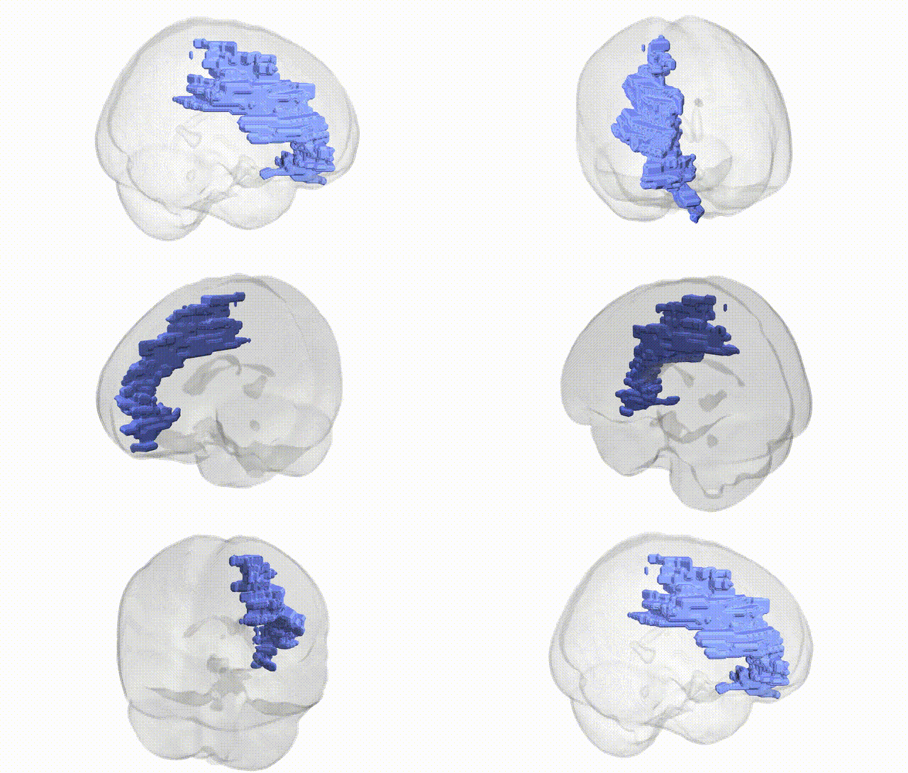
Full Quality Version: Download MP4
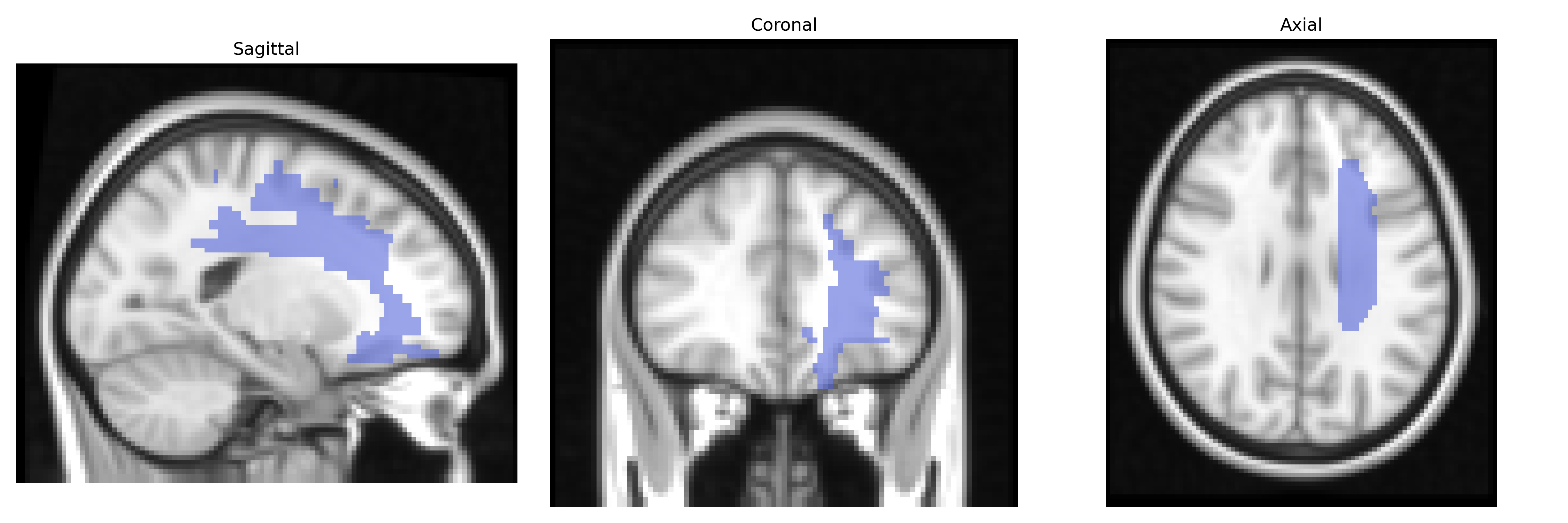
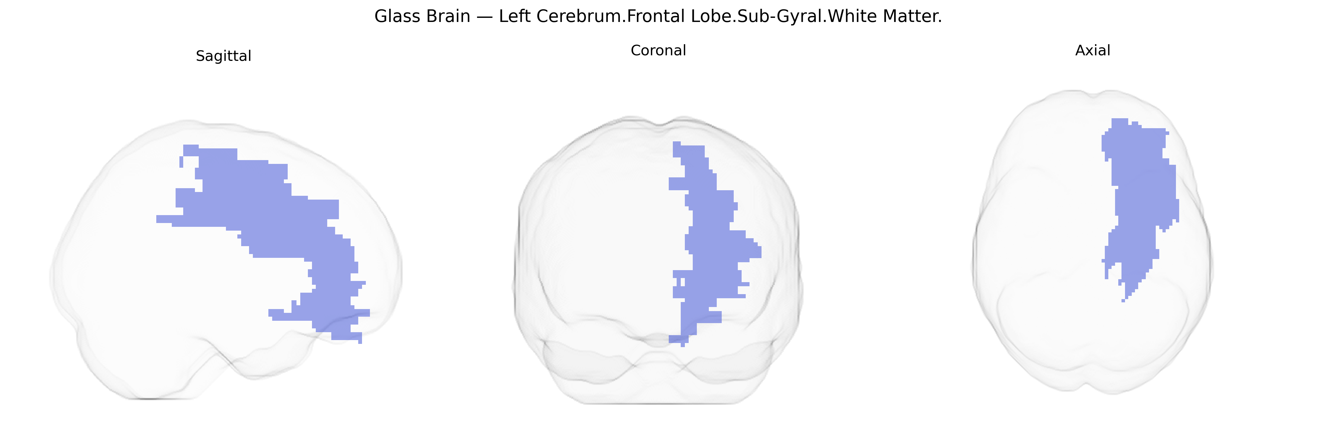
Wali Sidiqyar*, Gaurav Rudravaram*, Elyssa M. McMaster, Trent M. Schwartz, Adam M. Saunders, Kurt G. Schilling, Bennett A. Landman "Introducing SPINS: A Shared Public Visualization Library of Neuroanatomical Structures." Medical Imaging with Deep Learning- short paper
This resource is licensed under CC0 1.0 Universal (Public Domain).