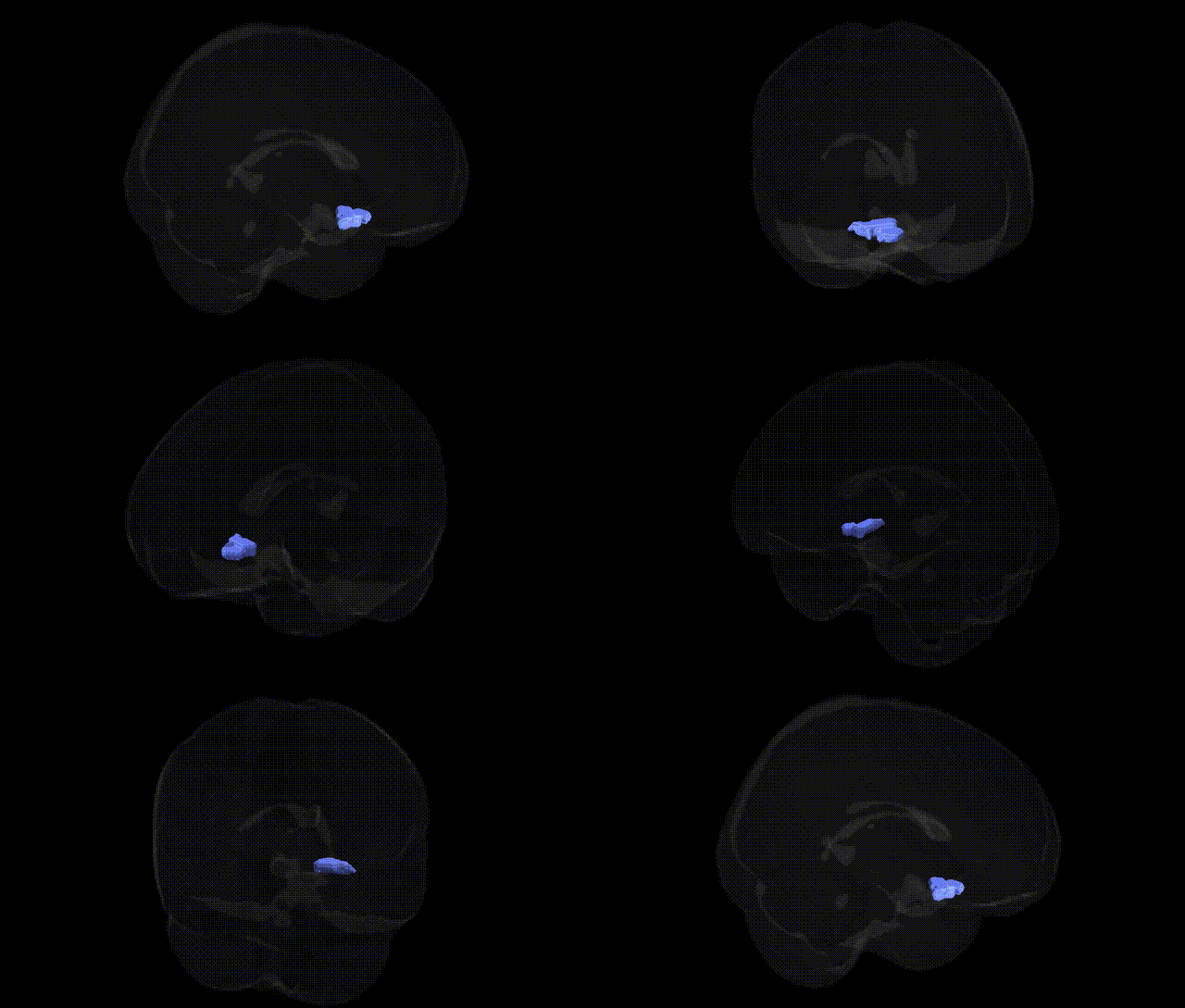
The bilateral Left Cerebrum.Frontal Lobe.Subcallosal Gyrus.White Matter corresponds to the deep frontal lobe fibers underlying the subcallosal area, a ventromedial region situated inferior to the genu of the corpus callosum and adjacent to the septal region and basal forebrain. Although the Talairach label refers specifically to white matter, this zone lies beneath the cortical subcallosal gyrus, which is part of the limbic system and closely connected with the cingulate cortex, orbitofrontal cortex, hypothalamus, and other structures involved in mood regulation, autonomic control, and reward processing. Functionally, the subcallosal region and its white matter tracts participate in fronto-limbic networks that integrate emotional and visceral information with higher-order executive functions, and the adjacent subcallosal cingulate white matter has been a target in neuromodulation approaches for treatment-resistant depression. There is no direct Wikipedia article for this specific Talairach white matter label; a closely related structure is the subcallosal cingulate region: Subcallosal cingulate.
The subcallosal (subgenual) frontal white matter beneath the corpus callosum, corresponding to the Left Cerebrum.Frontal Lobe.Subcallosal Gyrus.White Matter in the Talairach 2 mm atlas, has been repeatedly implicated in genetic studies of mood and related psychiatric traits, largely through its overlap with the subgenual anterior cingulate/medial orbitofrontal circuitry. Structural and functional variation in this region is influenced by common variants in genes regulating monoaminergic signaling (e.g., SLC6A4/5-HTTLPR, HTR1A/2A), neurotrophic and plasticity pathways (BDNF Val66Met), and stress-response systems (FKBP5, CRHR1), which have been associated with depression, anxiety, and stress-related disorders in imaging genetics and candidate gene studies. Large-scale GWAS of major depressive disorder, bipolar disorder, and neuroticism (e.g., Psychiatric Genomics Consortium, UK Biobank) have identified loci near genes involved in synaptic function and neuronal development (such as PCLO, CACNA1C, and others) that show downstream effects on subcallosal/ventromedial prefrontal morphology and connectivity, though these effects are typically polygenic and small. Variants in genes modulating glutamatergic and GABAergic transmission, myelination, and axonal integrity (including genes related to oligodendrocyte function) have been linked to white-matter microstructural changes (e.g., fractional anisotropy) in tracts traversing this subcallosal region in diffusion MRI GWAS, which in turn relate to risk for mood disorders, suicidality, and cognitive-affective traits. Overall, genetic influences on this subcallosal frontal white matter are highly polygenic, embedded within broader fronto-limbic and cingulo-orbitofrontal networks, and most robust associations arise from large GWAS of psychiatric phenotypes rather than region-specific locus discovery.
Overview generated by GPT-4o (2026).
Region ID: 290
Hemisphere: bilateral
Atlas: Talairach labels 2mm
Full Quality Version: Download MP4
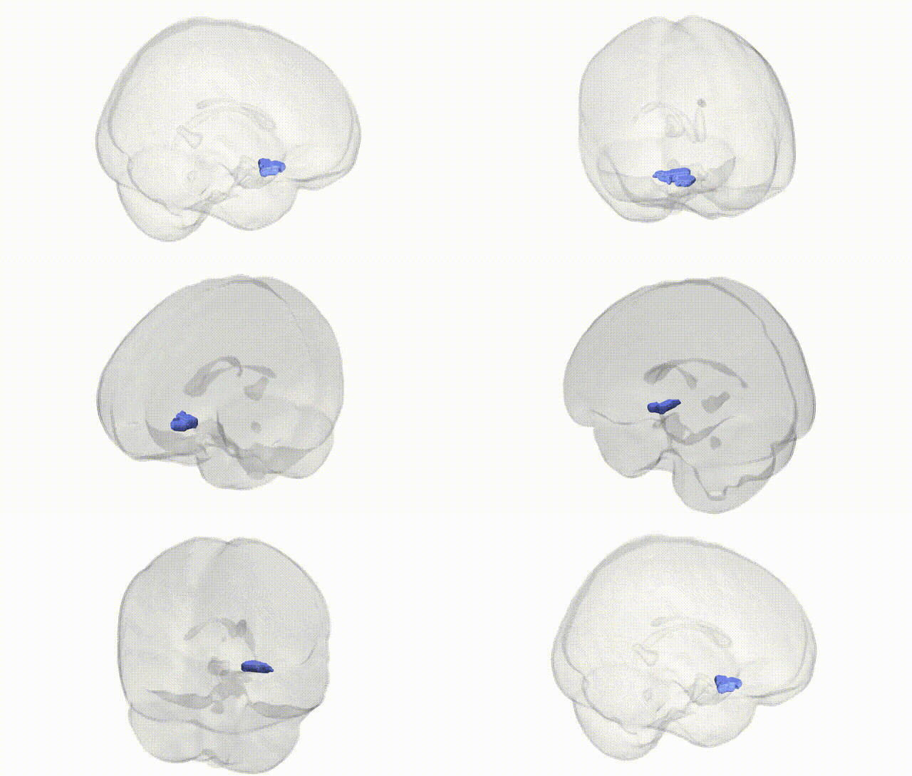
Full Quality Version: Download MP4
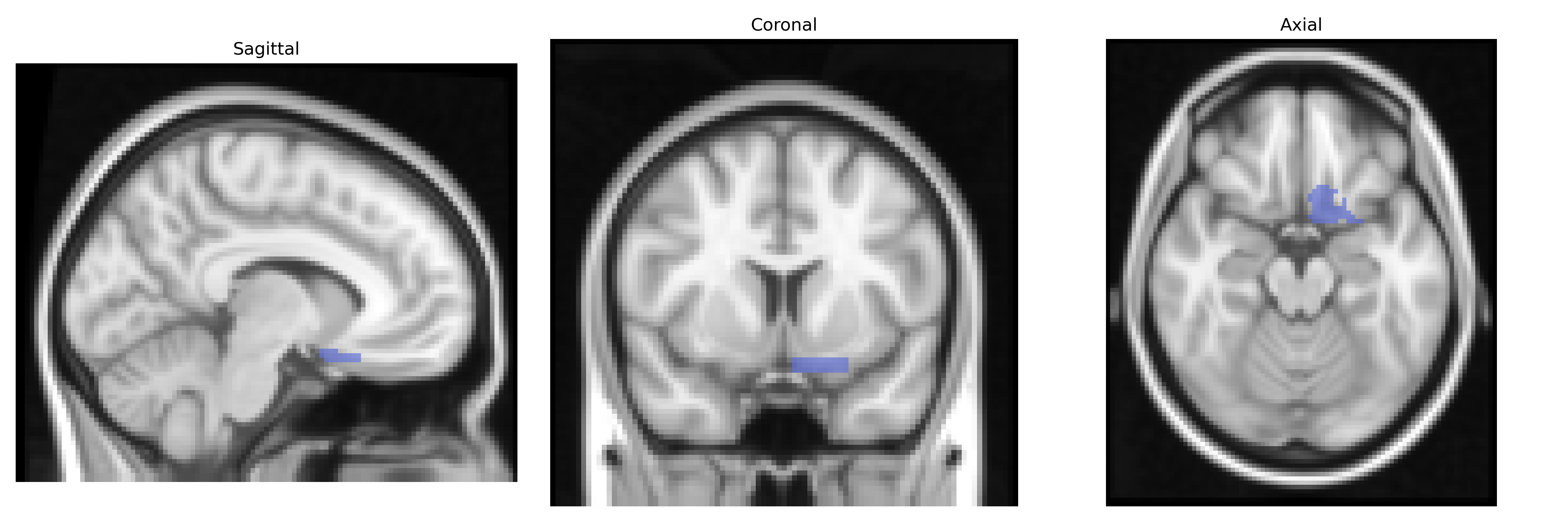
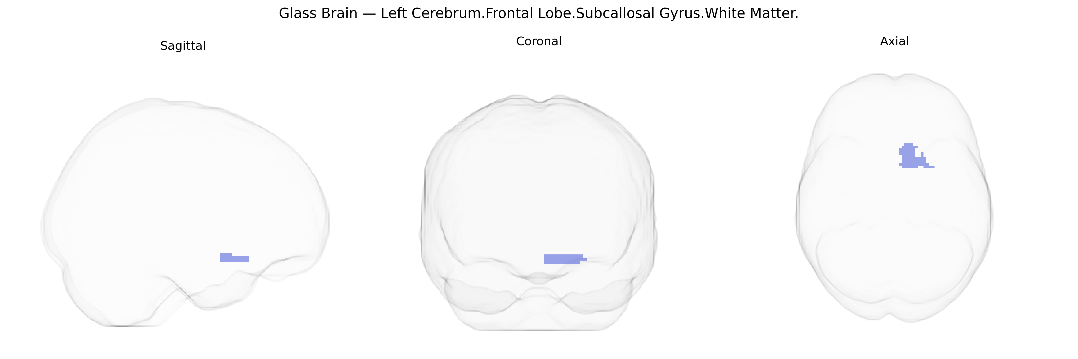
Wali Sidiqyar*, Gaurav Rudravaram*, Elyssa M. McMaster, Trent M. Schwartz, Adam M. Saunders, Kurt G. Schilling, Bennett A. Landman "Introducing SPINS: A Shared Public Visualization Library of Neuroanatomical Structures." Medical Imaging with Deep Learning- short paper
This resource is licensed under CC0 1.0 Universal (Public Domain).