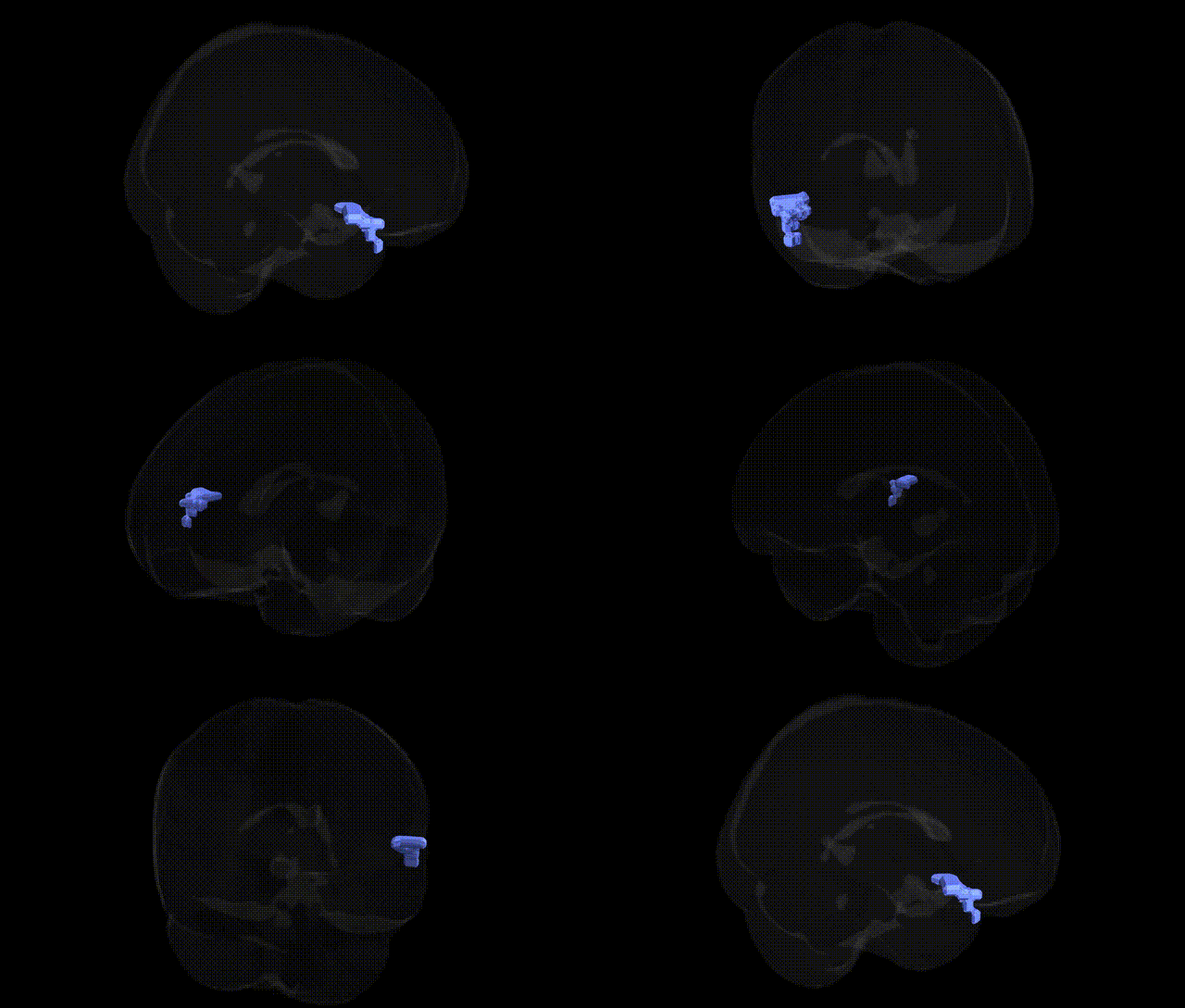
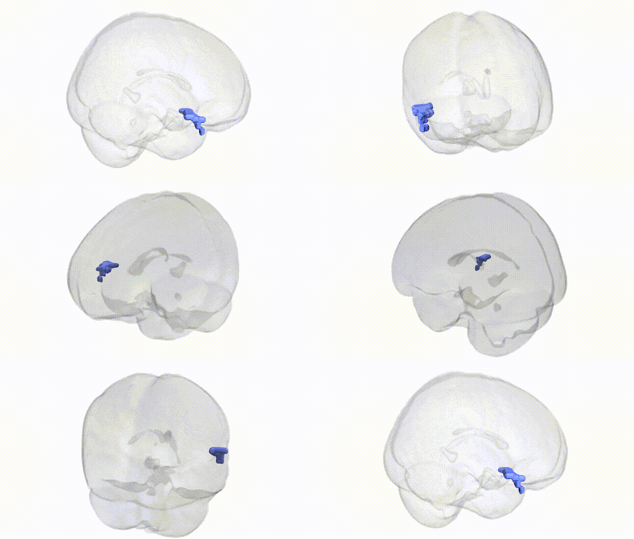
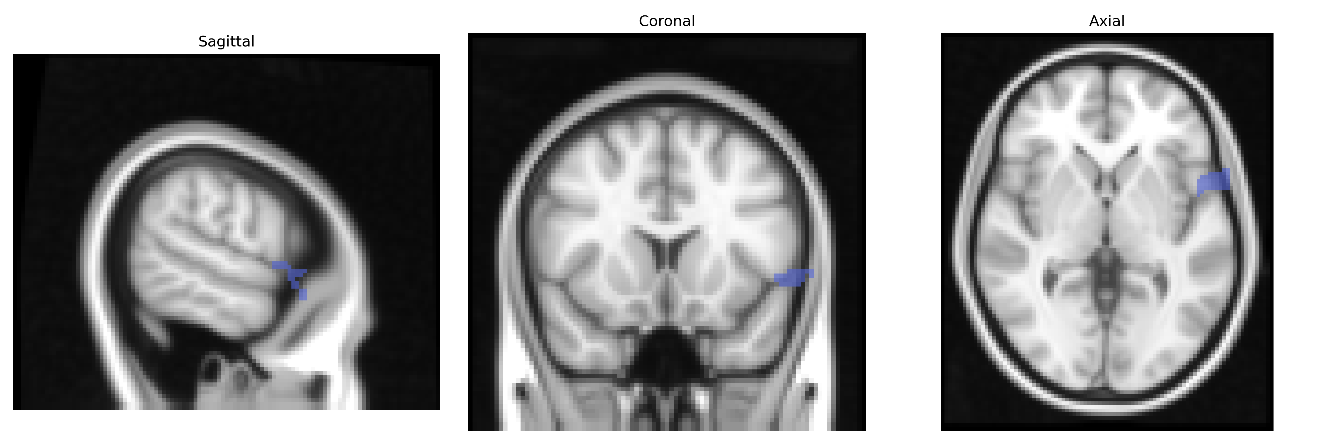
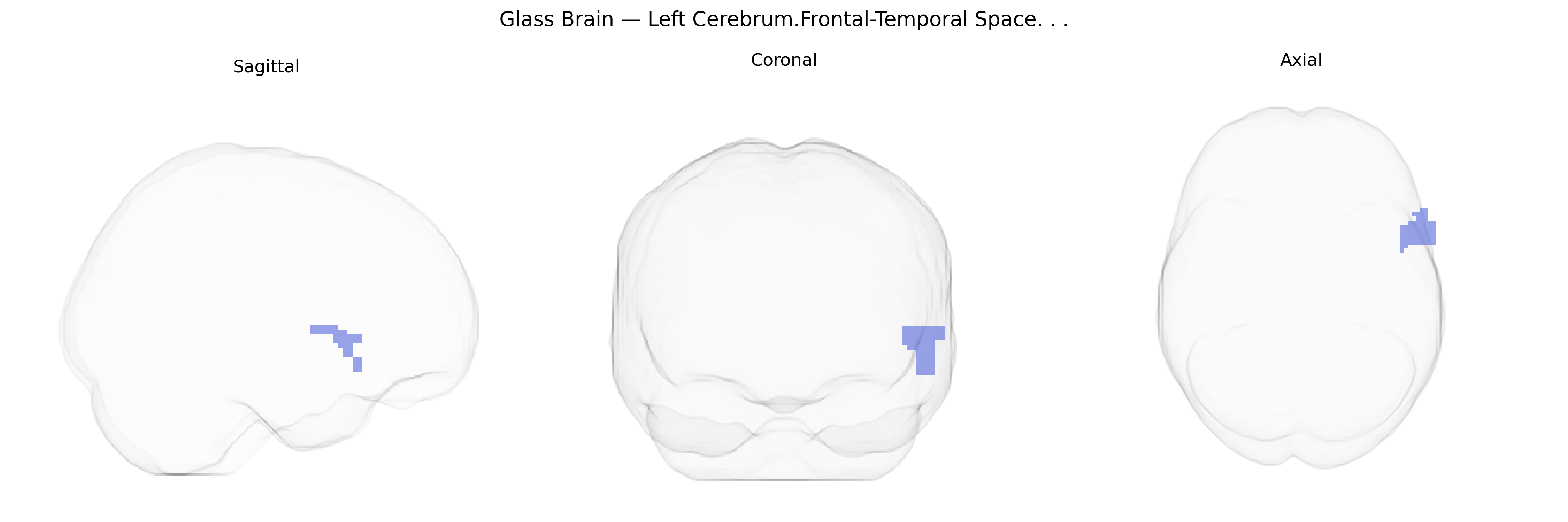
The bilateral Left Cerebrum.Frontal-Temporal Space, as designated in the Talairach 2 mm atlas, refers to a composite anatomical and functional territory spanning the frontal and temporal lobes of the left hemisphere, encompassing association cortices involved in higher-order cognition, language processing, working memory, and socio-emotional integration. This region includes portions of the inferior and middle frontal gyri, prefrontal association areas, and adjacent temporal structures such as the superior and middle temporal gyri, which collectively participate in speech production and comprehension, semantic processing, and multimodal sensory integration. Functionally, this frontal-temporal circuitry is critical for executive control over language, regulation of behavior, and integration of auditory and visual information into meaningful representations. There is no direct Wikipedia article for “Left Cerebrum.Frontal-Temporal Space”; a closely related structure is the Frontal lobe and the Temporal lobe.
The bilateral Left Cerebrum.Frontal-Temporal Space in the Talairach 2 mm atlas spans portions of the inferior frontal and anterior temporal cortices, regions repeatedly implicated in genetic studies of language, social cognition, and psychiatric risk. GWAS and imaging-genetics work have shown that common variants influencing cortical thickness, surface area, and gyrification in these frontal–temporal territories often map to genes involved in neurodevelopment, synaptic function, and axon guidance (e.g., CNTNAP2, FOXP2, DCC, and several glutamatergic and GABAergic pathway genes), with downstream effects on language ability, verbal intelligence, and social communication. Polygenic risk scores for autism spectrum disorder, schizophrenia, and major depressive disorder have been associated with structural and functional alterations in adjacent inferior frontal and superior temporal regions, including reduced gray matter volume or atypical activation during speech, emotion, and theory-of-mind tasks. In addition, large-scale consortia such as ENIGMA and UK Biobank have reported genome-wide significant loci for frontal and temporal lobe morphology that overlap risk loci for cognitive performance, educational attainment, and neurodegenerative disorders (e.g., Alzheimer’s disease), suggesting shared genetic architecture between this frontal–temporal space and diverse behavioral and clinical phenotypes. While no GWAS targets the Talairach “Frontal-Temporal Space” label specifically, convergent imaging-genetic evidence indicates that the bilateral frontal–temporal transition zone is a key substrate through which neurodevelopmental and psychiatric risk variants exert effects on language, cognition, and socio-emotional processing.
Overview generated by GPT-4o (2026).
Region ID: 394
Hemisphere: bilateral
Atlas: Talairach labels 2mm
Full Quality Version: Download MP4
Full Quality Version: Download MP4
Wali Sidiqyar*, Gaurav Rudravaram*, Elyssa M. McMaster, Trent M. Schwartz, Adam M. Saunders, Kurt G. Schilling, Bennett A. Landman "Introducing SPINS: A Shared Public Visualization Library of Neuroanatomical Structures." Medical Imaging with Deep Learning- short paper
This resource is licensed under CC0 1.0 Universal (Public Domain).