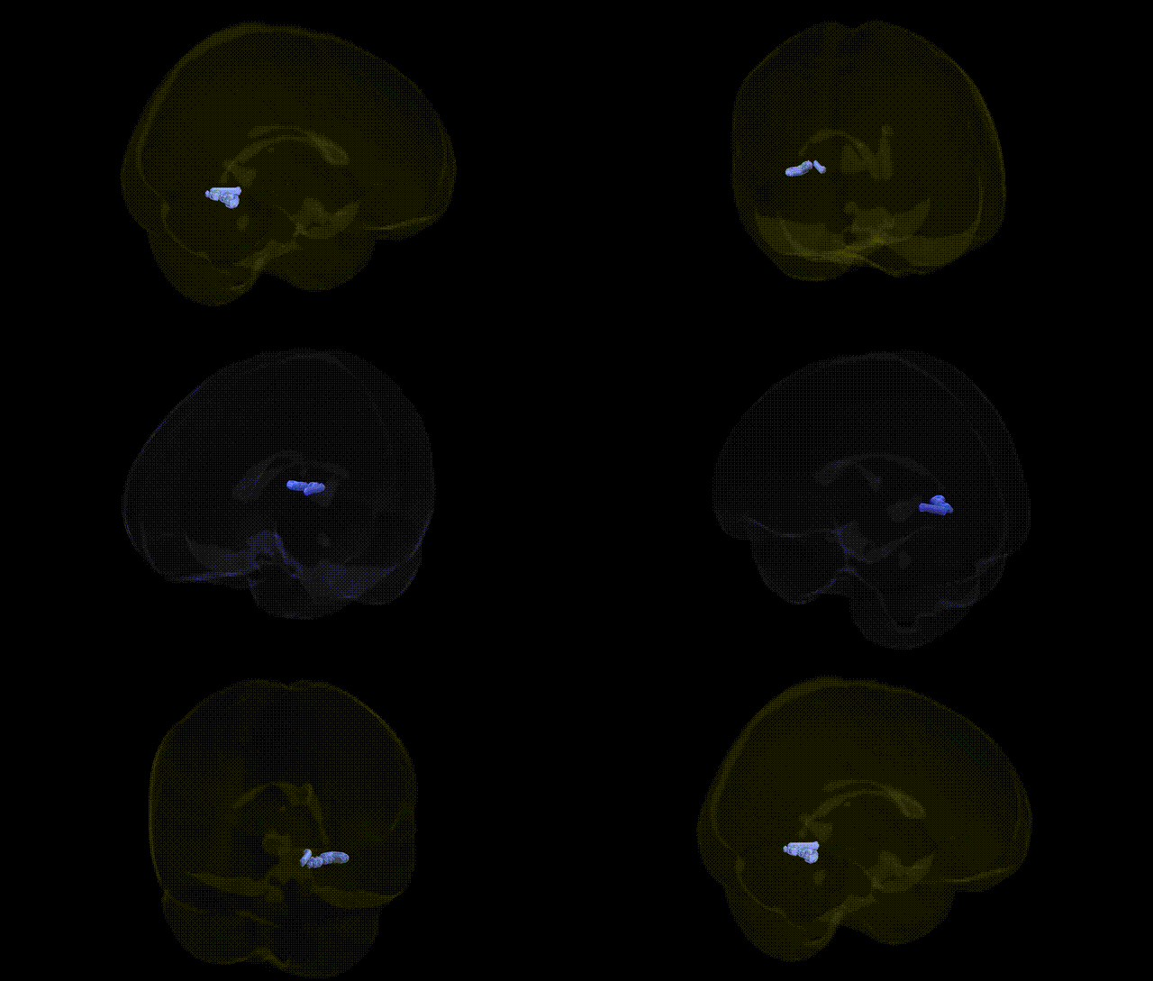
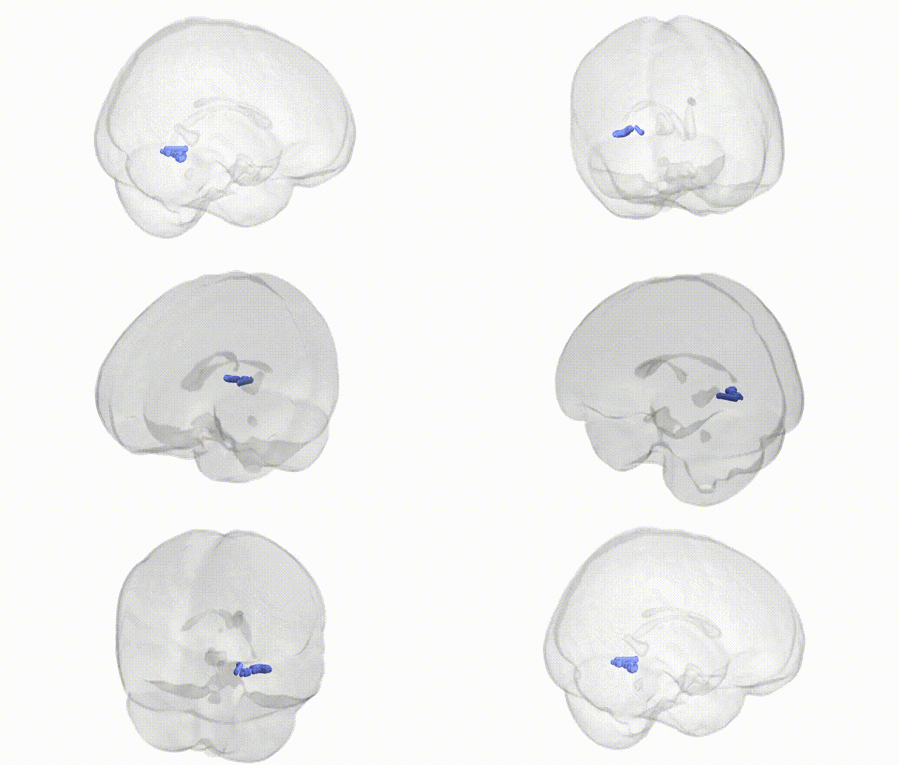
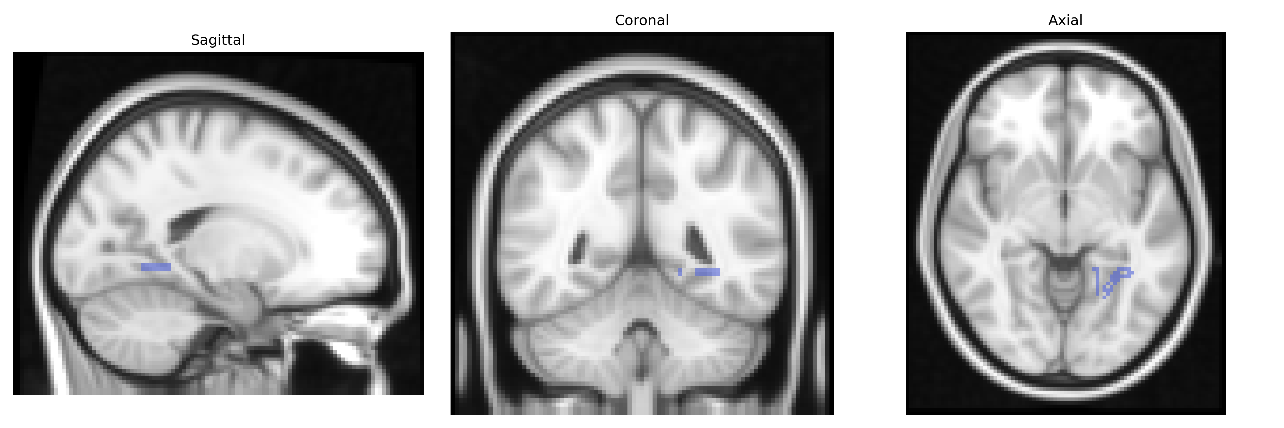
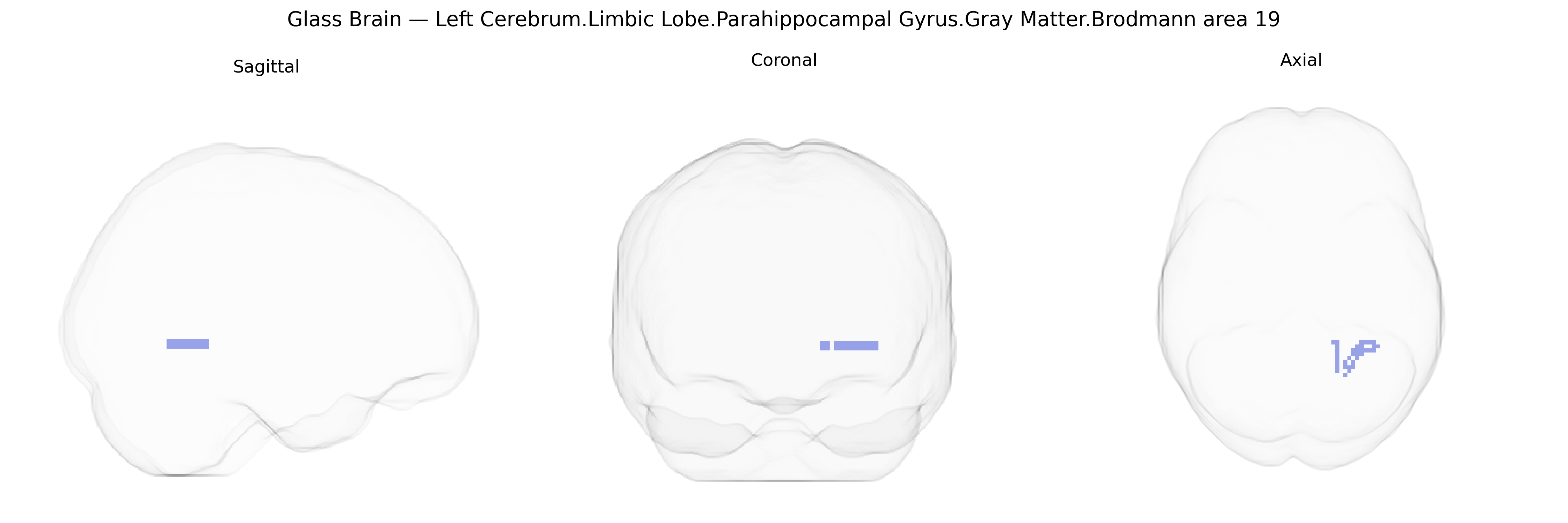
The bilateral Left Cerebrum.Limbic Lobe.Parahippocampal Gyrus.Gray Matter.Brodmann area 19 region in the Talairach labels 2 mm Atlas represents cortical tissue located at the interface of limbic and occipitotemporal territories, where the parahippocampal gyrus blends with adjacent visual association cortex. Brodmann area 19 is part of the extrastriate visual cortex, receiving processed input from primary and secondary visual areas and participating in higher-order visual integration, including complex pattern, scene, and spatial-context processing. In this parahippocampal sector, BA19 is functionally linked to visuospatial memory and contextual association, contributing to the encoding of environmental layout and the visual aspects of episodic memory. Anatomically, it lies deep on the medial temporal–occipital surface, adjacent to the hippocampal formation and retrosplenial cortex, and forms part of the broader network underlying visually guided navigation and scene recognition. There is no direct link for this exact composite region; a closely related structure is the Parahippocampal gyrus.
The bilateral parahippocampal gyrus gray matter region overlapping Brodmann area 19 (as defined in the Talairach 2 mm atlas) lies at the intersection of medial temporal and occipitotemporal cortex and has been implicated in genetic studies of memory, spatial navigation, and visual scene processing, as well as in several neuropsychiatric and neurodegenerative traits. GWAS of brain structure (e.g., ENIGMA and UK Biobank imaging genetics) have identified associations between parahippocampal and adjacent occipitotemporal cortical thickness/volume and common variants in genes involved in synaptic function and neurodevelopment, notably in and around APOE and TOMM40 (linked to Alzheimer’s disease risk and medial temporal atrophy), as well as loci near genes such as KCNK2, WNT3, and MAPT in broader temporal and occipital cortical measures. Imaging‑genetic studies of schizophrenia, major depression, and bipolar disorder frequently report genetically influenced volume or activation changes in the parahippocampal/lingual/BA19 region, including effects of risk variants in CACNA1C, ZNF804A, and other polygenic load indices on medial temporal and occipitotemporal activation during memory and emotional tasks. GWAS of PTSD, anxiety, and stress-related disorders have linked polygenic risk to altered parahippocampal and related limbic activation and structure, although most associations are at the level of networks rather than a single Brodmann area. In addition, genetic influences on scene- and face-selective visual cortices, including parahippocampal and fusiform/BA19 territories, have been observed in twin and SNP-heritability studies, with significant heritability of cortical thickness, surface area, and functional responses in this region, but specific single-locus GWAS hits uniquely and robustly tied to bilateral BA19 parahippocampal gray matter remain limited and are typically reported as part of larger temporal–occipital or limbic cortical clusters rather than as an isolated region.
Overview generated by GPT-4o (2026).
Region ID: 425
Hemisphere: bilateral
Atlas: Talairach labels 2mm
Full Quality Version: Download MP4
Full Quality Version: Download MP4
Wali Sidiqyar*, Gaurav Rudravaram*, Elyssa M. McMaster, Trent M. Schwartz, Adam M. Saunders, Kurt G. Schilling, Bennett A. Landman "Introducing SPINS: A Shared Public Visualization Library of Neuroanatomical Structures." Medical Imaging with Deep Learning- short paper
This resource is licensed under CC0 1.0 Universal (Public Domain).