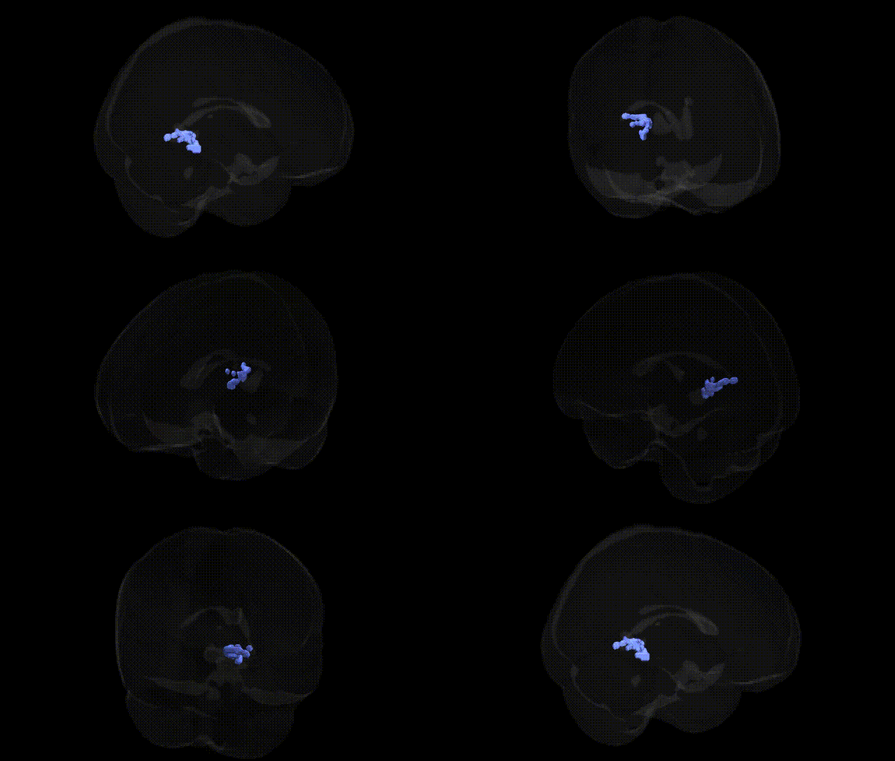
Bilateral Left Cerebrum Limbic Lobe Parahippocampal Gyrus Gray Matter Brodmann area 30 corresponds to a retrosplenial cortical region located in the medial temporal-limbic network, adjacent to the posterior cingulate and parahippocampal gyrus. This agranular cortex is implicated in episodic memory, spatial navigation, contextual association, and integration of visuospatial information with mnemonic processes, forming part of the broader default mode and memory-related circuitry. Functionally, BA30 participates in linking environmental cues with internal representations, contributing to scene processing and the consolidation and retrieval of autobiographical memories, and is interconnected with the hippocampal formation, posterior cingulate cortex, and other limbic structures. There is no direct link; see related area Retrosplenial cortex.
The bilateral parahippocampal gyrus gray matter in Brodmann area 30, a retrosplenial/medial limbic region involved in contextual memory, navigation, and emotional processing, has been implicated indirectly in genetic studies through imaging genetics and GWAS of brain structure and disease rather than through region-specific GWAS hits for the Talairach 2 mm BA30 label itself. Variants in genes associated with hippocampal–parahippocampal volume and cortical thickness—such as APOE (particularly ε4), CR1, BIN1, CLU, and other Alzheimer’s disease risk loci—have been linked to atrophy and altered activation in medial temporal and parahippocampal/retrosplenial areas in neuroimaging studies, often in the context of Alzheimer’s disease, mild cognitive impairment, and age-related memory decline. Large-scale GWAS of brain imaging phenotypes (e.g., ENIGMA, UK Biobank) have identified polygenic influences on medial temporal and limbic lobe volumes and thickness, including associations with loci near genes involved in synaptic function and neurodevelopment (e.g., BDNF, KIBRA/WFS1, and several novel loci), though these findings typically map to broader parahippocampal or medial temporal measures rather than narrowly to BA30. Imaging genetics work has also linked risk variants for schizophrenia (e.g., in CACNA1C, ZNF804A) and major depression (e.g., in SLC6A4, FKBP5) to functional and structural differences in medial limbic circuitry including parahippocampal and retrosplenial regions, supporting a role for this area in genetic vulnerability to psychosis and mood disorders. Additionally, GWAS of spatial navigation, memory performance, and PTSD have implicated loci that, in follow‑up imaging studies, show altered activation or connectivity in parahippocampal and adjacent limbic regions. Overall, genetic associations to Brodmann area 30 per se are inferred from broader medial temporal/limbic imaging phenotypes, with convergent evidence highlighting Alzheimer’s disease and cognitive aging loci, schizophrenia and depression risk genes, and memory- and navigation-related variants as key contributors to structural and functional variation in this parahippocampal/retrosplenial region.
Overview generated by GPT-4o (2026).
Region ID: 429
Hemisphere: bilateral
Atlas: Talairach labels 2mm
Full Quality Version: Download MP4
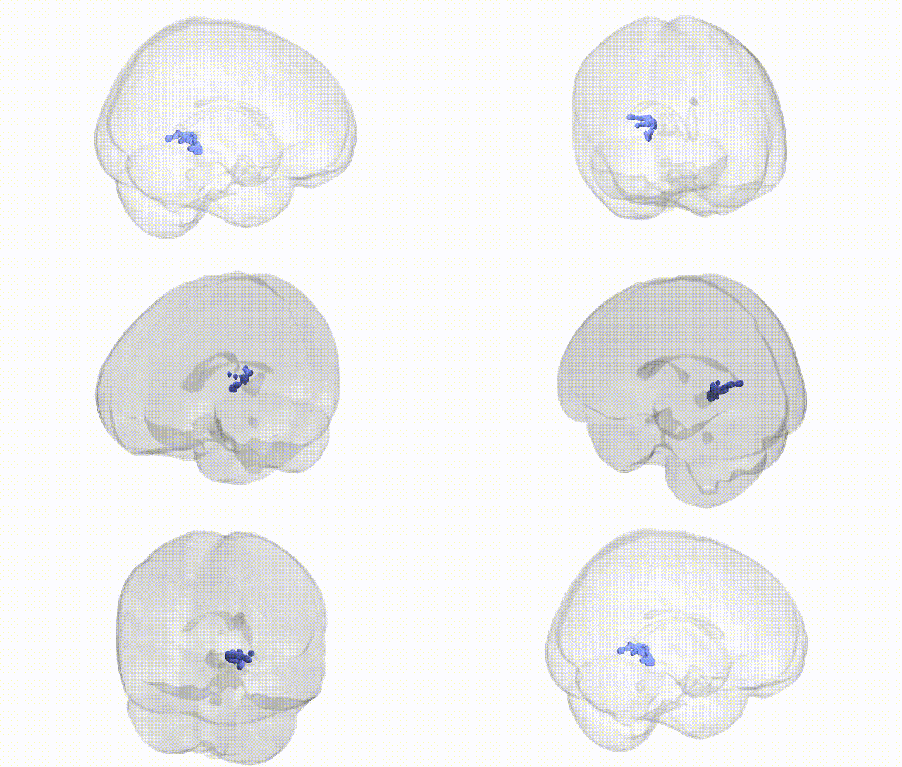
Full Quality Version: Download MP4
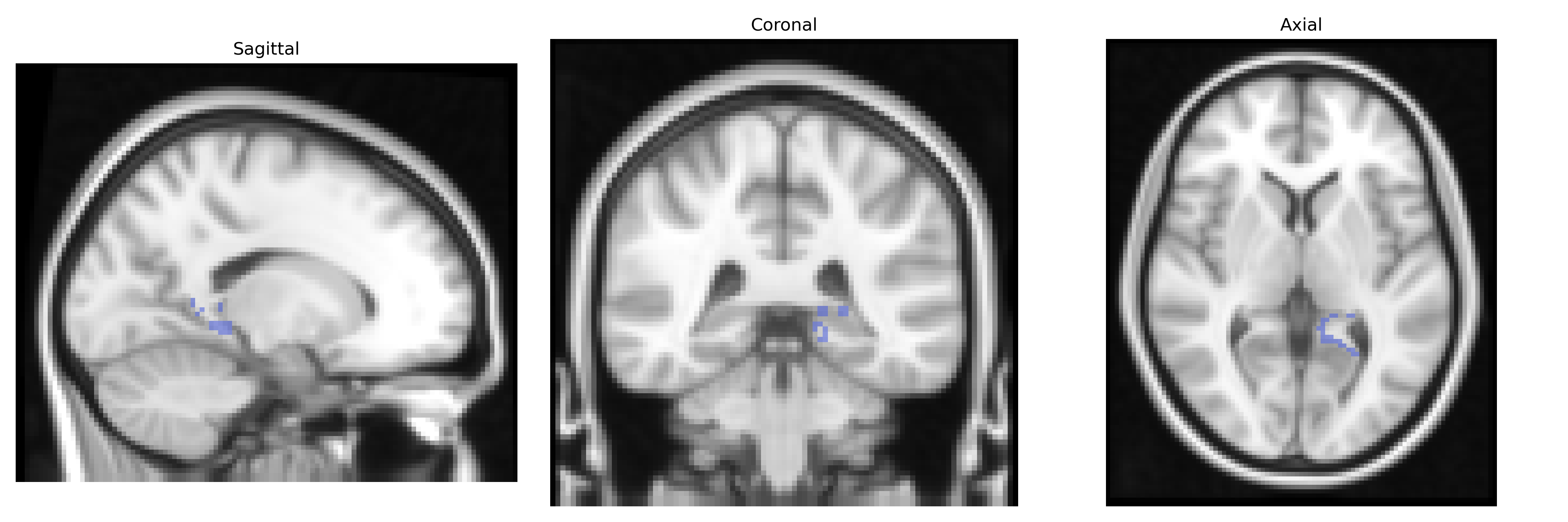
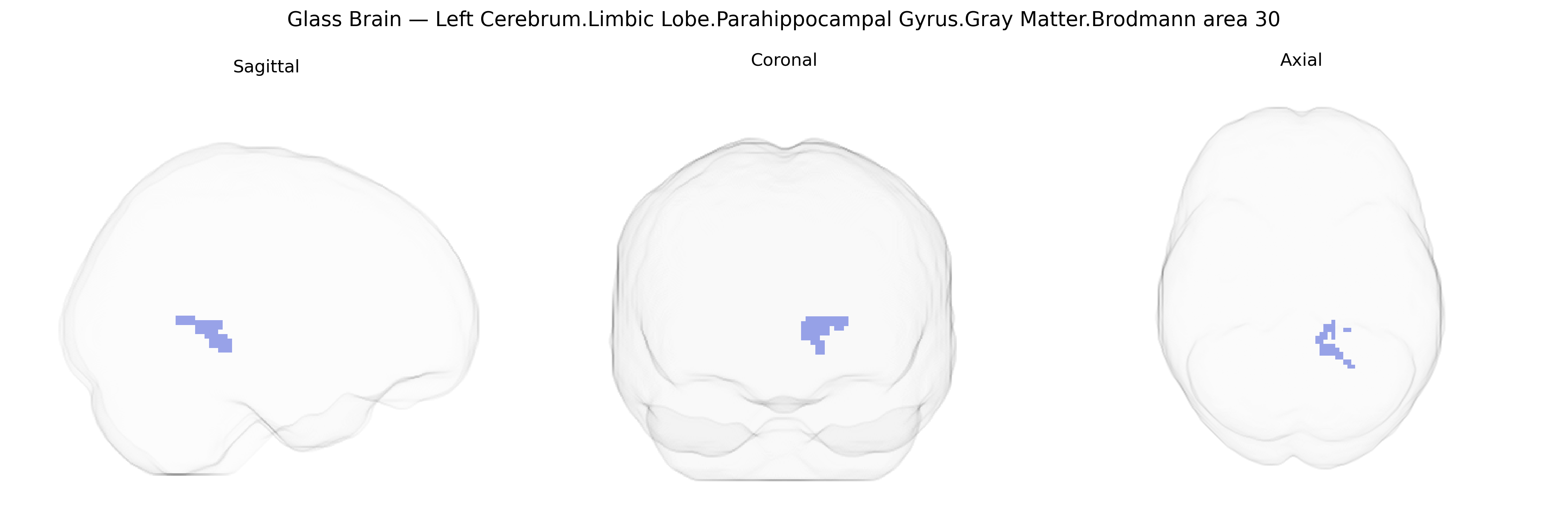
Wali Sidiqyar*, Gaurav Rudravaram*, Elyssa M. McMaster, Trent M. Schwartz, Adam M. Saunders, Kurt G. Schilling, Bennett A. Landman "Introducing SPINS: A Shared Public Visualization Library of Neuroanatomical Structures." Medical Imaging with Deep Learning- short paper
This resource is licensed under CC0 1.0 Universal (Public Domain).