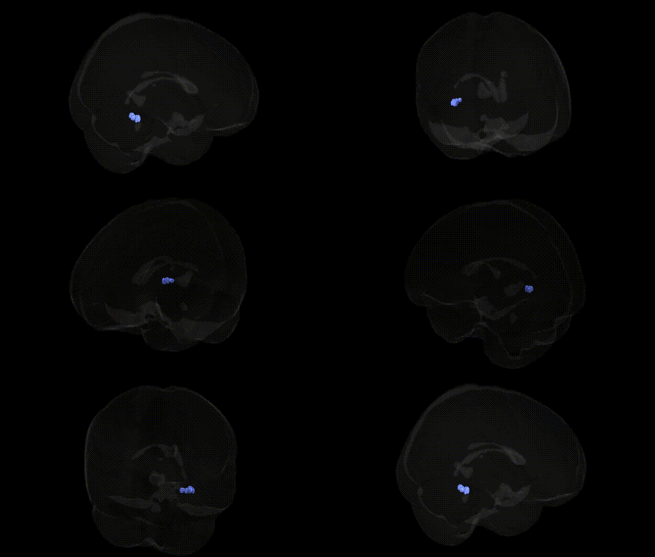
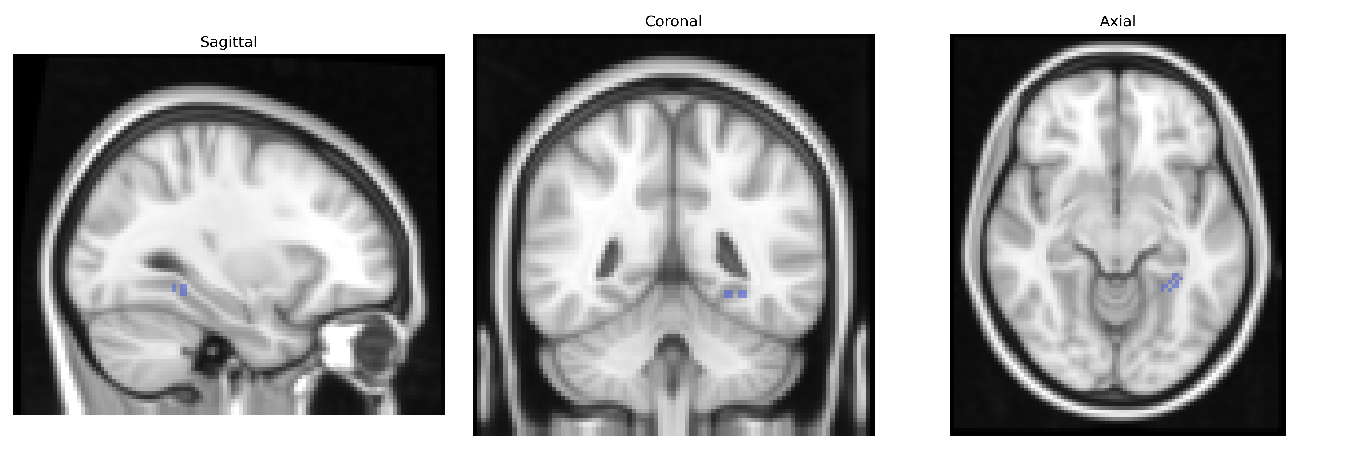
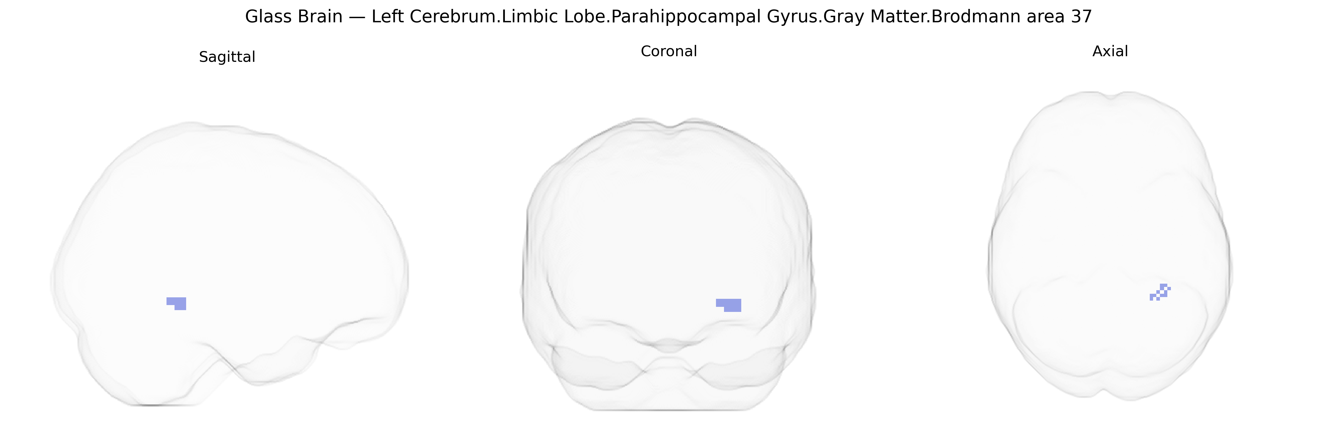
The bilateral Left Cerebrum.Limbic Lobe.Parahippocampal Gyrus.Gray Matter.Brodmann area 37 corresponds to cortical territory in the posterior parahippocampal region extending into the fusiform/occipitotemporal cortex, a multimodal association area implicated in high-level visual processing and memory-related functions. Brodmann area 37 is classically associated with the recognition of complex visual stimuli, including objects and faces, and participates in semantic processing and the integration of visual information with stored mnemonic representations. Cytoarchitectonically, it is a heteromodal association cortex with relatively dense granular layer IV and well-developed supragranular layers, receiving input from primary and secondary visual areas and projecting to limbic and frontal regions. Functionally, this territory supports visual memory encoding and retrieval, contributes to the ventral “what” pathway, and shows strong involvement in language-related semantic networks as well as category-specific processing of visual stimuli. There is no direct Wikipedia article for this exact composite region; a closely related structure is Brodmann area 37.
The parahippocampal gyrus (BA37, ventral temporal/occipitotemporal cortex) has been repeatedly implicated in genetic studies of memory, language, and neuropsychiatric and neurodegenerative disorders, although GWAS typically reference functional or structural measures rather than Brodmann areas per se. Imaging genetics and GWAS of cortical thickness and surface area have linked common variants in loci such as KIAA0586, SEMA3A, DACT1, and genes involved in neurodevelopmental pathways to variation in temporal and occipitotemporal cortical morphology that includes BA37. Functionally, BA37 contributes to visual word and object recognition and semantic processing, and genetic associations affecting these traits (including variants near FOXP2, CNTNAP2, and KIAA0319 in dyslexia and language-related phenotypes) have been related to altered activation in this region in fMRI studies. In Alzheimer’s disease, GWAS-identified risk loci including APOE, BIN1, CLU, and CR1 are associated with parahippocampal and medial temporal atrophy and hypometabolism, and these changes extend into BA37 in advanced stages, correlating with episodic memory impairment. Schizophrenia and bipolar disorder GWAS signals (for example in CACNA1C, ZNF804A, and GRIN2A) have been associated with altered connectivity and activation in limbic–temporal networks encompassing the parahippocampal gyrus and BA37 during emotional and semantic tasks. Autism spectrum disorder and social cognition–related genetic variants (e.g., in NRXN1, SHANK3, and other synaptic genes) are linked to abnormal development and functional responses in temporal-limbic regions including BA37, particularly in face, object, and social perception paradigms. Overall, convergent evidence from GWAS of brain structure, cognition, and major neuropsychiatric and neurodegenerative disorders indicates that common genetic variation in synaptic, neurodevelopmental, and lipid/immune pathways exerts measurable influence on the morphology and function of the bilateral BA37 parahippocampal/occipitotemporal region.
Overview generated by GPT-4o (2026).
Region ID: 270
Hemisphere: bilateral
Atlas: Talairach labels 2mm
Full Quality Version: Download MP4
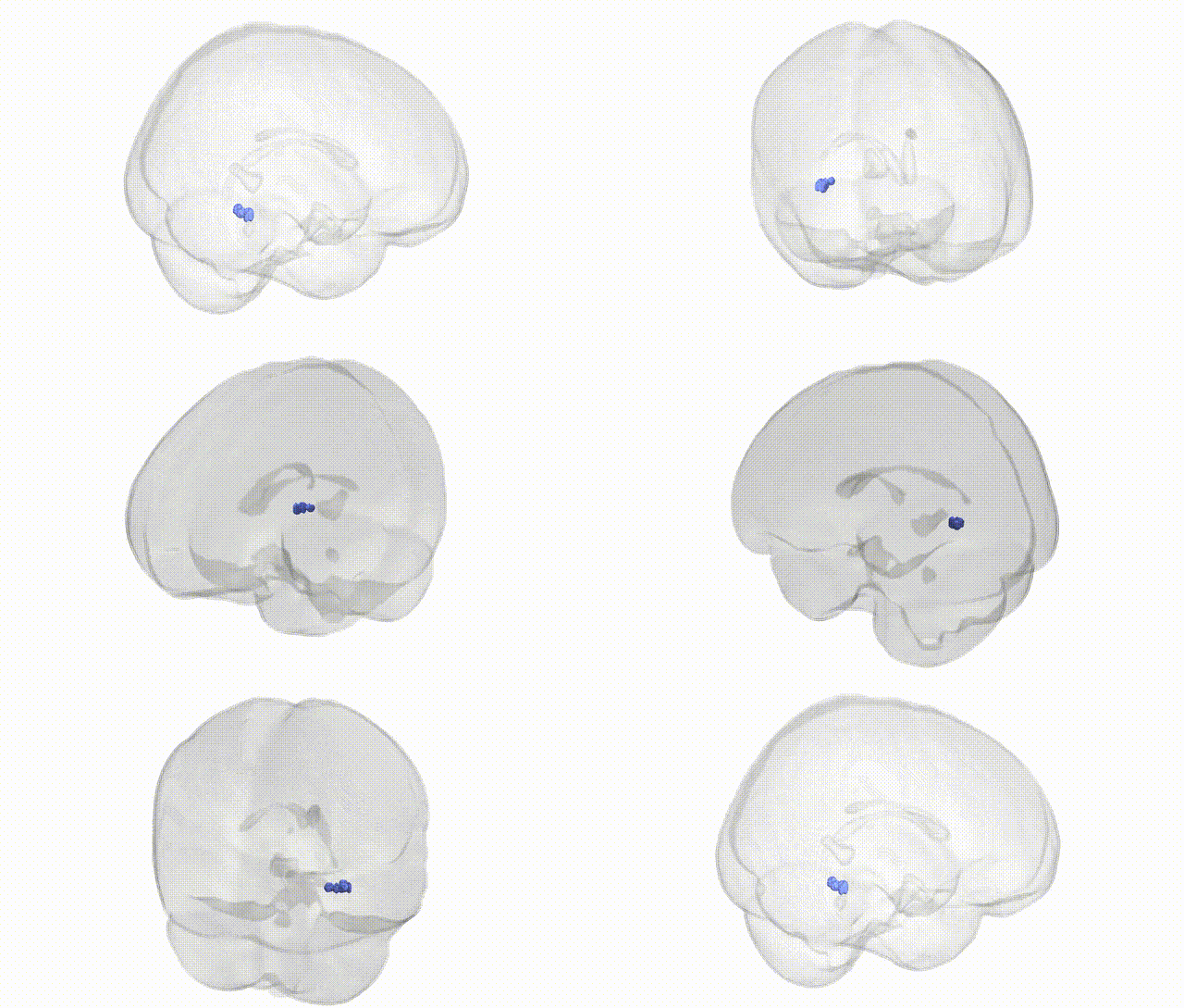
Full Quality Version: Download MP4
Wali Sidiqyar*, Gaurav Rudravaram*, Elyssa M. McMaster, Trent M. Schwartz, Adam M. Saunders, Kurt G. Schilling, Bennett A. Landman "Introducing SPINS: A Shared Public Visualization Library of Neuroanatomical Structures." Medical Imaging with Deep Learning- short paper
This resource is licensed under CC0 1.0 Universal (Public Domain).