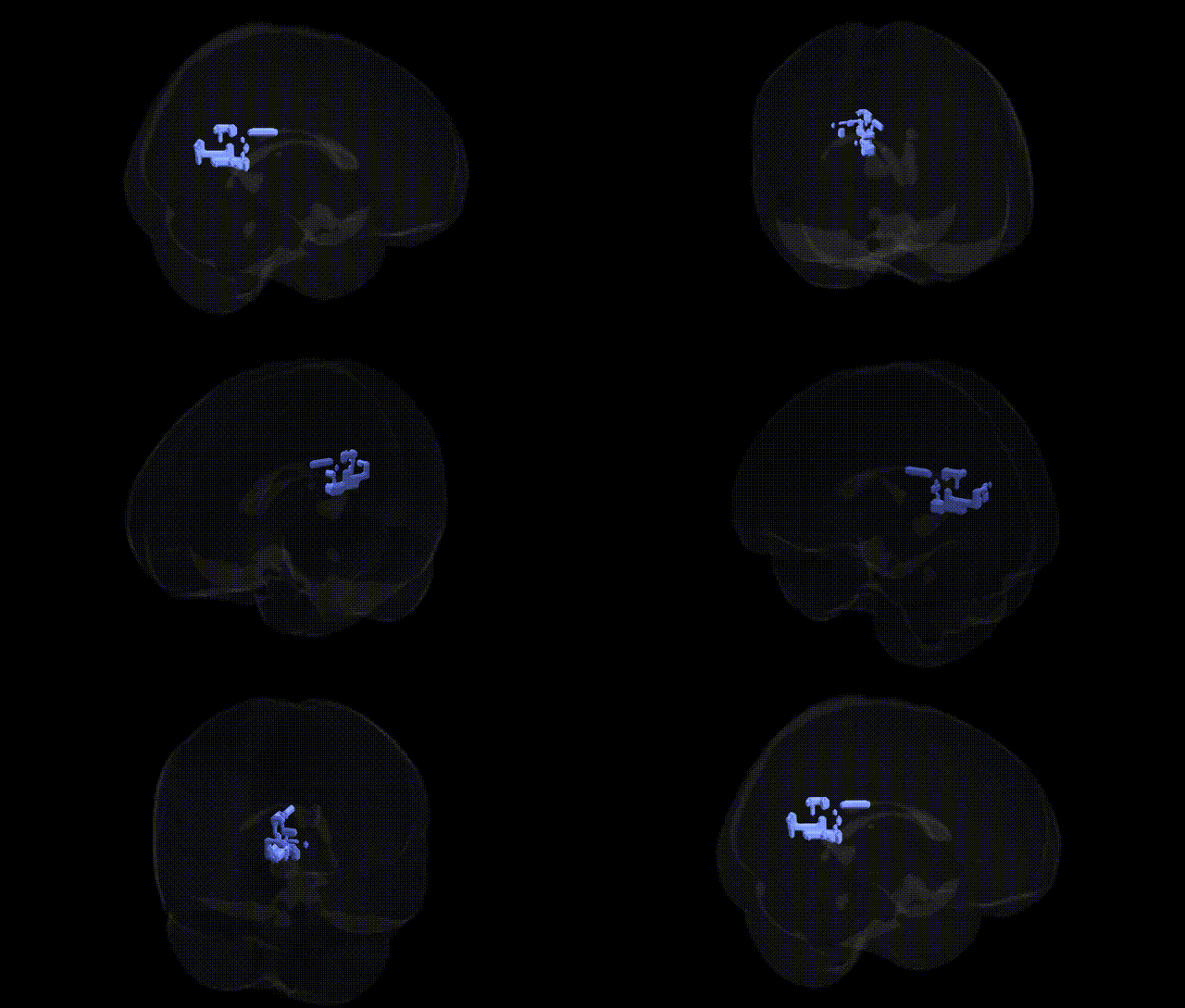
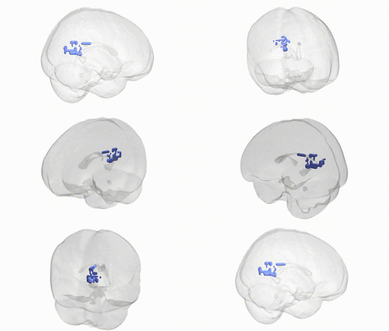
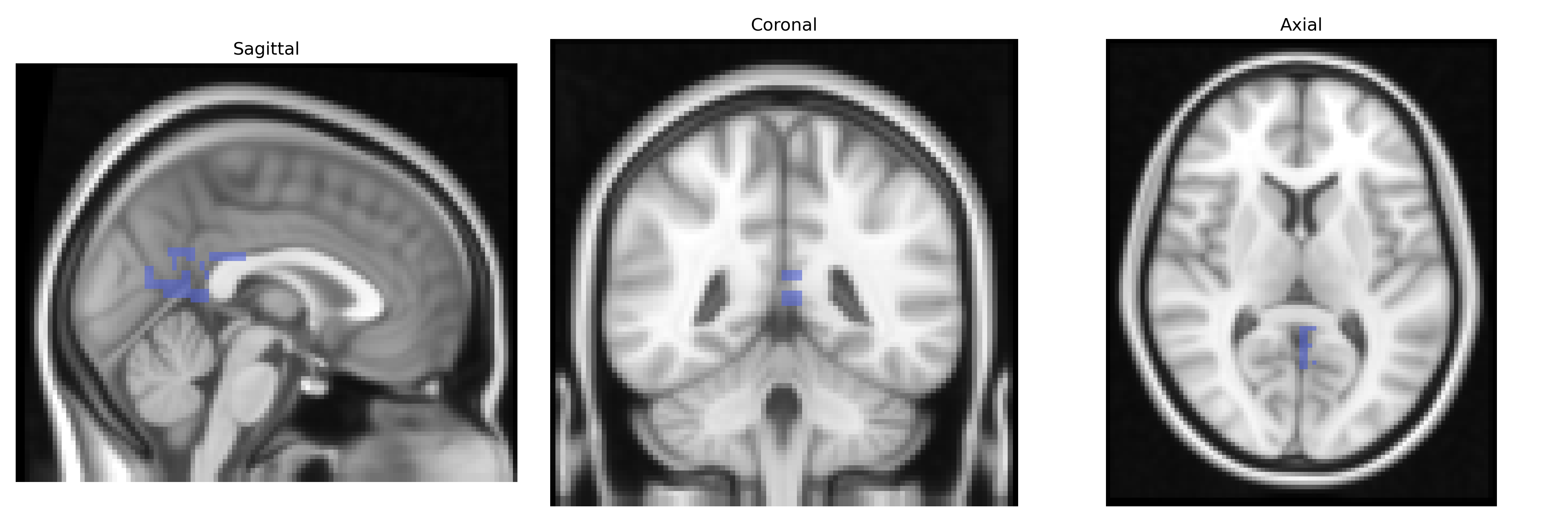
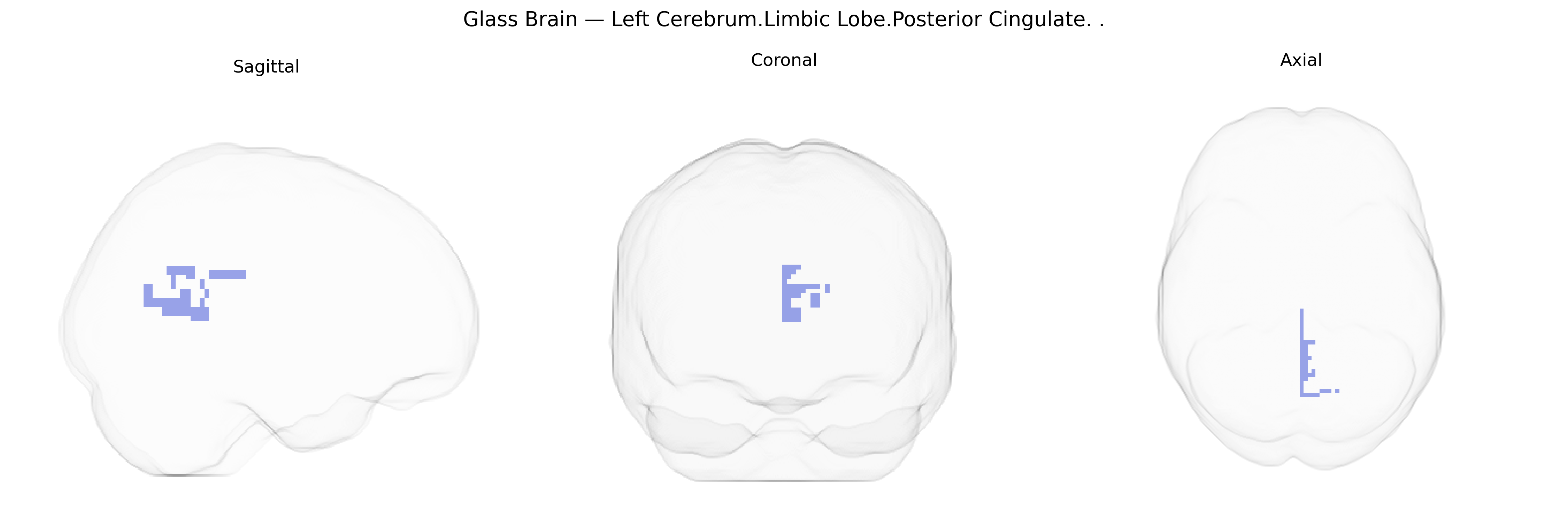
The bilateral Left Cerebrum.Limbic Lobe.Posterior Cingulate corresponds to the posterior portion of the cingulate gyrus, a key node of the limbic system located on the medial surface of the cerebral hemispheres, dorsal to the corpus callosum. This region is heavily interconnected with the precuneus, medial prefrontal cortex, and medial parietal areas, and is a core component of the default mode network. Functionally, the posterior cingulate is implicated in internally directed cognition, autobiographical and episodic memory retrieval, visuospatial orientation, and the integration of emotional and cognitive information. It also shows prominent resting-state metabolic activity and is often involved in neurodegenerative and psychiatric conditions, including Alzheimer’s disease and major depression.
Posterior cingulate cortex
The bilateral left posterior cingulate cortex (PCC), within the limbic lobe, has been repeatedly implicated in genetic studies of brain morphology, connectivity, and disease, although findings are typically reported at the regional or network level rather than specifically using Talairach 2 mm labels. Twin and family studies demonstrate high heritability of PCC volume and resting-state activity, especially as a hub of the default mode network, and GWAS of cortical thickness and surface area (e.g., ENIGMA and UK Biobank consortia) have identified variants in genes related to synaptic function, neurodevelopment, and myelination (such as those near MEF2C, DCC, and genes in Wnt and axon-guidance pathways) that influence PCC structure and connectivity. PCC morphology and functional connectivity show genetic correlations with cognitive traits including general intelligence, memory, and attentional control, and polygenic scores for educational attainment and cognitive performance have been associated with default mode network features that prominently involve the PCC. In psychiatric genetics, PCC structure and connectivity are altered in major depressive disorder, schizophrenia, bipolar disorder, and anxiety disorders, with GWAS-based imaging–genetics studies reporting that polygenic risk scores for these conditions relate to PCC volume or connectivity patterns, although no single locus is uniquely specific to this region. PCC involvement in Alzheimer’s disease is notable: APOE ε4 and other AD-risk variants are associated with metabolic and connectivity changes in the PCC and adjacent precuneus, and imaging–genetics studies link amyloid and tau pathology to PCC functional disruption. Additionally, GWAS of pain sensitivity, rumination, and self-referential processing—traits that engage the PCC—suggest overlapping genetic influences on limbic and default mode circuitry, reinforcing the PCC as a genetically influenced hub integrating emotion, memory, and internally directed cognition rather than a region with highly specific, isolated genetic associations.
Overview generated by GPT-4o (2026).
Region ID: 667
Hemisphere: bilateral
Atlas: Talairach labels 2mm
Full Quality Version: Download MP4
Full Quality Version: Download MP4
Wali Sidiqyar*, Gaurav Rudravaram*, Elyssa M. McMaster, Trent M. Schwartz, Adam M. Saunders, Kurt G. Schilling, Bennett A. Landman "Introducing SPINS: A Shared Public Visualization Library of Neuroanatomical Structures." Medical Imaging with Deep Learning- short paper
This resource is licensed under CC0 1.0 Universal (Public Domain).