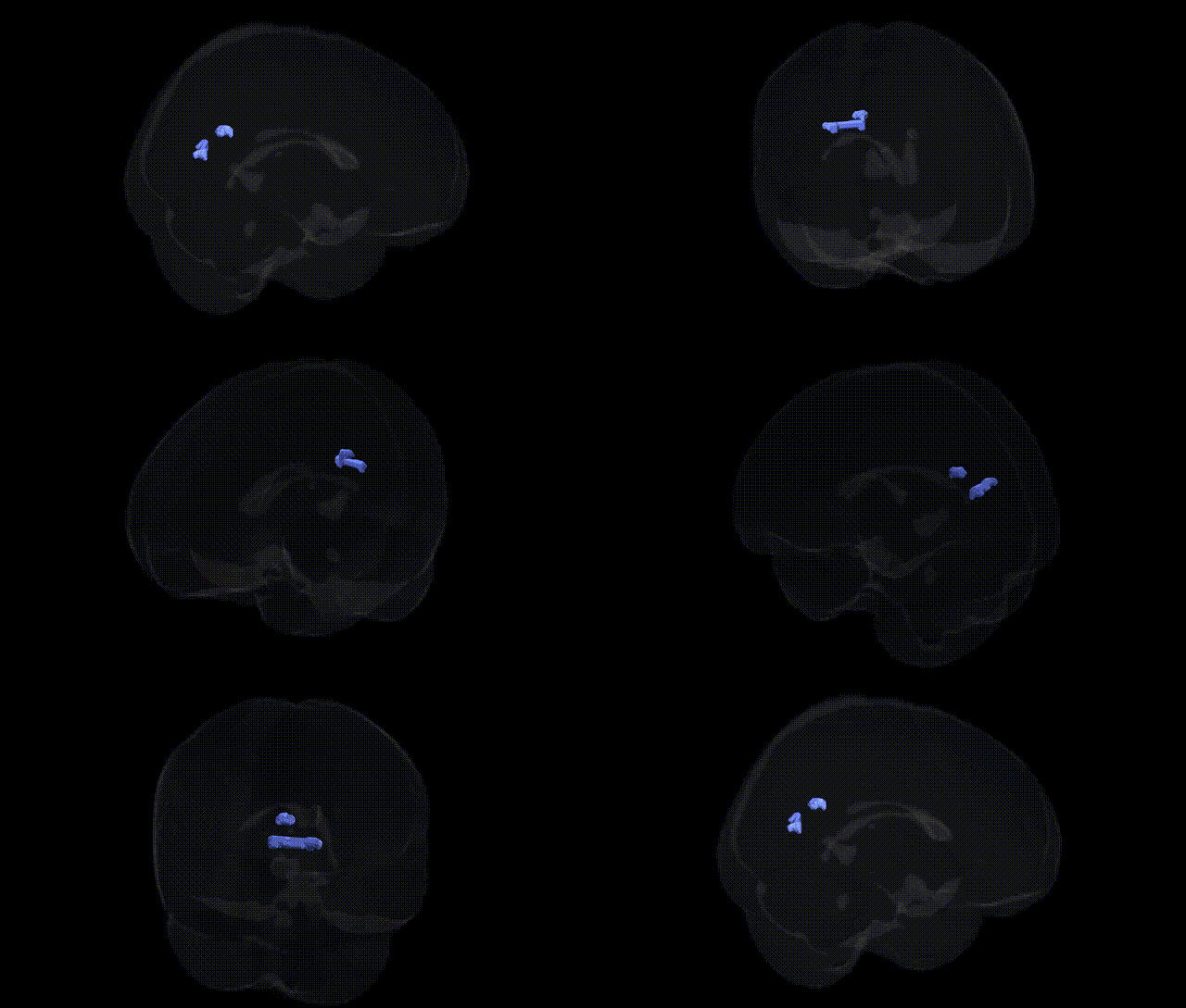
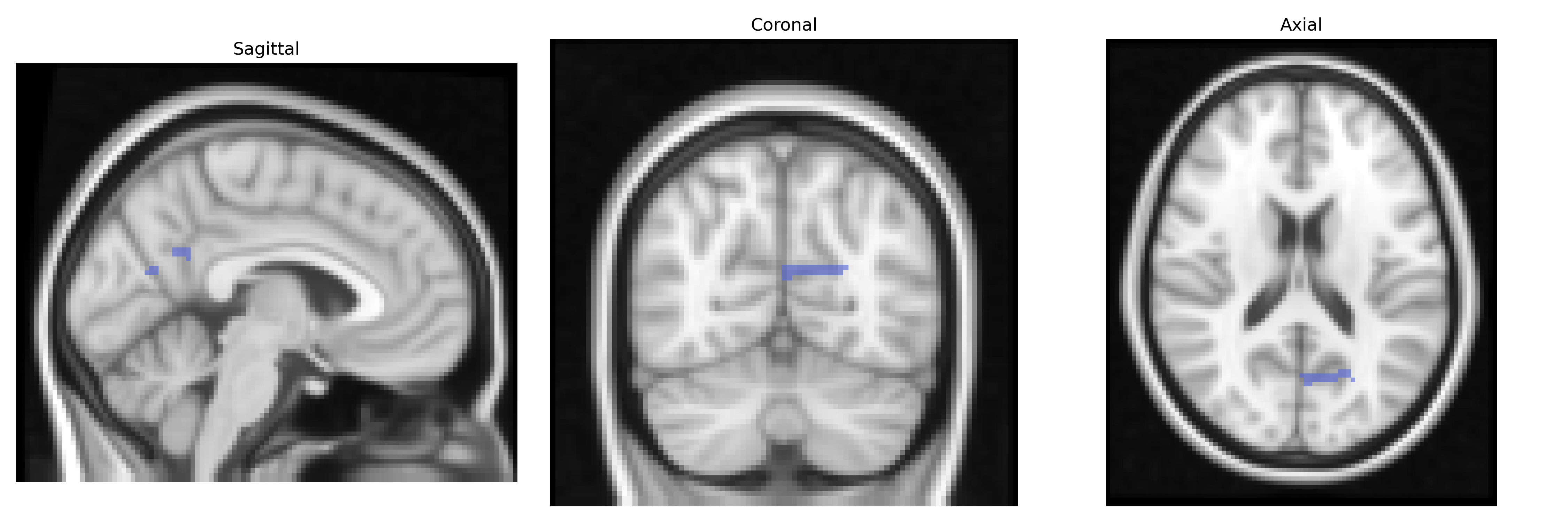
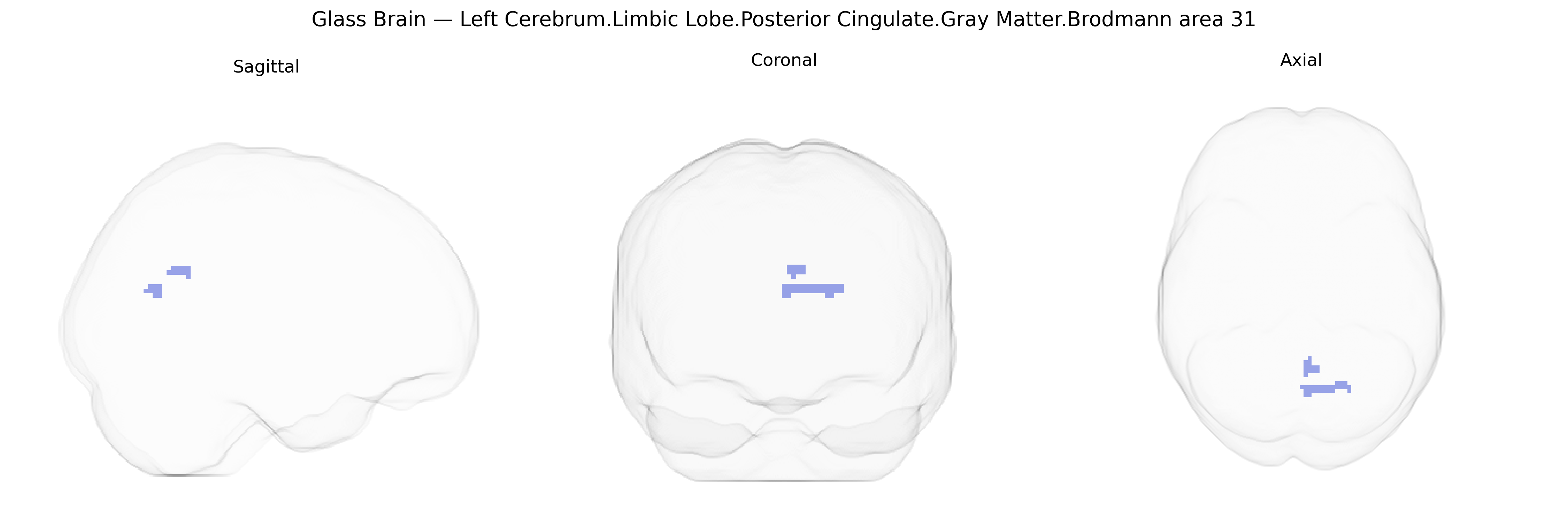
Bilateral Left Cerebrum Limbic Lobe Posterior Cingulate Gray Matter Brodmann area 31 corresponds to a subdivision of the posterior cingulate cortex situated in the medial parietal region of the limbic lobe, bordering the precuneus and retrosplenial cortex. This area is cytoarchitectonically defined by Brodmann’s criteria and is implicated in high-level associative and integrative functions, including internally directed cognition, autobiographical and episodic memory processing, and aspects of visuospatial and self-referential evaluation. Functionally, it forms a key node of the default mode network, exhibiting strong connectivity with medial prefrontal, hippocampal, and parietal regions, and shows characteristic metabolic and perfusion patterns that are altered in neurodegenerative and psychiatric disorders. The gray matter in Brodmann area 31 includes pyramidal neurons and interneurons organized in distinct cortical layers that support complex multimodal integration and modulation of attention and consciousness.
The posterior cingulate cortex (PCC), including Brodmann area 31 in the limbic lobe, shows multiple genetic associations primarily through imaging genetics and GWAS of brain structure and function rather than single-region sequencing studies. Structural MRI GWAS have linked PCC gray matter volume, thickness, and connectivity to variants in genes involved in synaptic function, neurodevelopment, and myelination, such as those near APOE (especially ε4), CLU, and CR1 in the context of Alzheimer’s disease, with PCC atrophy and hypometabolism serving as a hallmark of genetically driven AD risk. Polygenic risk scores for schizophrenia, major depressive disorder, bipolar disorder, and autism spectrum disorder have been associated with alterations in PCC connectivity within the default mode network, and several large-scale studies (e.g., ENIGMA, UK Biobank) report that higher psychiatric polygenic load correlates with structural and functional changes in this region. GWAS of cognitive traits (intelligence, memory, and attention) and personality (e.g., neuroticism) have identified variants in genes such as KIBRA (WWC1), CNTNAP2, and others that influence default mode network activity and PCC activation during memory and self-referential tasks, although these links are generally network-level rather than PCC-specific. Additionally, genetic variation in immune-related and microglial genes (e.g., in the complement pathway) has been implicated in PCC changes in neurodegenerative and neuroinflammatory conditions, while imaging genetics of mindfulness, rumination, and pain sensitivity highlight PCC as a key hub influenced by distributed polygenic architecture affecting limbic and default-mode systems. Overall, the bilateral BA31 PCC emerges as a genetically sensitive node within the default mode and limbic networks, mediating the impact of common risk alleles on cognition, affective regulation, and vulnerability to neuropsychiatric and neurodegenerative disorders.
Overview generated by GPT-4o (2026).
Region ID: 756
Hemisphere: bilateral
Atlas: Talairach labels 2mm
Full Quality Version: Download MP4
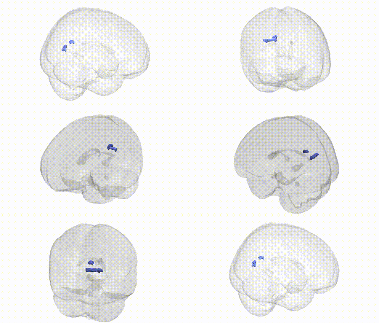
Full Quality Version: Download MP4
Wali Sidiqyar*, Gaurav Rudravaram*, Elyssa M. McMaster, Trent M. Schwartz, Adam M. Saunders, Kurt G. Schilling, Bennett A. Landman "Introducing SPINS: A Shared Public Visualization Library of Neuroanatomical Structures." Medical Imaging with Deep Learning- short paper
This resource is licensed under CC0 1.0 Universal (Public Domain).